
A patient's legs begin aching after a short walk. Sitting brings relief, but standing in a queue or walking through a shop quickly brings the symptoms back. An MRI may then show a narrowed spinal canal, yet that finding alone doesn't answer the most important question: is the narrowing causing the symptoms?
That distinction sits at the center of how is spinal stenosis diagnosed. A reliable diagnosis combines the symptom pattern, physical and neurological examination, and imaging that shows a plausible source of nerve compression. A scan supplies anatomy. The clinician must determine whether that anatomy matches the patient's lived experience.
Table of Contents
- Understanding How Spinal Stenosis Gets Diagnosed
- Taking a Detailed Patient History and Recognizing Symptom Patterns
- Physical Examination and Neurological Assessment Techniques
- Choosing the Right Diagnostic Imaging for Spinal Stenosis
- Electrodiagnostic Testing to Confirm Nerve Involvement
- Differentiating Spinal Stenosis from Other Causes of Leg Pain
- Next Steps and When to Seek Specialized Evaluation
Understanding How Spinal Stenosis Gets Diagnosed
Two patients can have nearly identical lumbar MRI findings. One develops leg pain, heaviness, numbness, or weakness while walking or standing and improves after sitting or bending forward. The other has no leg symptoms. The images may look similar, but only the first patient has a clinical pattern that supports symptomatic stenosis.
Diagnosis therefore uses a clinical-plus-imaging model. A clinical reference from the National Center for Biotechnology Information explains that clinicians bring together the patient's history, examination findings, and imaging evidence of narrowing. MRI is usually preferred because it displays the spinal canal, nerve roots, and surrounding soft tissues. CT or CT myelography can provide an alternative when MRI is unsuitable.
Why an abnormal scan may not explain pain
Imaging detects spinal narrowing frequently, including in people without related symptoms. A 2020 review of lumbar spinal stenosis reported imaging-based stenosis in 11% of asymptomatic people and 38% of the general population. This is why an MRI is not a useful screening test for every person with back discomfort.
A scan can show a narrowed canal, thickened tissue, or less space around a nerve. It cannot determine by itself how much pain the finding produces, whether another condition is responsible, or what happens to symptoms during walking and standing. Those questions require clinical assessment.
Practical rule: A convincing diagnosis requires a plausible match between the patient's symptoms, examination findings, and the level of narrowing shown on imaging.
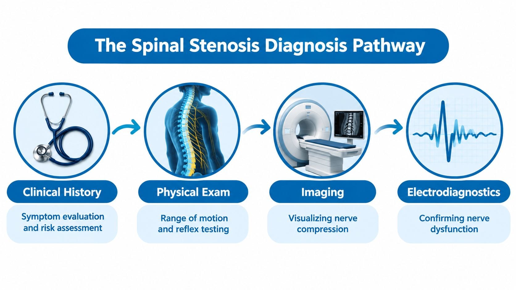
The diagnostic pathway commonly includes:
- Clinical history: The clinician assesses triggers, relieving positions, walking tolerance, pain distribution, numbness, and weakness.
- Physical examination: Strength, sensation, reflexes, gait, posture, and pulses help determine whether suspected nerve compression fits the patient's presentation.
- Imaging: MRI or an appropriate alternative confirms the location and anatomical severity of narrowing.
- Additional testing: Electrodiagnostic studies may help when symptoms and imaging do not align or when peripheral nerve disease is also possible.
Patients can review this spinal stenosis resource for a broader explanation. The central point remains straightforward: a bad-looking MRI isn't automatically a diagnosis. The finding matters when it explains the symptoms and examination, not just because the report sounds severe.
Taking a Detailed Patient History and Recognizing Symptom Patterns
The first diagnostic tool is the patient's account of what happens during ordinary activity. The clinician needs more than “the back hurts.” The useful details include where symptoms begin, how they travel, which activities trigger them, and what position brings relief.
A typical history explores:
- Walking and standing: Does leg discomfort appear after walking or standing, and does it ease after sitting?
- Posture: Does leaning forward over a shopping cart or counter make walking easier?
- Distribution: Does pain, tingling, or numbness follow one leg, both legs, the buttocks, or the feet?
- Function: Has leg weakness affected stairs, balance, foot clearance, or the ability to continue normal activities?
- Timing: Are symptoms intermittent with activity, constant at rest, or increasingly difficult to control?
Neurogenic claudication, the classic symptom pattern associated with lumbar stenosis, often involves leg pain, numbness, or weakness that worsens with walking or standing and improves with sitting or forward flexion. The posture matters because bending forward can increase available space around affected neural structures, while upright extension may aggravate compression.
Distinguishing the pattern from a simple back strain
A muscle strain may hurt with certain movements, but it doesn't usually produce a consistent activity-related pattern of leg symptoms that repeatedly improves with sitting. A nerve-root problem from a disc or another structural change may produce more localized radiating pain, sensory symptoms, or weakness. The history helps determine whether stenosis, another spinal condition, or a non-spinal problem deserves priority.
Patients should describe what happens during a real activity rather than relying only on pain scores. For example, “both legs become heavy after standing, and sitting helps” gives more diagnostic information than “the pain is severe.” A symptom diary can record walking distance qualitatively, positions that help, and whether symptoms affect one or both legs.
Some symptoms require prompt medical assessment rather than routine diagnostic scheduling. New bowel or bladder dysfunction, rapidly progressing weakness, severe loss of sensation, or a major change in walking ability can indicate serious nerve compromise. Symptoms that are unexplained, steadily worsening, or present at night also deserve a clinician's evaluation instead of being attributed to a scan result.
The most useful history often describes what the legs do during movement, not simply what the back looks like on an image.
Physical Examination and Neurological Assessment Techniques
The physical examination tests whether the suspected spinal narrowing has a functional effect. It begins with observation. Clinicians assess how the patient stands, walks, turns, rises from a chair, and manages heel or toe walking when appropriate. A guarded gait, reduced stride, balance difficulty, or weakness may support the history, although no single gait finding confirms stenosis.
The examiner then evaluates spinal movement and symptom response. Extension may reproduce leg symptoms in some patients, while forward flexion may reduce them. A standing and flexion-based maneuver, sometimes described as the Shapiro test, can support the suspected pattern when symptoms appear upright and improve with bending forward. It remains one piece of the assessment, not a stand-alone diagnostic test.
What the neurological examination measures
A focused neurological assessment looks for objective evidence of nerve dysfunction:
- Strength: The examiner compares major muscle groups in the hips, knees, ankles, and feet. Weakness may suggest involvement of a particular nerve root or a broader neurological problem.
- Sensation: Light touch and other sensory checks help identify patterns of altered feeling and whether they correspond to a plausible nerve distribution.
- Reflexes: Knee and ankle reflexes can become reduced or asymmetric with certain nerve-root problems, though age and other neurological conditions also affect reflexes.
- Coordination and gait: Balance, foot placement, and walking mechanics may reveal deficits that the patient hasn't noticed clearly.
- Peripheral circulation: Foot pulses help assess whether walking-related leg pain could arise from reduced arterial blood flow rather than spinal nerve compression.
The examination has limits. Some patients with meaningful symptoms have few abnormal findings at rest because their symptoms appear only after standing or walking. Others have mild neurological changes that don't correspond neatly to one spinal level. Expert guidance in a peer-reviewed review of lumbar stenosis diagnosis emphasizes that physical examination and plain radiography have limited sensitivity and specificity, reinforcing the need to interpret findings together.
A normal examination doesn't automatically exclude stenosis, and an abnormal reflex doesn't prove it. The findings guide the next step, including whether imaging is appropriate, whether another diagnosis needs attention, and whether electrodiagnostic testing could clarify nerve involvement.
Choosing the Right Diagnostic Imaging for Spinal Stenosis
A patient may have severe-looking narrowing on an MRI yet walk comfortably, while someone with less dramatic changes may develop predictable leg pain after standing or walking. Imaging identifies anatomy. Diagnosis requires matching that anatomy with the symptom pattern and examination.
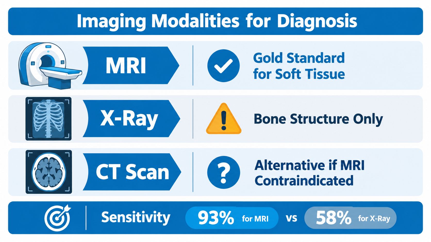
MRI is usually the preferred imaging study because it displays the structures most relevant to nerve compression. It shows the spinal canal, dural sac, nerve roots, discs, ligaments, and other soft tissues that can contribute to narrowing. It can also show whether several levels are involved. Those findings help determine whether the visible anatomy plausibly explains the patient's symptoms.
MRI has limits. A review in the American Journal of Roentgenology reported sensitivity ranging from 77% to 90% and specificity ranging from 72% to 100% across studies. Differences in study design, diagnostic definitions, and image interpretation affect those results. Incidental age-related findings are common, so an abnormal scan does not establish the pain source by itself.
How the modalities differ
| Imaging method | What it shows | Practical role |
|---|---|---|
| MRI | Nerves, spinal canal, discs, ligaments, and other soft tissues | Preferred confirmatory test when clinically indicated |
| CT | Bone detail and the shape of the spinal canal | Useful when MRI isn't suitable or when bony anatomy needs closer definition |
| CT myelography | CT anatomy after contrast outlines the dural sac and nerve pathways | Alternative when MRI is contraindicated or does not answer the clinical question |
| Plain X-ray | Alignment, bone changes, and some structural features | Helpful for selected questions, but insufficient by itself to confirm nerve compression |
Radiologists and clinicians may use anatomical measurements when describing central lumbar narrowing. A commonly cited anteroposterior canal diameter is under 10 millimeters. Another criterion sometimes used in diagnosis is a cross-sectional canal area below 70 square millimeters. These measurements are described in the NCBI clinical reference on spinal stenosis. They provide context, not automatic treatment triggers.
The practical rule is simple: review the actual images, not only the report, and compare them with the patient's history and examination. Imaging supplies structural detail, but it cannot determine symptom severity or prove that a particular level causes pain. The clinical pattern remains decisive.
Electrodiagnostic Testing to Confirm Nerve Involvement
Electrodiagnostic testing adds physiological information when the clinical picture remains uncertain. Nerve conduction studies measure how electrical signals travel along peripheral nerves. Electromyography, or EMG, evaluates electrical activity in selected muscles and can identify changes associated with nerve-root dysfunction.
These tests can be useful when an MRI shows several abnormalities and the responsible level isn't clear. They can also help distinguish a spinal radiculopathy from peripheral neuropathy, an entrapment neuropathy, or another disorder affecting the nerves outside the spine.
What EMG and nerve conduction studies can clarify
An electrodiagnostic evaluation may support:
- The presence of nerve dysfunction: Abnormal electrical findings can provide objective evidence that a nerve pathway is affected.
- The likely location: Results may help distinguish a nerve-root problem from a peripheral nerve disorder.
- The distribution: Testing can show whether the process appears focal, widespread, or related to more than one nerve.
- The timing pattern: Findings may suggest a recent injury, a longstanding process, or a mixture of acute and chronic changes.
The test doesn't show spinal narrowing directly. A normal result also doesn't eliminate stenosis, especially when symptoms are mild, early, intermittent, or primarily related to walking. Conversely, an abnormal result doesn't prove that every finding on an MRI is clinically important.
Clinical perspective: EMG confirms nerve dysfunction more directly than it confirms the anatomical cause. The MRI still has to provide a plausible explanation, and the symptoms still have to fit.
Testing is particularly relevant when a patient has weakness, persistent numbness, or a pattern that could arise from either the spine or a peripheral nerve. Progressive weakness involving the foot, for example, merits focused evaluation, and information about foot drop assessment can help patients understand why clinicians may investigate nerve function rather than relying on imaging alone.
The procedure can be uncomfortable because it may involve small electrodes and brief muscle sampling, but the findings can prevent an incorrect assumption that all leg symptoms come from spinal stenosis. It's generally a complementary test, not the first test ordered for every patient.
Differentiating Spinal Stenosis from Other Causes of Leg Pain
Leg pain during walking doesn't automatically indicate a spinal problem. Vascular claudication can also cause exertional leg discomfort, and hip disease, peripheral neuropathy, and disc-related nerve irritation can create overlapping symptoms. A careful clinician compares the trigger, relieving factor, distribution, examination, and overall medical context before assigning the pain to spinal stenosis.
The most useful distinction is often positional. Neurogenic symptoms commonly improve with sitting or bending forward, including leaning over a shopping cart or riding a bicycle. Vascular symptoms may relate more consistently to exertion and circulation, regardless of whether the torso is flexed.
A practical comparison
| Possible source | Clues that raise suspicion | Evaluation that may follow |
|---|---|---|
| Neurogenic claudication | Leg pain, numbness, or heaviness with standing or walking, improved by sitting or flexion | Neurological examination and clinically indicated MRI or CT |
| Vascular claudication | Activity-related leg discomfort with concerning circulation findings or less positional relief | Foot pulse assessment, ankle-brachial index testing, and possible vascular referral |
| Hip osteoarthritis | Groin or hip-centered pain, restricted hip movement, pain with loading | Hip examination and targeted imaging when indicated |
| Peripheral neuropathy | More diffuse or symmetric sensory symptoms, often involving the feet | Neurological assessment and possible electrodiagnostic testing |
| Disc-related radicular pain | Radiating pain or neurological symptoms following a more focal nerve-root pattern | Spine examination and imaging matched to the suspected level |
Foot pulses should be checked when vascular disease is plausible, particularly if symptoms don't change with posture. An ankle-brachial index may be considered when the history or circulation examination raises concern about reduced arterial flow. Recent spine imaging guidance recognizes that spinal imaging can help distinguish neurogenic from vascular claudication, but imaging doesn't replace a vascular assessment.
A disc bulge can also confuse the picture. A patient may have stenosis and a herniated or bulging disc at the same time, yet the disc may be the more relevant source of a focal radiating symptom. The correct diagnosis is the one that best explains the patient's actual limitations, not the one represented by the most striking phrase in a radiology report.
Next Steps and When to Seek Specialized Evaluation
After the history, examination, and imaging are complete, the clinician should be able to answer three practical questions. Is there anatomical narrowing? Do the symptoms and examination indicate nerve involvement? Does the suspected level plausibly explain the patient's functional limitations?
If those answers align, treatment planning can proceed with greater confidence. If they don't, the next step may be additional evaluation rather than an immediate procedure. That can include reviewing the actual images, assessing vascular or hip causes, obtaining electrodiagnostic testing, or monitoring symptoms with structured follow-up.
When specialist review adds value
A multidisciplinary spine evaluation is especially useful when symptoms are progressive, imaging shows abnormalities at several levels, weakness is present, or conservative care hasn't clarified the diagnosis. Board-certified physicians in pain management, anesthesiology, orthopedics, and spine surgery can assess non-invasive care, image-guided procedures, and surgical options according to the clinical findings.
Patients in New Jersey and Staten Island can prepare for an appointment by bringing:
- Prior imaging: Include the image files when available, not just the written report.
- A symptom record: Note walking and standing triggers, relieving positions, numbness, weakness, and changes over time.
- Medication information: List current medicines and previous treatments, including the response to each.
- Specific functional examples: Explain how symptoms affect stairs, shopping, work, sleep, or balance.
Diagnosis is more than naming a narrowed space. It's the process of establishing whether that narrowing is the pain generator and whether the clinical evidence supports a particular treatment path.
Interventional Pain Management evaluates spinal stenosis through coordinated history, neurological examination, imaging review, and additional testing when needed. Patients in New Jersey and Staten Island can visit Interventional Pain Management to request an evaluation and discuss non-invasive, interventional, or surgical options suited to the findings.