

A shoulder can become so painful that lifting a shirt sleeve, reaching behind the back, or sleeping on one side feels impossible. A patient may assume the tendon has torn, especially after seeing “rotator cuff tear” on an MRI report. Yet a shoulder that hurts with movement can be suffering from a tight joint capsule, tendon failure, pain-related inhibition, or more than one problem at the same time.
The central question in frozen shoulder vs rotator cuff tear isn't which diagnosis sounds worse. It's whether the shoulder is mechanically stiff, functionally weak, or both. The most useful early clue is the relationship between active motion, which the patient performs, and passive motion, which a clinician performs for the patient.
Table of Contents
- Why These Two Shoulder Problems Get Confused
- What Each Condition Actually Is
- Causes, Risk Factors, and Who Each One Affects
- How a Clinician Tells Them Apart on Examination
- What Imaging Can and Cannot Tell You
- Treatment Pathways Side by Side
- Recovery Timelines and What to Expect
- How to Use This Comparison When You See a Doctor
Why These Two Shoulder Problems Get Confused
A typical patient arrives unable to lift an arm overhead. Reaching into a cupboard is difficult, putting on a coat is awkward, and lying on the affected side may disturb sleep. If the problem began gradually, the patient may describe stiffness. If it followed a fall or a sudden pull, the same loss of function may feel like tendon damage.
Both conditions can produce pain, limited use, and difficulty with overhead movement. A frozen shoulder, also called adhesive capsulitis, tightens the capsule surrounding the glenohumeral joint. A rotator cuff tear disrupts one or more tendons that help center and lift the shoulder. The symptoms overlap because pain itself can reduce movement, while weakness can make a shoulder appear stiff.
The first clinical task is therefore not to label the condition from one symptom. It's to determine what happens when the patient stops trying to move and the examiner moves the arm.
| Clinical question | Frozen shoulder | Rotator cuff tear |
|---|---|---|
| What is primarily affected? | The joint capsule becomes contracted and restricts motion | A cuff tendon becomes partially or completely disrupted |
| Active motion | Reduced | Often reduced, especially with elevation or rotation |
| Passive motion | Also restricted across multiple planes | Often relatively preserved in a substantial tear |
| Main functional pattern | Global stiffness and loss of reach | Weakness, loss of active elevation, or pain with loading |
| Typical imaging role | Excludes competing causes and may support the diagnosis | Shows tendon integrity, retraction, atrophy, or degeneration |
| Main diagnostic caution | Pain can make stiffness look worse | An MRI tear may be incidental rather than the pain generator |
Practical rule: If someone else can't move the shoulder much farther than the patient can, capsular stiffness moves higher on the list. If passive movement is relatively preserved but active lifting is disproportionately weak, tendon failure deserves closer attention.
This distinction isn't absolute. Partial tears can be painful enough to restrict passive movement, and a frozen shoulder can make strength testing appear poor because the joint won't allow the required position. The useful comparison is a structured examination, not a home test or an isolated radiology phrase.
What Each Condition Actually Is
Adhesive capsulitis is a disorder of the glenohumeral joint capsule. The capsule thickens, contracts, and loses its normal ability to permit movement. The resulting restriction affects both active and passive motion, often across several directions. Plain radiographs may remain otherwise normal after arthritis, fracture, and other structural causes have been excluded.
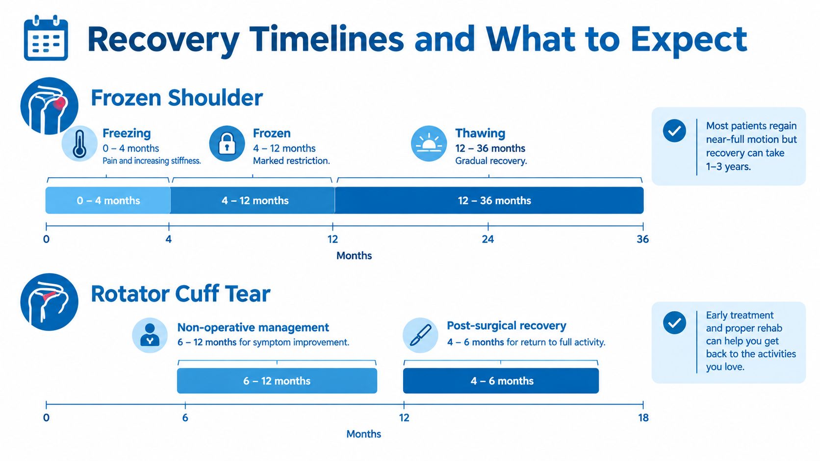
Patients often describe a progression from increasing pain and movement loss to a period of marked stiffness, followed by gradual return of motion. The labels “freezing,” “frozen,” and “thawing” describe this clinical pattern, but real patients don't always move through the stages neatly. Pain can remain prominent even when the dominant problem has become restricted mobility.
A rotator cuff tear is a discontinuity in one or more of the tendons that help stabilize and move the shoulder. The defect may be partial, involving only part of the tendon thickness, or full-thickness, extending through the tendon. A major tear can reduce active elevation and strength while leaving passive movement comparatively available.
The biology is different. Adhesive capsulitis is dominated by capsular contracture and inflammatory fibrosis. A cuff tear reflects mechanical tendon damage, whether from an acute injury, chronic degeneration, or both. Tendon irritation without a tear, bursal pain, arthritis, neck-related symptoms, and labral problems can imitate either pattern, which is why a broader shoulder assessment may include conditions discussed in shoulder tendinitis and bursitis care.
One patient can have both a tear and capsular stiffness. The most accurate one-sentence summary is this: frozen shoulder is a contracted joint capsule that limits movement even passively, while a rotator cuff tear is tendon disruption that more often causes weakness and loss of active movement while passive motion remains available.
Causes, Risk Factors, and Who Each One Affects
The patient's age, medical history, and onset of symptoms change the starting probability before any scan is ordered. Frozen shoulder is commonly associated with diabetes and thyroid disease, although many cases are idiopathic. It may also follow a period of reduced shoulder use, injury, or surgery.
Rotator cuff disease follows a different pattern. Tears often develop through age-related tendon degeneration, while a younger patient may sustain one after a fall, traction injury, or forceful lifting event. That doesn't mean a younger adult can't have degenerative disease or that an older adult's tear must be symptomatic. It means the same complaint deserves different initial questions in different patients.
Age has a particularly strong effect on the chance of seeing a full-thickness supraspinatus tear on MRI in patients with atraumatic shoulder pain. One study reported full-thickness tearing in 0% of patients aged 30 or younger, 1.6% of those aged 31 to 40, 8.1% aged 41 to 50, 14.8% aged 51 to 60, 37.7% aged 61 to 70, and 50% of patients older than 70. The study also reported full-thickness tearing in 42% of patients over age 60, as described in this age-related review of rotator cuff pathology.
That finding changes how an MRI is interpreted. In an older gardener with shoulder pain and a visible tear, the scan may show the cause, a contributor, or an unrelated age-associated finding. In a younger desk worker with gradual pain and pronounced stiffness, the examination may point more strongly toward capsular restriction even when no dramatic tendon defect is visible.
Risk profiles that shift the evaluation
- Medical associations: Diabetes and thyroid disease increase clinical suspicion for adhesive capsulitis, but they don't establish the diagnosis.
- Injury history: Sudden weakness immediately after trauma raises concern for a clinically important tear or another structural injury.
- Age: The probability of an MRI-visible tear rises substantially with age, so correlation with strength and function becomes more important.
- Immobilization: Reduced use after surgery or injury can contribute to capsular stiffness.
- Functional demand: A small tear may be manageable for one person and disabling for someone whose work requires repeated overhead activity.
A diagnosis should explain the pattern, not merely fit one risk factor. An older patient can have frozen shoulder, and a younger patient can have a tear. The history narrows the field, but the motion examination determines whether the leading problem is stiffness or tendon-related weakness.
How a Clinician Tells Them Apart on Examination
The examination starts before special tests. The clinician observes how the patient removes a jacket, raises the arm, and compensates with the shoulder blade. Pain behavior matters, but it doesn't reveal whether the capsule or tendon is primarily responsible.
The next step is to measure motion separately. Passive forward elevation is tested with the patient relaxed. External rotation at the side and internal rotation are recorded as distinct findings, rather than summarized as “limited range of motion.” The same movements are then assessed actively, followed by resisted testing of elevation and rotation.
The active and passive comparison
In adhesive capsulitis, passive glenohumeral motion is restricted across multiple planes. External rotation is often especially informative, but the diagnosis depends on the overall pattern and exclusion of arthritis, fracture, and other structural disease. The examiner may feel a firm limitation rather than seeing the patient stop because of pain.
A substantial full-thickness cuff tear more often preserves passive movement. The patient may be unable to actively lift the arm to the same level, yet the examiner can move it farther. Marked weakness, an external-rotation lag, or a positive drop-arm test can strengthen concern for a significant tear.
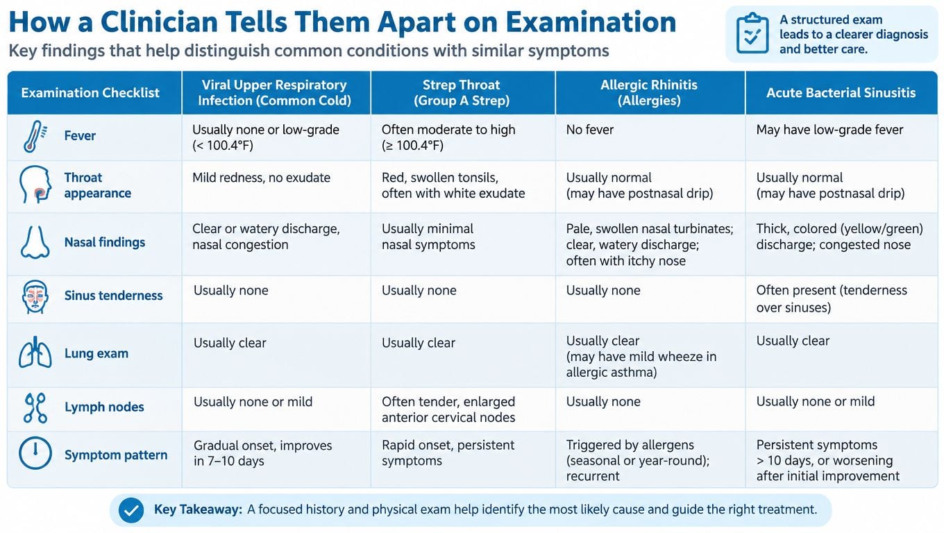
Pain complicates the distinction. A partial-thickness tear can cause guarding that reduces both active and passive motion. Conversely, a severely stiff shoulder can make resisted strength testing look weak because the patient cannot position the arm normally. Bedside cuff tests are useful, but guidance emphasizes that they generally help rule in rather than rule out a full-thickness tear. A normal or equivocal test doesn't settle the question.
A useful question for a clinic visit is simple: “Can the examiner move the arm farther than the patient can move it alone?”
The same reasoning separates cuff disease from other shoulder conditions, including labral pathology. A broader assessment may be needed when clicking, instability, a catching sensation, or a specific athletic injury is part of the story. Information about labral tear evaluation can be relevant when the motion pattern doesn't fit either classic diagnosis.
What Imaging Can and Cannot Tell You
Imaging works best when it answers a defined question. A plain radiograph can show arthritis, fracture, calcific tendinitis, or other bone-related explanations for pain. It is often normal in adhesive capsulitis, and a normal X-ray doesn't reliably exclude a rotator cuff tear.
Ultrasound can assess tendon continuity dynamically and may be useful when the clinical question is whether a cuff tendon is intact. MRI provides a broader view, including tendon discontinuity, retraction, muscle atrophy, fatty degeneration, and associated joint findings. For adhesive capsulitis, MRI can support the diagnosis by showing capsular or synovial abnormalities, particularly around the rotator interval and axillary recess, but the diagnosis remains clinical.
The important trade-off is that a more detailed scan can reveal more abnormalities without clarifying which abnormality causes the symptoms. A systematic review of MRI studies reported rotator cuff abnormalities in 26.2% of asymptomatic shoulders and 49.4% of symptomatic shoulders, with full-thickness tears accounting for 11.75% and partial-thickness tears for 18.49% in the pooled MRI sample, as reported in this systematic review of asymptomatic and symptomatic shoulders. Those findings don't make a tear unimportant. They show why the scan must be matched to weakness, motion loss, trauma, and functional limitation.
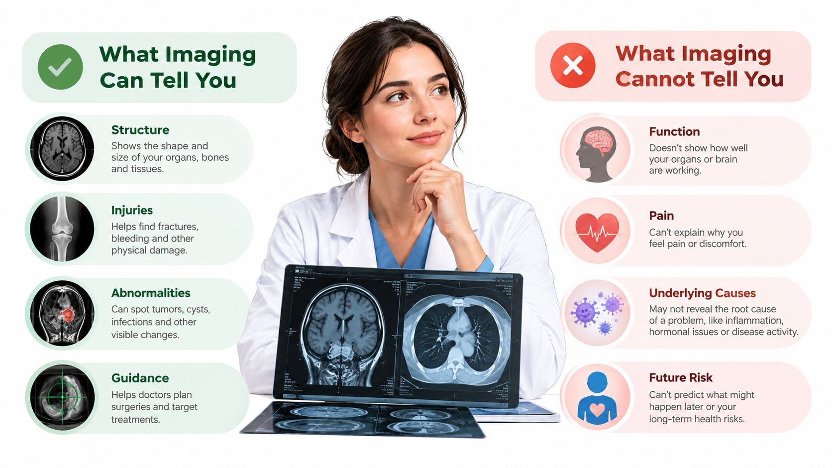
MRI is more likely to change management when there is acute traumatic weakness, persistent functional loss, or a possible tear that could require surgical planning. In chronic pain, clinical assessment and sometimes ultrasound may come first, with MRI reserved for a focused unresolved question.
A 2025 meta-analysis found that axillary capsular enhancement for adhesive capsulitis had 95% pooled sensitivity, a diagnostic odds ratio of 107, and an area under the curve of 0.96, while the shoulder-shrug sign showed only 46.4% sensitivity and 48.1% specificity for rotator cuff disease, according to the published MRI feature analysis. Even a promising imaging feature doesn't replace the examination. The scan should confirm or refine a clinical question, not create one.
Treatment Pathways Side by Side
Treatment should follow the dominant problem. A patient with a contracted capsule needs a mobility strategy, while a patient with tendon failure and meaningful weakness may need a strength-preservation strategy and, in selected cases, surgical review.
| Treatment stage | Frozen shoulder | Rotator cuff tear |
|---|---|---|
| First priorities | Control pain and restore capsular mobility gradually | Modify aggravating activity and preserve usable strength |
| Rehabilitation | Guided stretching and progressive range-of-motion work | Scapular control, remaining cuff strength, and functional retraining |
| Injection role | An intra-articular corticosteroid injection can help during the painful phase | A subacromial or intra-articular injection may reduce pain enough to participate in rehabilitation |
| Additional procedure | Hydrodilatation may be considered in selected patients | Imaging-guided planning may be appropriate when weakness or structural damage persists |
| Surgical or procedural escalation | Manipulation under anesthesia or arthroscopic capsular release for refractory stiffness | Repair consideration for selected acute traumatic or large retracted tears, or persistent functional loss |
For adhesive capsulitis, aggressive stretching through severe pain can backfire by increasing guarding. Physical therapy should aim to recover motion without turning every session into a flare. An intra-articular corticosteroid injection may be most useful when pain dominates the early course, while hydrodilatation is an option for selected patients. Persistent, function-limiting stiffness despite appropriate nonoperative care may prompt discussion of manipulation under anesthesia or arthroscopic capsular release.
For a cuff tear, rehabilitation focuses on the muscles that remain functional, scapular mechanics, and gradual loading. Pain relief can make therapy possible, but an injection doesn't reattach a disrupted tendon. A patient with sudden weakness after trauma, a large full-thickness tear with retraction, or ongoing loss of useful function may need an orthopedic surgical opinion rather than prolonged trial-and-error care.
Treatment principle: An injection can reduce pain, but it shouldn't be used to conceal progressive weakness or postpone evaluation of a potentially repairable traumatic tear.
The decision is individualized. A manual worker, an older adult with low overhead demand, and a competitive athlete may reasonably choose different paths even when their scans look similar. The role of corticosteroid injections in musculoskeletal care is to support a diagnosis-specific plan, not to substitute for one.
Recovery Timelines and What to Expect
Frozen shoulder is usually prolonged rather than permanently destructive. Symptoms often move through painful restriction, a period of substantial stiffness, and gradual improvement. The overall course can last 1 to 3 years, and recovery doesn't always proceed at a steady pace.
That long timeline can create two opposite mistakes. Some patients abandon therapy because the shoulder isn't normal quickly, while others assume every persistent restriction is harmless because adhesive capsulitis can improve eventually. A plateau, worsening function, or an examination that shows significant weakness should trigger reassessment rather than automatic reassurance.
Rotator cuff recovery depends on tear size, tissue quality, demands, and treatment choice. Some partial tears and smaller full-thickness tears improve with activity modification and rehabilitation, while a repaired tendon requires a carefully staged period of protection, motion recovery, strengthening, and return to loading. Surgery may address the tendon defect, but it doesn't create an instant return to normal function.
Reasons to seek earlier review
- Progressive weakness: Increasing difficulty lifting the arm, especially when passive motion remains available, needs reassessment.
- New or worsening night pain: Night pain can occur with either condition, but a change in pattern may indicate that the working diagnosis needs revision.
- A traumatic onset: Sudden inability to raise the arm after an injury warrants timely evaluation.
- Failure to improve: Symptoms that don't follow the expected response to self-care or supervised rehabilitation deserve a fresh examination.
- Unexpected stiffness: New global loss of passive motion may indicate capsular contracture, arthritis, or another competing cause.
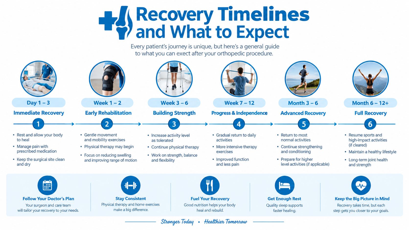
The better question isn't whether recovery is fast or slow. It's whether the shoulder is moving in the expected direction, with pain becoming more manageable and function gradually expanding. If the clinical trajectory and the original diagnosis no longer match, the plan should change.
How to Use This Comparison When You See a Doctor
A productive appointment starts with function, not the MRI report. The patient should describe when the problem began, whether a specific injury preceded it, which movements fail, and whether another person can move the arm farther than the patient can move it alone.
A shoulder that remains restricted when the examiner moves it points toward adhesive capsulitis or another joint-level restriction. A shoulder with relatively preserved passive motion but clear weakness raises concern for a clinically important cuff tear. Neither pattern proves the diagnosis by itself, but each tells the clinician which questions and tests deserve priority.
Before the visit, it helps to record:
- Onset: Gradual, sudden, or linked to a fall or lifting event.
- Motion: Reaching overhead, behind the back, across the body, and outward rotation.
- Strength: Whether the arm feels painful, weak, or impossible to hold up.
- Night symptoms: Whether pain is constant, position-dependent, or newly worsening.
- Function: Dressing, grooming, work tasks, exercise, and sleep disruption.
Don't let an MRI label make the treatment decision by itself. A visible cuff abnormality may be incidental, while a patient with substantial weakness after trauma may need prompt specialist review even before every imaging detail is available. Progressive weakness, sudden inability to lift the arm, increasing night pain, or persistent symptoms after several weeks of reasonable self-care should prompt an in-person evaluation.
The practical conclusion in frozen shoulder vs rotator cuff tear is straightforward. Passive stiffness favors a capsular problem, while preserved passive motion with disproportionate weakness favors tendon dysfunction. A careful clinician then uses the history, examination, and targeted imaging to decide whether rehabilitation, an injection, further diagnostic work, or surgical consultation makes sense.
Interventional Pain Management evaluates shoulder pain by combining history, range-of-motion and strength testing, imaging review, and non-surgical or orthopedic treatment planning when appropriate. Patients in New Jersey and Staten Island can visit Interventional Pain Management to request an evaluation for persistent stiffness, weakness, or suspected shoulder tendon injury.