
A sharp sting across the top of the foot can make an ordinary step feel suddenly unsafe. It may begin after stepping off a curb, tightening a pair of laces, increasing walking distance, or finishing a long day on the feet. Because the painful area is easy to point to but harder to identify, many adults wonder whether they're dealing with a tendon, a joint, a nerve, or something that needs prompt attention.
Bridge of foot pain refers to symptoms around the top and central part of the foot, often called the dorsal midfoot. The same location can reflect joint wear, tendon overload, a stress injury, ligament damage, nerve irritation, or inflammation. Most causes are manageable, but the correct next step depends on the story behind the pain, the examination findings, and sometimes targeted imaging.
Table of Contents
- What Bridge of Foot Pain Really Feels Like
- The Anatomy of the Foot Bridge
- The Most Common Causes of Pain on the Bridge
- How a Clinician Evaluates Midfoot Pain
- Choosing the Right Imaging and Diagnostic Tests
- Treatment Options From Conservative to Interventional
- When to See a Specialist About Midfoot Pain
- Your Next Steps and What to Expect at a Visit
What Bridge of Foot Pain Really Feels Like
Pain across the foot's “bridge” can feel different from one person to another. You may notice a sharp spot during push-off, a dull ache after walking, burning or tingling beneath the laces, or tenderness where a shoe presses the top of the foot. Some symptoms stay in one small area; others seem to spread across the middle of the foot.
The location is useful, but it cannot identify the cause by itself. The dorsal midfoot contains joints, tendons, ligaments, nerves, and small bones that help transfer weight from the ankle toward the toes. Because these structures work close together, irritation in one can make the wider bridge feel painful.
The first sorting questions
Start with the story around the symptom:
- Did the pain follow an injury? A twist, fall, awkward landing, or force running along the foot can raise concern about a ligament injury or fracture.
- Did activity or footwear change? Longer walks, a new training load, or tight laces may overload a tendon or bone.
- What does the pain feel like? Deep aching and stiffness follow a different pattern from burning, tingling, or electric pain.
A gradual increase during activity often fits repetitive loading. Sudden pain with swelling or bruising calls for more caution, especially when walking becomes difficult.
Practical rule: Timing can be as informative as location. Tell a clinician what happened before the pain appeared, not only where it hurts now.
A 2025 clinical review describes dorsal midfoot symptoms associated with osteoarthritis, stress injury, tendon or ligament problems, nerve-related pain, inflammatory arthritis, infection, and fracture. It also notes uncertainty around some commonly used examination findings review of midfoot osteoarthritis and clinical assessment. This uncertainty is the reason this guide sorts symptoms into next steps instead of asking you to match yourself to a diagnosis list.
The Anatomy of the Foot Bridge
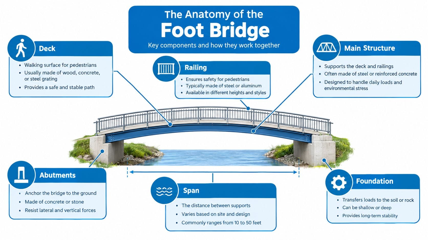
The foot works less like a rigid plank and more like a suspension bridge that changes shape under load. The longitudinal arch, especially the medial arch, forms the curved deck. Its main bony supports include the calcaneus, talus, navicular, medial cuneiform, and first metatarsal.
Ligaments and the plantar aponeurosis act like tensioning structures beneath the bridge. The long plantar ligament and short plantar ligament also contribute to stiffness and help the foot store and return energy during walking. On top of the foot, the extensor tendons help lift and control the toes. At the center of the midfoot, the Lisfranc ligament complex stabilizes the tarsometatarsal joints, which connect the long metatarsal bones to the bones closer to the ankle.
Why arch shape doesn't tell the whole story
Reduced arch height, known as pes planus or flatfoot, is common. A systematic review covering 12 studies and 16,000 participants reported an overall flatfoot prevalence of 15.6% systematic review of flatfoot prevalence and foot structure. Other adult samples commonly report pes planus in 19% to 37% of adults, although only about 11% of those people are symptomatic and seek medical care, according to the same review.
That distinction matters. Flexible flatfoot changes shape when the person stands or moves and is much more common. Rigid flatfoot remains stiff and is less common, but it's more likely to cause substantial pain or disability. A visible low arch doesn't automatically explain a painful bridge, and a normal-looking arch doesn't rule out a midfoot problem.
The foot also changes dynamically throughout gait. Hicks's windlass mechanism describes how toe extension tensions the plantar fascia and influences arch behavior. Modern biomechanics therefore treats the arch as a mobile system, not a structure that locks into a rigid lever at push-off.
The Most Common Causes of Pain on the Bridge
Several conditions can affect the same small area yet need different treatment. Identify the tissue first, because bone, tendon, ligament, nerve, and joint problems follow different patterns and timelines. The key questions are where the pain is, what triggered it, whether weight-bearing is possible, and which movement reproduces it.
Joint, tendon, bone, and ligament causes
Midfoot osteoarthritis commonly produces aching or stiffness over the upper inner or central midfoot. Pain may appear during push-off, stair climbing, or walking on uneven ground. A firm dorsal prominence can develop, but a lump alone does not establish arthritis. The relationship between a dorsal exostosis and midfoot osteoarthritis can remain uncertain, so the surrounding symptoms and examination matter more than appearance alone.
Extensor tendon irritation usually causes pain across the top of the foot. Moving the toes or ankle against resistance may reproduce it. Tight laces, repetitive walking, running, or a recent increase in activity can place extra pressure on these tendons. An explanation of tendinitis and bursitis can help distinguish irritated soft tissue from a joint or bone problem.
A stress fracture or stress reaction often develops gradually rather than after one obvious injury. Pain becomes more activity-related and may ease with rest, while swelling or very specific tenderness over a bone can occur. An early X-ray may appear normal because the injury can begin at a microscopic level. Persistent, sharply localized pain needs reassessment and may require MRI, which is more sensitive to early changes in the bone marrow. See this information on stress fractures of the foot and ankle.
Lisfranc injury deserves attention after a twist, fall, or force transmitted through the foot. Swelling, difficulty bearing weight, pain when the forefoot is gently stressed, and bruising on the sole are important clues. The Lisfranc ligament complex supports the tarsometatarsal joints. If the injury is unstable, normal load transfer can change and later joint damage may develop. This review of Lisfranc injuries explains why these injuries can be missed when the initial appearance seems mild.
Nerve and inflammatory causes
Nerve irritation may feel burning, tingling, electric, or numb rather than only achy. Shoe pressure or a particular position may aggravate it more than direct loading of a joint. Inflammatory arthritis can bring swelling, warmth, stiffness, and pain that does not follow a predictable activity pattern. Symptoms in other joints, skin changes, fever, or a known systemic illness can change the level of concern.
| Cause | Typical mechanism | Hallmark symptom | Key clue |
|---|---|---|---|
| Midfoot osteoarthritis | Joint wear or altered loading | Aching, stiffness, painful push-off | Dorsal medial or central tenderness |
| Extensor tendon irritation | Repetitive use or shoe pressure | Pain with toe or ankle motion | Pain follows a tendon course |
| Stress injury | Repetitive loading exceeds bone recovery | Focal pain that worsens with activity | Point tenderness and possible swelling |
| Lisfranc injury | Twist, fall, or axial loading | Midfoot pain with swelling | Plantar bruising or difficulty bearing weight |
| Nerve irritation | Compression or traction of a nerve | Burning, tingling, or numbness | Symptoms change with pressure or position |
| Inflammatory arthritis | Joint inflammation | Swelling, warmth, and stiffness | Symptoms may involve more than one joint |
These patterns overlap, which is why a symptom list cannot replace an examination. The most useful clue may be the injury mechanism, the ability to bear weight, bruising, or the exact structure that reproduces pain during testing. Sudden inability to bear weight, marked swelling, plantar bruising, or rapidly increasing redness and warmth warrants prompt medical assessment rather than routine self-care.
How a Clinician Evaluates Midfoot Pain
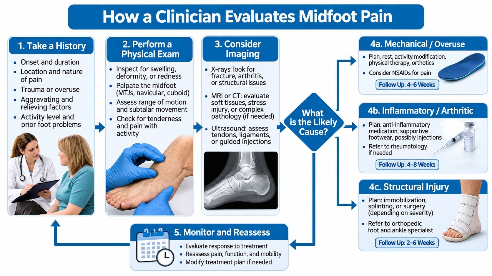
A specialist usually begins with the timeline. The person may be asked when the pain started, whether it followed trauma, which activities aggravate it, whether training or walking volume changed, and whether shoes or laces affect the symptoms. Morning stiffness lasting under or over 30 minutes can influence the differential, although it isn't diagnostic on its own.
The clinician also asks what the foot can still do. Can the person walk normally? Can they bear weight? Does the pain limit stairs, work, exercise, or ordinary household movement? These answers show functional impact and help determine whether imaging or protection is needed quickly.
What the examination adds
The foot is compared with the other side. The clinician looks for swelling, bruising, redness, a bony prominence, altered alignment, or a change in walking pattern. Palpation then maps the tender area, including bones, joint lines, tendon paths, and nearby nerve locations.
Movement testing helps separate tissues. If resisted toe or ankle motion reproduces pain, tendon involvement becomes more plausible. If pressure over a joint line or movement through the midfoot causes pain, a joint problem moves higher on the list. If a gentle forefoot abduction or pronation stress maneuver causes marked midfoot pain, the clinician considers Lisfranc injury.
Plantar bruising deserves particular attention. It can be a strong clue to a midfoot ligament injury, especially after trauma. Inability or marked difficulty bearing weight also changes the urgency because it may indicate a significant fracture or unstable injury rather than uncomplicated tendon irritation.
During the visit: A painful dorsal lump is a finding to investigate, not a diagnosis to assume. It may relate to a joint, bone, tendon, footwear pressure, or another structure.
If there was trauma and weight bearing is difficult, the clinician considers fracture or ligament instability. If there was no trauma but pain followed increased activity, tendon overload or stress injury becomes more relevant. If burning or numbness dominates, the examination shifts toward nerve assessment. Patients can make the visit more useful by bringing a list of symptom triggers, recent activity changes, and the shoes worn when the pain occurs. Information about fractures and their evaluation may also help patients understand why protection and reassessment can matter when bone injury is suspected.
Choosing the Right Imaging and Diagnostic Tests
A scan should answer a specific clinical question. The same area of midfoot pain may need different testing depending on whether the concern is joint alignment, bone injury, tendon disease, ligament damage, or nerve irritation.
Matching the test to the structure
Weight-bearing X-rays are useful when the clinician needs to assess alignment, joint spaces, degenerative change, or how the midfoot behaves while you stand. A non-weight-bearing image can miss changes that appear only under load. Current guidance supports weight-bearing radiographs when assessing the presence or extent of degenerative disease, while MRI is not required for every acute pain presentation or degenerative joint complaint 2025 clinical guideline on foot and ankle imaging.
MRI answers many questions about tendons, ligaments, bone marrow, and injuries hidden from an X-ray. It may show an early stress reaction when the first radiograph looks normal and can display soft tissues that plain films cannot.
CT gives a detailed view of cortical bone. It may clarify a fracture line, displacement, joint-surface involvement, or healing when the main concern is bony structure.
Ultrasound can assess superficial tendons and selected nerve problems while the foot moves, which may reveal an issue a still image misses. Bone scanning has a narrower role in selected cases. The choice depends on the suspected tissue, examination findings, and results from earlier tests.
Where injections fit
For chronic foot pain, imaging options include radiography, ultrasound, MRI, CT, and bone scanning. Image-guided anesthetic or corticosteroid injection is another option in selected cases. A targeted anesthetic injection may help show whether a particular joint is producing the pain. A corticosteroid injection may also treat inflammation in carefully chosen situations.
An injection is not a universal test or cure. Ask, “Which structure is this evaluating?” and “How will the result change treatment?” Clear answers keep testing focused rather than automatic.
Treatment Options From Conservative to Interventional
Treatment works best when it matches the tissue and the person's functional goals. A tendon that is overloaded needs a different plan from an unstable Lisfranc injury, and a stress fracture shouldn't be treated like ordinary soreness.
Reducing load while preserving function
Early care may include temporarily reducing impact activity, changing footwear, and avoiding laces or shoe pressure over a tender dorsal area. Elevation and cold therapy may help swelling when used safely. A clinician may recommend protected weight bearing or immobilization when a fracture or significant sprain is suspected.
Rehabilitation often addresses calf flexibility, ankle control, intrinsic foot strength, and the way force travels through the foot. Orthotics or supportive inserts may help selected people by redistributing load, but they should support a diagnosis rather than substitute for one. A gradual return to walking, work, or sport is generally safer than a sudden return to full intensity after symptoms settle.
Targeted medical options
Anti-inflammatory medication may be appropriate for some adults, but medical history, kidney function, stomach health, blood pressure, and other medicines affect safety. Image-guided corticosteroid injections can be considered for selected inflamed joints or tendons. Their value comes from targeting a suspected pain generator, not from treating every case of bridge of foot pain. More information about corticosteroid injections can help patients discuss indications and limitations with a clinician.
Regenerative procedures may be discussed in some practices, but they shouldn't be presented as guaranteed repairs. Evidence and suitability vary by diagnosis, and a procedure should never replace assessment for fracture or instability.
When surgery enters the conversation
Most uncomplicated stress fractures are treated without surgery when loading is controlled early. An unstable Lisfranc injury may require fixation or stabilization to protect alignment. Advanced painful midfoot arthritis may lead to a discussion about fusion when nonoperative care no longer provides acceptable function.
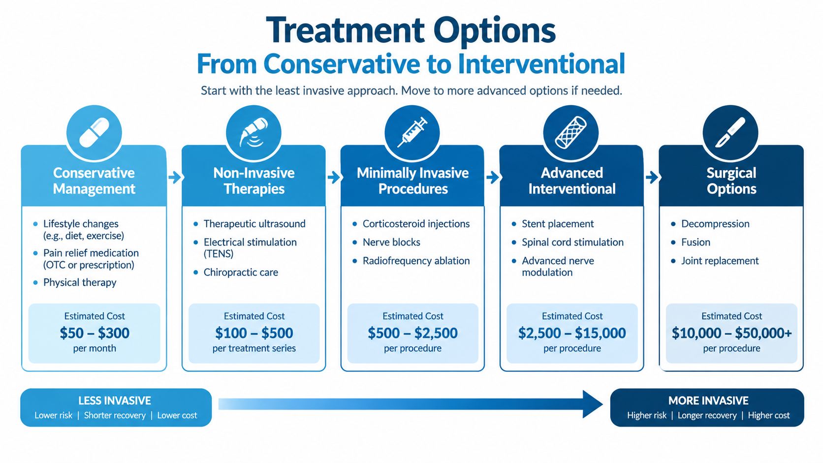
The timeline depends on the diagnosis. Mild tendon irritation may improve with load changes and rehabilitation, while bone healing and recovery from ligament injury require stricter protection. Surgery is not the next automatic rung on a ladder. It is one tool for a structural problem that cannot be safely or effectively managed with less invasive care.
When to See a Specialist About Midfoot Pain
Some symptoms justify same-day assessment rather than a period of home care. These include:
- Acute trauma: Pain follows a fall, twist, crush, or awkward landing.
- Rapid swelling or visible deformity: The foot changes shape or enlarges quickly.
- Inability to bear weight: Walking is impossible or markedly difficult.
- Neurologic symptoms: New numbness, weakness, or significant tingling appears.
- Possible infection: Fever accompanies redness, warmth, swelling, or an open wound.
Plantar bruising after an injury deserves particular caution because it can signal a midfoot ligament injury. A person shouldn't attempt to train through focal pain that continues to worsen, especially when a stress injury is possible.
Symptoms that aren't urgent still deserve a scheduled evaluation when they persist despite rest, recur with ordinary walking, produce repeated swelling, or alter the way a person moves. Mild tenderness without trauma may be reasonable to monitor briefly, but persistent, progressive, or activity-limiting pain warrants a clinical opinion.

A multidisciplinary practice can coordinate pain management, orthopedic assessment, examination, and imaging when the cause isn't immediately clear. That coordination may reduce the risk of treating a ligament injury as simple tendinitis or assuming a dorsal lump represents arthritis without confirming the source.
Your Next Steps and What to Expect at a Visit
A person with bridge of foot pain can start with three practical questions:
- Did symptoms begin with trauma? If yes, especially with swelling, bruising, deformity, or difficulty bearing weight, prompt assessment is appropriate.
- Have symptoms persisted or progressed despite reducing activity? If yes, schedule an examination instead of repeatedly restarting the same routine.
- Has the pain changed walking, work, stairs, or exercise? If yes, the problem deserves evaluation even when the pain seems tolerable.
A first visit generally includes a focused history, comparison of both feet, palpation, movement and stability testing, and a discussion of whether weight-bearing imaging is appropriate. The plan may include footwear changes, protected activity, rehabilitation, advanced imaging, or referral for a targeted procedure. A useful appointment should leave the patient knowing what structure is suspected, what warning signs matter, and what would trigger reassessment.

Frequently asked questions
Can tight shoes cause pain on top of the foot?
Yes. Shoe pressure can irritate extensor tendons or compress superficial nerves. Persistent symptoms still need examination because footwear pressure can coexist with joint, bone, or ligament problems.
Does a flat arch explain the pain?
Not necessarily. Reduced arch height is common, and many people have no symptoms. The important question is whether the foot structure, movement pattern, or another condition matches the pain pattern.
Is a normal X-ray reassuring?
It can be useful, but it doesn't exclude every problem. Early stress injuries, some ligament injuries, and soft-tissue conditions may require clinical reassessment or different imaging.
Can an injection identify the painful joint?
A carefully selected image-guided anesthetic injection may help test a suspected joint, but it should follow an examination and be interpreted with the rest of the clinical evidence.
Persistent foot pain can feel worrying because every step keeps reminding the person that something is wrong. Once the responsible structure is identified, the problem becomes a treatment decision rather than an unanswered question.
Interventional Pain Management evaluates musculoskeletal and foot-related symptoms through coordinated pain management, orthopedic assessment, examination, and imaging when appropriate. Adults with persistent bridge of foot pain can visit Interventional Pain Management to request an evaluation and discuss a plan matched to the suspected cause.