
Disc height loss is a narrowed space between vertebrae visible on MRI or X-ray, not a diagnosis by itself. In asymptomatic adults, its prevalence rises from approximately 24% in the youngest age group to 84% in the oldest, so the finding becomes nearly universal with age even without back pain.
A report mentioning “disc-space narrowing” can make a routine imaging finding sound like proof that a damaged disc is causing every ache, twinge, or limitation. The more accurate interpretation is calmer and more useful: the disc has become thinner at a particular spinal level, and the clinical importance depends on whether that change matches the person's pain pattern, examination, nerve findings, and functional problems.
Table of Contents
- Understanding Disc Height Loss at a Glance
- How a Healthy Disc Works and Why It Loses Height
- Common Causes Behind Disc Height Loss
- Symptoms That May Relate to Disc Narrowing
- How Doctors Evaluate and Measure Disc Height
- Treatment Options from Conservative Care to Surgery
- Prognosis, Prevention, and Living With the Finding
Understanding Disc Height Loss at a Glance
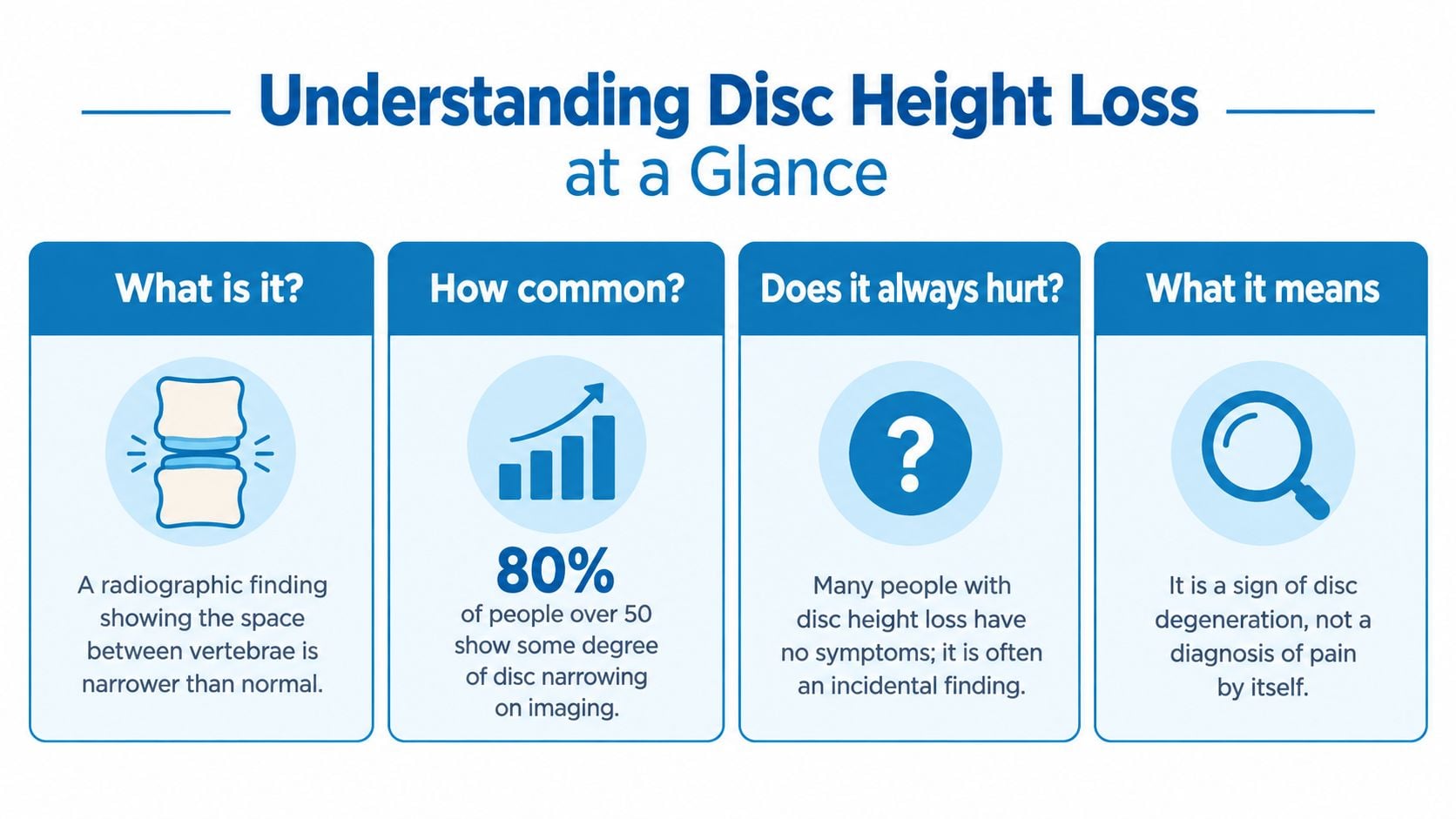
The spine contains stacked vertebrae separated by intervertebral discs. Each disc acts as a flexible cushion between two bones. When the disc becomes thinner, the space between those vertebrae appears narrower on an X-ray or MRI. That visible change is disc height loss, also called disc-space narrowing.
The phrase describes anatomy. It doesn't, by itself, identify a pain generator, establish a diagnosis of symptomatic degenerative disc disease, or determine whether treatment is needed. A scan can show a narrowed disc in someone who has no pain at all.
Practical rule: A scan describes structure. A clinical evaluation determines whether that structure matters.
The difference is important because disc-height loss is common across the adult lifespan. A systematic review of imaging in people without symptoms found prevalence estimates of approximately 24%, 34%, 45%, 56%, 67%, 76%, and 84% across progressively older age groups, with prevalence increasing by roughly one percentage point per year of age (systematic review of spinal degeneration imaging). The finding often represents structural aging rather than a disease causing pain.
What the report is really describing
A radiologist may mention reduced height alongside disc desiccation, a bulge, annular fissuring, osteophytes, endplate changes, foraminal narrowing, or spinal stenosis. These are related but distinct observations. Disc desiccation refers mainly to reduced water content, while height loss describes the physical reduction in the disc space.
The finding becomes more clinically relevant when several details line up:
- The level matches the symptoms. Lower-lumbar changes deserve closer attention when symptoms are centered in the corresponding back or leg distribution.
- The examination supports the scan. Reflex, strength, sensation, movement, and nerve-tension findings can help determine whether a nerve is involved.
- Function is affected. Difficulty walking, sitting, lifting, sleeping, or working carries more clinical weight than an isolated phrase in a report.
- A mechanism is visible. Foraminal narrowing, facet overload, instability, or nerve-root compression may explain symptoms more directly than height loss alone.
Disc height loss can contribute to pain indirectly by reducing the opening where a nerve exits or by shifting loads toward facet joints. Still, a narrowed space is a starting clue, not a final answer.
How a Healthy Disc Works and Why It Loses Height
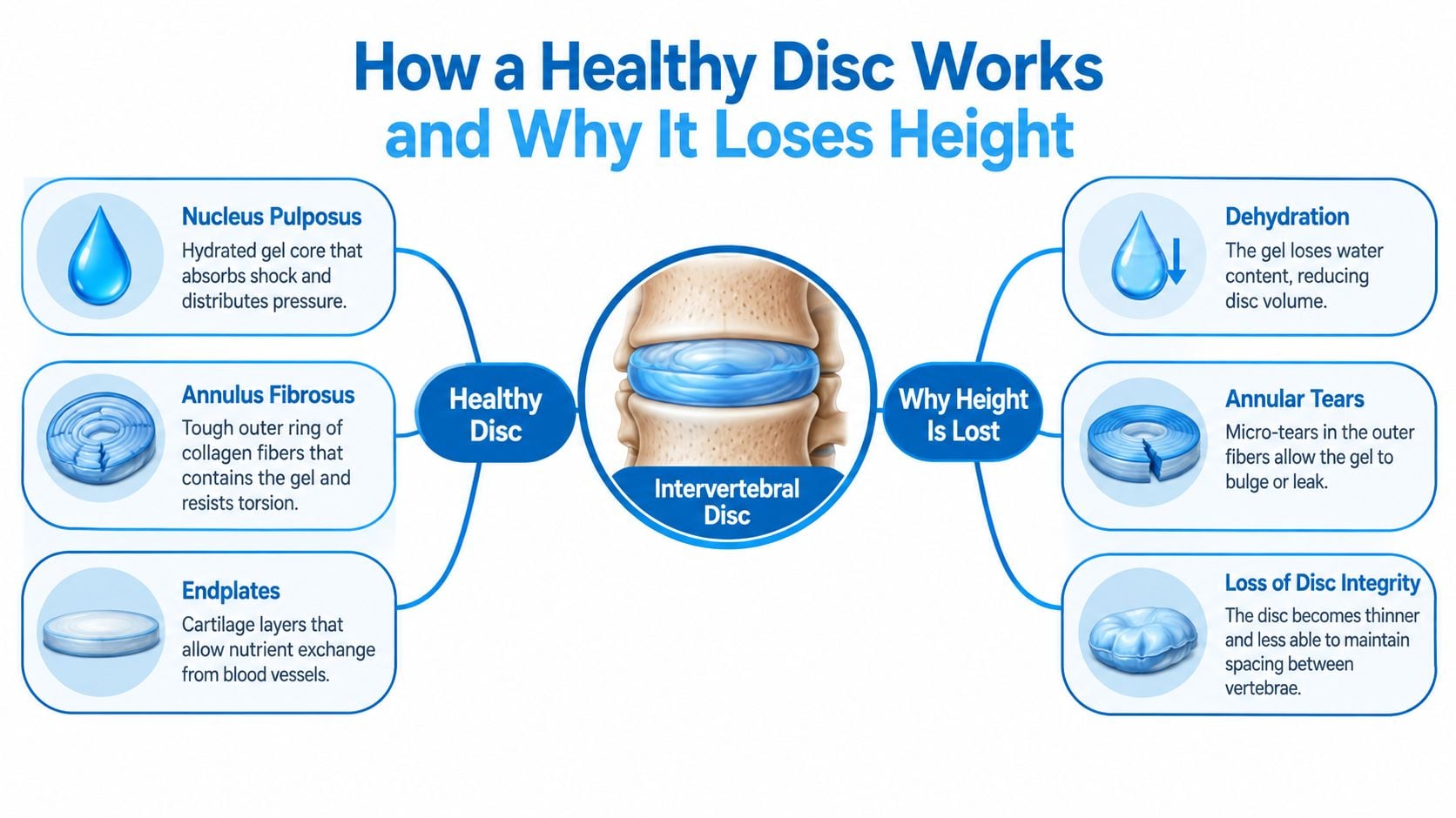
A healthy intervertebral disc keeps neighboring vertebrae apart while helping the spine carry and transfer loads. Its nucleus pulposus is a hydrated, gel-like center that spreads pressure as the spine bends, rotates, or bears weight. Surrounding it, the annulus fibrosus forms a layered ring of collagen fibers. This ring contains the nucleus and helps resist twisting.
Thin cartilage endplates lie above and below the disc. They support movement of nutrients into disc tissue, which has limited direct blood supply. Together, these structures create a flexible cushion that absorbs load and preserves space between the vertebrae.
Why hydration affects height
Disc height depends partly on the nucleus retaining water and maintaining pressure inside the disc. Degeneration alters the disc's extracellular matrix, making it harder to hold fluid. The annulus and the endplate environment may also weaken. Over time, the disc can lose volume, reducing the space between the vertebrae.
Unlike a bone collapse, this process involves water content, collagen structure, mechanical integrity, and the tissues around the disc. Reduced hydration may appear before substantial height loss. A disc with reduced height may also show signal loss, fissures, bulging, osteophytes, or endplate changes.
Different biological patterns
Degeneration follows different biological routes. A review of intervertebral-disc degeneration describes an endplate-driven pattern that may begin before age 30, has relatively high heritability, and appears more often in the upper lumbar and thoracic regions. An annulus-driven pattern generally progresses after age 30, is associated with radial fissures or disc prolapse, and more often affects the lower lumbar spine (review of disc degeneration mechanisms).
The same report label can therefore reflect different underlying processes. Spinal level, tissue pattern, mechanical stresses, and examination findings help determine what the image means for an individual.
As disc integrity declines, pressure may distribute less evenly. Segmental motion can change, facet joints may carry more load, and the opening around a nerve can become smaller. These effects may matter clinically, but disc height on an image remains a finding that must be matched with symptoms and examination results.
Common Causes Behind Disc Height Loss
A scan may show a narrowed disc space even when the cause is not immediately clear. The more useful question is which pattern fits: gradual degeneration, injury, mechanical overload, or change related to prior treatment. That distinction helps clinicians decide how closely the image should be matched with symptoms, examination findings, and medical history.
Gradual degeneration and cumulative loading
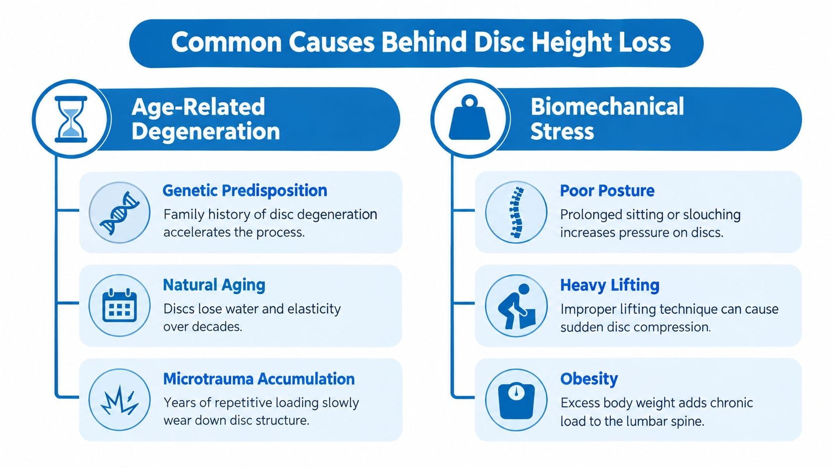
Disc biology reflects inherited factors, time, smoking exposure, repetitive vibration, sustained loading, and previous injury. Repeated stress does not mean one lifting event caused the finding. It may reflect years of occupational loading, sports activity, prolonged static posture, or ordinary movement acting on tissue with a particular biological susceptibility.
Different discs can change at different rates. Longitudinal evidence has associated faster height loss with degenerated discs, approximately 3% per year, compared with less than 1% per year in morphologically normal discs. Once the disc's structure has weakened, narrowing may progress more quickly (review of disc degeneration and height loss). These figures describe group patterns, not a timetable for one person.
Injury and acquired change
A vertebral compression fracture can alter the relationship between the bones and the disc space. Repetitive microtrauma may also contribute to structural change when work or sport involves frequent loading, vibration, bending, or rotation. Previous spinal surgery can change disc height or force distribution, depending on the procedure and the spinal level involved.
A 152-participant MRI and CT study found that mean disc height declined from 12.65 ± 0.69 millimeters in degeneration grade I to 7.34 ± 1.31 millimeters in grade V, an approximate 42% reduction (MRI and CT study of disc height and degeneration). The result illustrates how advanced degeneration can narrow the disc space. It does not establish that every person with a similar measurement has the same pain or functional limitations.
Questions that clarify the cause
A clinician may ask whether symptoms began after trauma, whether discomfort remains in the back or travels into a limb, and whether surgery affected the same level. Smoking, repetitive occupational stress, body habitus, conditioning, posture, and movement habits add context, but none proves a single cause by itself.
For a patient-friendly explanation of the broader condition associated with disc narrowing, see degenerative disc disease and its clinical context. The imaging finding becomes clinically meaningful when its pattern fits the history and examination.
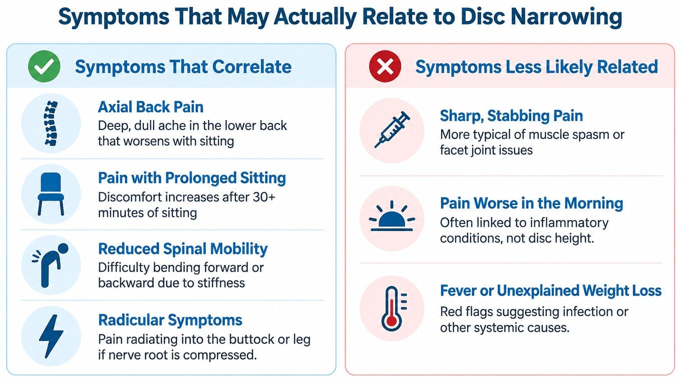
Symptoms That May Relate to Disc Narrowing
A scan may show disc height loss even when a person has no pain. The finding becomes clinically relevant only when its location, mechanical effects, symptoms, and examination fit together. The wording on an imaging report cannot establish the pain source by itself.
Pain that stays in the back or neck can arise from mechanical stress around a degenerated motion segment. Sitting, standing, bending, or repeated activity may increase it, while changing position or reducing the load may ease it. Stiffness after inactivity can occur too, although stiffness alone does not identify the disc as the source.
Indirect effects can matter more than the measurement
As the disc space narrows, the opening through which a nerve root passes may become smaller. Radiating pain, tingling, numbness, or weakness can follow, even when the report mentions narrowing rather than a large disc herniation. The facet joints may also carry more load, and altered movement between the vertebrae can produce another mechanical pain pattern.
A practical symptom history includes:
- Location: Pain limited to the back or neck differs from pain extending into a buttock, leg, shoulder, or arm.
- Distribution: A consistent one-sided path can suggest nerve irritation.
- Triggers: Sitting, walking, bending, coughing, lifting, and standing may offer different clues.
- Neurologic change: Weakness, reduced sensation, or altered reflexes makes a focused nerve examination more important.
- Functional effect: Reduced walking tolerance or difficulty using an arm may matter more than alarming scan language.
The distinction between isolated spinal aching and radiating symptoms is explained in this clinical guide to sciatica and pinched nerves.
Symptoms that need prompt attention
Progressive weakness, new difficulty controlling the bladder or bowel, or numbness in the saddle region requires urgent medical assessment. Fever, unexplained weight loss, significant trauma, or a history of serious systemic illness also changes the evaluation.
These warning signs do not prove that disc height loss is responsible. They require clinicians to consider nerve compression, infection, fracture, cancer, or another condition instead of assuming routine degeneration.
The MRI's appearance matters less than whether the level, mechanism, and examination explain what the patient is experiencing.
How Doctors Evaluate and Measure Disc Height
No single imaging test answers every question. Each modality shows a different part of the problem, and clinicians interpret the images alongside the history and physical examination.
| Modality | What it shows best | Main limitation |
|---|---|---|
| X-ray | Disc-space narrowing, alignment, bone spurs, and broad structural relationships | Limited detail of discs, nerves, and soft tissues |
| CT | Bone detail, endplate integrity, fractures, osteophytes, and postoperative anatomy | Uses radiation and provides less soft-tissue detail than MRI |
| MRI | Disc hydration and signal, annular fissures, nerve-root compression, stenosis, and endplate changes | Supine positioning may not reproduce symptoms under standing load |
What the measurements mean
A radiologist may compare disc height with adjacent levels or assess a normalized disc-height index. Measurements can vary with spinal level, age, body habitus, posture, imaging position, and technique. For that reason, repeat comparisons are most useful when the same level, modality, positioning, and measurement approach are used.
MRI also shows more than height. Darker T2 signal can indicate reduced water content. Annular fissures, bulges, protrusions, endplate changes, foraminal narrowing, and nerve-root contact may alter the clinical interpretation. A grading system such as Pfirrmann helps describe degeneration severity, but a grade isn't a diagnosis of pain.
A 2022 MRI study of 85 patients with low-back pain found a dose-response relationship between degeneration grade and lower disc height. After adjustment for age, sex, and body mass index, each higher degeneration level was associated with approximately 1.25 to 1.76 millimeters lower disc height depending on the lumbar level (MRI study of degeneration grade and disc height).
When imaging adds value
Imaging is more useful when symptoms persist, neurological findings appear, trauma raises concern for structural injury, or a procedure is being considered. Repeat imaging shouldn't be automatic because a prior scan showed age-related changes. A meaningful change in symptoms, examination, or treatment planning is usually the reason to reassess anatomy.
Treatment Options from Conservative Care to Surgery
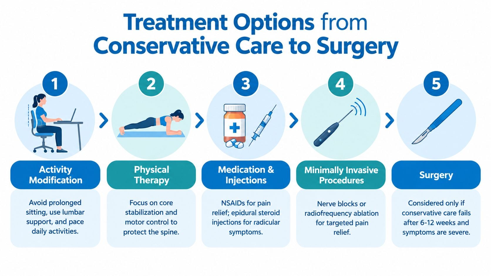
Treatment decisions should rest on the clinical problem rather than the measurement alone. Painless narrowing often calls for explanation, monitoring, and sensible health maintenance rather than a procedure aimed at changing an image. Nerve compression, instability, or disabling mechanical pain may require a more structured plan.
Start with function and load tolerance
Activity modification can reduce repeated aggravation while preserving useful movement. A physical therapist may address trunk and hip strength, movement control, lifting mechanics, walking tolerance, and positions that ease symptoms. Medication may provide short-term symptom control when medically appropriate.
The treatment target should match the suspected pain source. An epidural injection may be considered for nerve-root irritation with radiating symptoms. Facet or medial-branch blocks can help assess facet-mediated pain, while trigger-point treatment may address a myofascial contributor. These procedures do not rebuild a disc. They may manage or clarify one specific pain pathway.
Match the procedure to the objective
| Clinical problem | Possible treatment direction | Structural goal |
|---|---|---|
| Mechanical pain without neurological compromise | Rehabilitation, activity changes, medication, selected procedures | Improve function and loading tolerance |
| Radiating pain from nerve irritation | Targeted injection or decompression when indicated | Reduce nerve pressure or inflammation |
| Persistent facet-mediated pain | Diagnostic blocks and, in selected cases, radiofrequency treatment | Reduce pain from facet nerves |
| Collapse with instability or deformity | Reconstruction, which may include fusion | Restore alignment and stabilize the segment |
| Carefully selected disc-level disease | Motion-preserving surgery such as artificial disc replacement | Preserve motion while addressing the diseased level |
Biologic and regenerative treatments remain uncertain. Claims that a treatment will reliably restore a degenerated disc's original hydration, structure, or height deserve caution. Symptom improvement and structural regeneration are separate outcomes.
Surgery becomes a consideration when a mechanical or neurological problem is clearly defined, symptoms remain disabling after appropriate nonsurgical care, or progressive weakness and instability alter the risk-benefit balance. Decompression can relieve pressure on a nerve without biologically restoring disc height. Fusion can stabilize a painful, collapsed, or unstable segment. Motion-preserving surgery may suit only selected patients whose alignment, facet integrity, spinal level, and other clinical factors are appropriate.
For information about one motion-preserving surgical option, see artificial disc replacement surgery. The final choice should reflect examination findings, imaging correlation, overall health, spinal level, and the patient's functional goals.
Prognosis, Prevention, and Living With the Finding
Disc height loss doesn't equal inevitable disability or surgery. The imaging change may continue as part of structural aging while symptoms remain stable, improve, or never appear. The smaller group with progressive collapse, nerve compression, instability, deformity, or persistent function-limiting symptoms needs more active follow-up.
Prevention focuses on modifiable contributors rather than trying to make an aged disc look young again. Regular movement supports conditioning. Strengthening the trunk and hips can improve load control. Smoking cessation protects overall tissue health, while weight management can reduce sustained lumbar loading. Ergonomic adjustments, sensible lifting technique, and avoiding long periods in one position may also improve tolerance.
A practical plan after a report
- Read the full report: Identify the spinal level, associated narrowing, nerve findings, and any mention of fracture or instability.
- Map the symptoms: Note where pain travels, what triggers it, and whether numbness or weakness is changing.
- Get an examination: Strength, reflexes, sensation, gait, and movement findings help connect anatomy with function.
- Avoid treating the image alone: A structural abnormality without matching symptoms may need observation rather than intervention.
- Act quickly for red flags: Progressive weakness, saddle numbness, or bowel and bladder changes require urgent assessment.
A 2025 retrospective cervical-surgery study found that 24.1% of patients had more than 1 millimeter of intervertebral disc-height loss at a median 61-month follow-up, showing that postoperative structural change can occur even when the original procedure addressed another problem (study of postoperative cervical disc-height change). That finding reinforces an important distinction: maintaining measured height, restoring height, relieving nerve pressure, and improving symptoms are separate goals.
Interventional Pain Management evaluates back and neck symptoms by combining medical history, physical examination, and imaging, with options that may include rehabilitation, image-guided injections, minimally invasive procedures, and spine-surgery consultation when indicated. Patients with disc height loss can visit Interventional Pain Management to request an evaluation focused on matching the scan with the symptoms and choosing an appropriate next step.