
A patient with scoliosis rarely asks only, “Which operation corrects the curve?” More often, the central question is harder: Is the curve still growing, is pain coming from nerve compression, how much function could surgery restore, and what risks can the patient reasonably accept?
A growing child, a nearly mature adolescent, a healthy adult with stenosis, and a frail older adult may all carry the same diagnosis but need entirely different treatment plans. The right scoliosis surgery option depends on age, skeletal maturity, curve flexibility, symptoms, bone quality, and the patient's definition of success.
Table of Contents
- When Surgery Enters the Conversation
- How Scoliosis Surgery Evolved
- Comparing the Main Surgical Approaches
- Correction Magnitude and Complication Risk
- The Frailty Question Most Articles Skip
- Matching the Right Option to the Right Patient
- What Robotic and Navigation Assistance Change
- Choosing Your Next Step With Confidence
When Surgery Enters the Conversation
A middle-aged woman with a progressive lumbar curve, worsening leg pain, and unsuccessful physical therapy may reasonably wonder whether surgery is now the only path. It usually isn't. The first task is to identify what is really driving the disability. A curve may be visually striking but relatively painless, while a smaller degenerative curve may narrow the spaces around the nerves and make walking intolerable.
Surgery enters the discussion when the expected benefit becomes greater than the burden of reconstruction. Common triggers include:
- Documented progression: A curve that continues to worsen after skeletal maturity deserves closer surgical evaluation, particularly when alignment and function are deteriorating.
- Persistent nerve-related symptoms: Leg pain, numbness, weakness, or walking limitation linked to imaging-confirmed compression may justify decompression, fusion, or both.
- Severe thoracic effects: A major thoracic deformity can affect breathing and physical endurance.
- Functional or personal impact: Visible deformity matters when it limits movement, clothing choices, work, sleep, or self-image.
- Failure of appropriate nonoperative care: Exercise-based rehabilitation, medication management, injections, and other targeted treatments may help some patients, but persistent structural compression changes the conversation.
The four surgical families
The main options fall into four broad groups:
- Posterior spinal fusion, the established workhorse for definitive correction.
- Anterior, lateral, or combined approaches, which reach the disc spaces from the front or side and may be paired with posterior fixation.
- Growth-preserving procedures, including expandable growing rods and other systems designed for young children with substantial growth remaining.
- Fusionless procedures, including vertebral body tethering, which attempt to guide growth while retaining motion in selected adolescents.
For adolescent idiopathic scoliosis, spinal fusion is generally considered when a curve reaches approximately 45 to 50 degrees, or when substantial progression is likely after skeletal maturity, according to the Scoliosis Research Society's surgical treatment guidance. That threshold is not a stand-alone command to operate. Curve flexibility, balance, symptoms, growth remaining, and progression all matter.
Practical rule: The operation should solve the patient's dominant problem, not merely produce an impressive postoperative image.
How Scoliosis Surgery Evolved
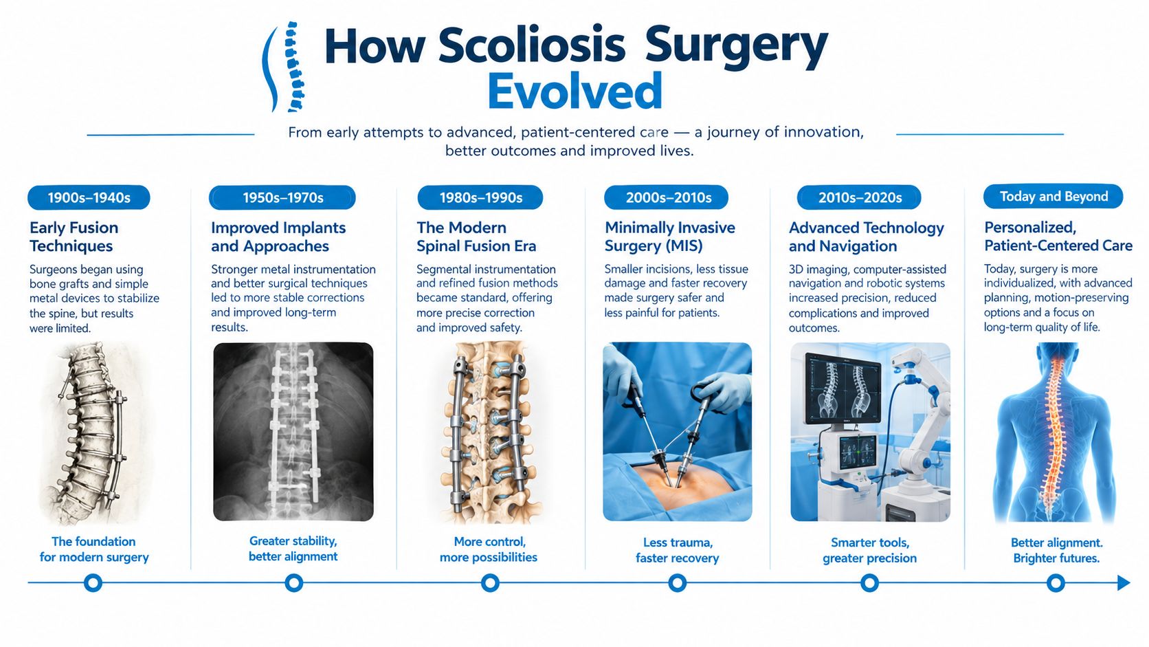
Early scoliosis surgery focused on one achievable goal, stopping the curve from worsening by joining vertebrae together. Russell Hibbs reported spinal fusion for three patients with Pott's disease, and his institution later treated 59 scoliosis cases between 1914 and 1919 using fusion techniques, as documented in the historical timeline from the Scoliosis Research Society. Fusion established that a curved spine could be stabilized, but it offered limited mechanical correction.
Instrumentation changed the field. In 1963, John Moe presented results from 66 prospectively collected patients treated with Harrington's method. Rods and attached implants gave surgeons the mechanical advantage to correct the spine rather than immobilize it. The technique improved correction, supported earlier mobilization, reduced pseudarthrosis compared with uninstrumented fusion, and lowered reoperation rates. It also exposed new problems, including long rigid constructs and postoperative alignment concerns.
From rigid correction to three-dimensional planning
Later systems, including segmental fixation approaches, spread corrective forces across more levels. Surgeons gained better control of rotation, coronal balance, and sagittal alignment instead of treating the spine as a simple side-to-side bend. Modern posterior fusion still uses that basic principle, but with more detailed imaging, stronger fixation strategies, and more deliberate attention to the levels that should remain mobile.
Historical results also explain why technique matters. One review of early deformity surgery recorded pseudarthrosis in 54 of 180 correction-and-fusion cases, while 29% of 214 patients lost all correction, according to the same Scoliosis Research Society historical source. Contemporary planning aims to avoid those failures through better patient selection, fixation, bone-healing strategy, and alignment targets.
The pediatric branch of scoliosis surgery developed in response to a different problem. A very young child may need correction but cannot safely lose the growth of a large portion of the spine or chest. Expandable growing rods span the curve while avoiding injury to spinal growth tissues. Vertebral body tethering uses implants and a flexible cord on the convex side to restrain growth there while the opposite side catches up.
Fusionless treatment remains selective. A pooled analysis cited by the Scoliosis Research Society reported an 8.66% unplanned reoperation rate after vertebral body tethering across 23 studies, reinforcing that motion preservation can involve a different form of surgical uncertainty. Each generation solved a specific limitation, and each created a new set of trade-offs.
Comparing the Main Surgical Approaches
A growing child, a mature adolescent, and an adult with painful deformity should not receive the same surgical plan. The right choice depends on growth remaining, the main goal of treatment, curve flexibility, and whether the priority is progression control, pain relief, or three-dimensional correction. Technique follows that decision.
| Approach | Surgical Access | Main Role | Best-Fit Patient |
|---|---|---|---|
| Posterior spinal fusion | Incision through the back with screws, hooks, rods, and bone graft | Definitive correction when reliable fixation and fusion are needed | Mature adolescent or adult with a progressive, symptomatic, or structurally significant curve |
| Anterior or combined fusion | Access through the front, side, or staged front-and-back routes | Disc-level correction and selected strategies to limit posterior exposure or fusion levels | Patient whose curve pattern or alignment benefits from direct disc access |
| Growth-preserving techniques | Implants placed along the curve, with planned or externally controlled expansion | Progression control while allowing continued spinal and thoracic growth | Young child with substantial growth remaining and a progressive curve |
| Fusionless techniques | Usually anterior or lateral access with a tether or similar implant | Growth modulation while preserving motion | Skeletally immature adolescent meeting strict curve and flexibility criteria |
Posterior spinal fusion
For a mature adolescent with a substantial progressive curve, posterior instrumented fusion remains the most established option. Screws, hooks, or other implants connect to rods, and bone graft encourages the treated vertebrae to fuse in the corrected position. The main advantage is dependable structural control. The trade-off is permanent loss of motion across the fused levels, along with a substantial rehabilitation period.
Before discussing implant designs, patients should understand what spinal fusion involves in scoliosis correction. The number of levels fused matters because it affects both correction and the motion that remains afterward.
Anterior and lateral approaches
These operations reach the disc spaces from outside the spinal canal. That access can help a surgeon release a stiff segment, place an interbody device, or build correction across several levels without relying on a single posterior exposure.
A staged front-and-back plan may suit a patient who needs correction from both directions. It can distribute the reconstruction between procedures, but staging may mean two operations, additional anesthesia, and a longer overall treatment period. Lateral surgery alone is not a substitute for posterior reconstruction when the deformity includes major sagittal imbalance, severe rigidity, extensive central stenosis, or fixed pelvic compensation. Those findings may require direct posterior decompression, osteotomies, and longer fixation.
In adults with flexible degenerative scoliosis, lateral interbody fusion approaches and posterior approaches generally produce similar functional and radiographic outcomes, according to a comparative review of adult scoliosis surgery.
Growth preservation and fusionless correction
Growing rods are intended for young children whose spine and thorax still have substantial development ahead. They control the curve while allowing continued growth, usually through planned or externally controlled expansion.
Tethering has a narrower role. NICE describes a representative target group for minimally invasive fusionless correction as children aged 8 to 17 with skeletal immaturity, a flexible major curve up to 60 degrees, and correction to 30 degrees or less on side-bending radiographs, as summarized by the Scoliosis Research Society. These criteria show why tethering is not a less invasive version of fusion. It requires remaining growth, suitable flexibility, and a curve pattern that can respond to growth modulation. When the primary goal is reliable correction in a mature spine, posterior fusion is usually the more predictable choice.
Correction Magnitude and Complication Risk
The right correction depends on the patient's age, remaining growth, and goal. A younger patient may prioritize preserving growth, while an adult may seek pain relief, improved balance, or visible deformity correction. The operation should deliver the correction that goal requires, not the largest possible change.
Flexible curves may respond to interbody positioning with posterior fixation. A rigid deformity may require releases or a spinal osteotomy, including a three-column osteotomy, to restore alignment. Greater correction demands more surgical disruption, which raises concern about blood loss, neurologic injury, wound healing, bone quality, and rehabilitation.
| Surgical Approach | Typical Coronal Correction | Major Complication Rate | Average Blood Loss | Average Hospital Stay |
|---|---|---|---|---|
| Lateral interbody fusion with or without posterior fixation | Often meaningful for flexible deformity, with lateral-plus-posterior correction reported at approximately 40% to 75% | Varies by staging, fixation, patient health, and deformity complexity | Often lower than open posterior reconstruction, but patient-specific | Varies by approach and staging |
| Posterior instrumented fusion | Depends on flexibility, levels fused, and alignment targets | Varies substantially with complexity | Varies substantially with complexity | Varies by reconstruction and medical risk |
| Three-column osteotomy | Larger correction for rigid or severely malaligned deformity | Up to 66% in the highest-risk category reported in a review | Often substantial | Often prolonged and rehabilitation-intensive |
For an adult whose priority is coronal deformity correction, lateral interbody fusion combined with posterior percutaneous screws can provide substantial correction. Lateral-only surgery usually produces less correction and limited improvement in global sagittal balance. A meta-analysis of 29 studies involving 1,228 patients reported pooled fusion of 95.9% for anterior or lateral minimally invasive approaches. Construct or hardware-related complications were 4.4% for anterior or lateral techniques versus 5.2% for posterior techniques, as reported in the adult scoliosis review.
The risk changes sharply when the goal requires major reconstruction. A review of adult scoliosis surgery found complication rates reaching 66% with three-column osteotomies, compared with about 45% for non-three-column procedures in the cited series. Across 4,980 Scoliosis Research Society cases, at least one perioperative complication occurred in 10.5%, and mortality was 0.3%, according to the review of complication risk in adult scoliosis surgery.
Ask two direct questions before accepting a proposed operation: What correction is needed to improve my function or appearance, and does that benefit justify the surgical burden? A larger correction is not automatically a better result.
The Frailty Question Most Articles Skip
Curve size dominates online discussions because Cobb angles are easy to measure. For older adults, however, frailty can change risk more than age or curve magnitude alone. Frailty reflects reserve, nutrition, strength, medical complexity, bone health, and the ability to recover from a major physiologic insult.
In one adult spinal-deformity cohort undergoing long-segment corrective surgery, major postoperative complications occurred in 29.8% of frail patients versus 10.5% of non-frail patients, and frailty independently predicted complications with an adjusted odds ratio of 2.77, according to the study of frailty and adult spinal deformity outcomes. The same evidence links frailty with greater mortality, transfusion, thromboembolism, overall complications, and reoperation.
What the consultation should measure
A serious adult evaluation should address more than standing radiographs:
- Medical reserve: Cardiopulmonary disease, kidney function, diabetes, medication burden, and prior surgical tolerance.
- Bone strength: Osteoporosis can compromise fixation and fusion, so bone-density assessment may change the plan.
- Nutrition and muscle: Poor intake, low strength, and sarcopenia can delay wound healing and rehabilitation.
- Functional goals: Walking farther, sleeping, standing, working, and maintaining independence may matter more than a perfectly straight image.
- Support after discharge: A long reconstruction can require substantial help, therapy, equipment, and temporary changes in living arrangements.
The frailty evidence does not mean frail patients cannot benefit. Frail patients in the cited cohort achieved meaningful improvements in alignment and health-related quality of life, with no statistically significant difference in those improvements compared with non-frail patients. It does mean the operation must be justified by a clear functional goal, not by radiographic ambition alone.
A candid surgical recommendation may be rehabilitation, targeted decompression, pain treatment, or medical optimization before any fusion is considered.
Matching the Right Option to the Right Patient
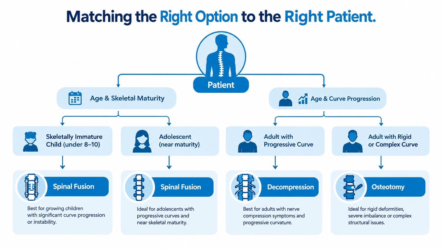
The surgical menu should follow the patient, not the other way around. Age and skeletal maturity establish the first branch, but curve flexibility, progression, symptoms, and long-term goals determine the final recommendation.
The young child with growth remaining
A young child with a progressive curve presents a growth problem as much as a curvature problem. Traditional growing rods or other growth-preserving systems may control the deformity while allowing the spine and chest to develop. Some systems require planned expansions, while newer designs can reduce the need for repeated lengthening procedures.
These techniques aren't appropriate for every child. The team must consider the underlying diagnosis, thoracic development, curve progression, skin and soft-tissue condition, and the family's ability to manage repeated follow-up.
The adolescent near skeletal maturity
A nearly mature adolescent with a substantial, progressive curve is the clearest candidate for posterior spinal fusion. Fusion is more established in this setting because the main growth-related reason to preserve motion has diminished, while the need for durable correction remains.
Tethering or another fusionless strategy may fit a carefully selected patient with skeletal immaturity, a flexible curve, and a strong preference for preserving motion. The family must also accept that eligibility is narrow and later procedures may be necessary. A motion-preserving label doesn't remove surgical risk.
The adult with degeneration and stenosis
An adult with leg pain, foraminal narrowing, and a flexible degenerative curve may need decompression with short-segment fusion rather than a long deformity reconstruction. Lateral interbody fusion can be useful when disc collapse and foraminal narrowing are central problems, particularly when posterior muscle disruption should be limited.
Severe rigidity, major sagittal imbalance, fixed pelvic compensation, or extensive central stenosis changes that calculation. Those findings may require direct posterior decompression, releases, or a longer construct.
The frail older adult
A frail patient with osteoporosis, sagittal imbalance, and limited walking tolerance needs a different conversation. A staged or less invasive plan may be safer, but only if it addresses the actual pain generator and alignment problem. In some cases, nonoperative care is the more responsible recommendation.
The decisive question is not whether an operation can straighten the spine. It is whether the patient can recover enough function to make the burden worthwhile.
What Robotic and Navigation Assistance Change
Robotic and navigation systems change how implants are planned and placed. They do not simplify a complex deformity or replace surgical judgment. The patient's age, remaining growth, pain pattern, and correction goal still determine whether surgery is appropriate and which levels require treatment.
A navigation workflow may use preoperative imaging or intraoperative scans to create a three-dimensional map. The system tracks the patient's anatomy as positioning changes and guides the surgeon while each screw pathway is prepared. Robotic assistance can help execute a planned spinal instrumentation trajectory, but the surgeon still decides which levels to treat, how to correct the curve, and whether the anatomy makes that trajectory unsafe.
A 2025 systematic review and Bayesian network meta-analysis in adolescent idiopathic scoliosis found better pedicle-screw accuracy with robot-assisted placement than with freehand fluoroscopic and navigation-assisted techniques. It found no significant differences in blood loss, Cobb-angle correction, or hospital stay, while operative time was longer, as reported in the published evidence review on robotic assistance.
| Technique | Pedicle Breach Rate | Operative Time Impact | Radiation Exposure | Learning Curve |
|---|---|---|---|---|
| Freehand placement | Depends heavily on anatomy and surgeon experience | Often avoids registration and setup time | Imaging requirements vary | Familiar technique, but demanding in rotated anatomy |
| Navigation-assisted placement | Accuracy may improve in complex anatomy | Can add setup and imaging time | Intraoperative imaging may increase exposure | Requires reliable registration and workflow |
| Robotic-assisted placement | Published evidence supports improved placement accuracy | Often longer than freehand techniques | Depends on the imaging and registration protocol | Requires team experience and consistent planning |
Accuracy matters most when pedicles are small, rotated, or difficult to identify. It does not automatically produce less pain, faster recovery, a higher fusion rate, or better curve correction. A separate umbrella review associated robotic assistance with improved screw precision, fewer revisions, shorter length of stay, and lower blood loss, while also noting longer operations, as summarized in the same published review.
Patients should ask:
- Which outcome is the system expected to improve?
- Does the team use it for planning, navigation, robotic guidance, or all three?
- How does the system affect imaging and radiation exposure?
- What happens when the planned trajectory conflicts with the anatomy?
- Are the surgeon's results comparable across assisted and non-assisted cases?
Choosing Your Next Step With Confidence
A useful consultation produces a recommendation that can be explained in plain language. The surgeon should be able to state the primary problem, the proposed operation, the expected functional benefit, the main risks, and the reason less extensive options may not be enough.
The following questions deserve direct answers:
- Case experience: How often does the surgeon perform the proposed operation and similar deformity cases?
- Personal outcomes: What complications has the surgeon seen with this approach, and how are those complications managed?
- Technical plan: Which levels will be treated, which approach will be used, and what implant strategy is planned?
- Recovery demands: What should the patient expect at approximately three, six, and twelve months?
- Fallback plan: What happens if pain persists, fusion does not occur, or the correction changes?
- Nonoperative alternative: What is the likely course if surgery is deferred?
When no surgery is the right answer
Adults with stable, moderate curves and tolerable symptoms may reasonably choose structured exercise, targeted pain treatment, activity modification, and periodic imaging. A decision against surgery isn't neglect when the symptoms are manageable and the risks outweigh the expected functional gain.
The decision should also account for whether pain comes from the curve itself, stenosis, a disc, a hip, or another condition. Treating the wrong pain generator can produce an impressive reconstruction without meaningful relief.
The strongest recommendation is simple: match the patient profile to the procedure family first, then choose the surgeon. A mature adolescent with a progressive curve may fit posterior fusion. A growing child may need growth preservation. An adult with stenosis may need targeted decompression and limited fusion. A frail older adult may need optimization or nonoperative care before any reconstruction is considered.
Interventional Pain Management evaluates spine-related pain through coordinated pain, orthopedic, and surgical services, including nonoperative treatment, image-guided procedures, decompression, fusion, and scoliosis correction when clinically appropriate. Patients in New Jersey and Staten Island can review their options and request an individualized evaluation through Interventional Pain Management.