
A patient may start with a stiff lower back, keep working, and manage the discomfort with over-the-counter medication. Months later, the complaint may sound completely different: burning along the outer edge of one foot, numb toes, heel pain after walking, or a sudden limp. The back and foot can appear to be separate problems, but the symptoms may share one neurological or mechanical pathway.
That distinction matters. Low back pain affected 619 million people globally in 2020, and the World Health Organization projects that the number could reach 843 million by 2050. The condition is common enough that clinicians can easily focus on the loudest symptom instead of tracing the full chain from lumbar spine to pelvis, leg, ankle, and foot. The available global back-pain data supports a practical conclusion: persistent back and foot symptoms deserve an integrated assessment rather than two disconnected appointments.
Table of Contents
- Why a Patient With Back Pain Ends Up Complaining About the Foot
- The Four Ways the Back and the Foot Are Clinically Connected
- Nerve Pathways That Send Back Pain Down to the Foot
- How Foot Mechanics and Gait Change Load on the Lower Back
- How Specialists Actually Work Up Combined Back and Foot Symptoms
- Foot Diagnoses That Are Commonly Mistaken for Spine Problems
- Integrated Treatment Options Across Spine and Podiatric Specialties
- Choosing the Right Starting Point for Your Symptoms
Why a Patient With Back Pain Ends Up Complaining About the Foot
A warehouse supervisor may first report lower back pain after long shifts spent standing, lifting, and walking across concrete floors. The patient takes anti-inflammatory medication, changes nothing about the work routine, and returns months later because the outer edge of the right foot burns during walking. A foot examination may suggest local irritation, while the history still contains clues pointing toward the lumbar spine.
The common mistake is to treat the back and foot as separate compartments. One clinician may order lumbar imaging without examining foot posture or gait. Another may recommend inserts or local foot treatment without checking sensation, reflexes, strength, or nerve tension. The patient then receives reasonable care for the wrong dominant mechanism.
The loudest symptom isn't always the source
Pain often announces itself at the point where tissue or nerve irritation becomes most noticeable. A compressed lumbar nerve can produce symptoms in the calf, ankle, or foot even when the back itself feels relatively calm. Conversely, a painful heel, unstable arch, or restricted ankle can alter walking enough to aggravate the pelvis and lower back.
This is why the first complaint doesn't always identify the origin. The foot may be the site of referred nerve pain, the back may be reacting to altered gait, or both regions may be contributing at the same time.
Practical rule: A back-and-foot assessment should ask not only where pain is felt, but also what movement, posture, nerve pathway, or local tissue reproduces it.
A single chain with two directions
The lower limb operates as a connected kinetic chain. Foot contact influences tibial rotation, knee alignment, pelvic motion, and lumbar loading. The lumbar nerves influence muscle activation, sensation, and foot placement. When one link changes, another link may compensate.
That doesn't mean every case of flat feet causes back pain, or that every foot-burning complaint comes from sciatica. It means the workup should test both possibilities. A patient whose symptoms are unilateral, electric, and associated with weakness needs a different pathway from a patient with symmetrical heel pain that is worst during local loading.
The useful question isn't, “Is the problem in the back or the foot?” It is, “Which mechanism is driving the current disability, and which findings would change treatment?”
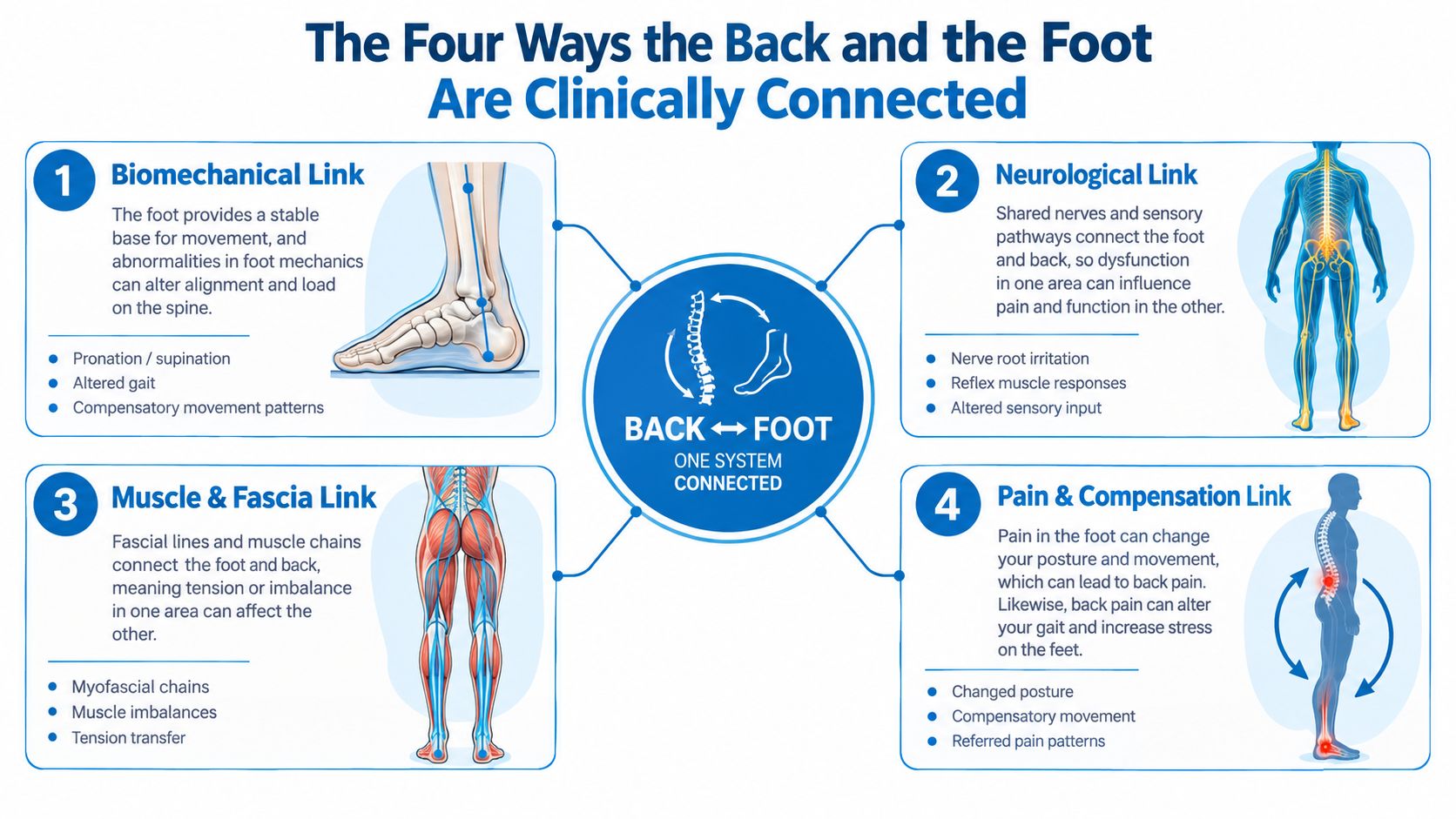
The Four Ways the Back and the Foot Are Clinically Connected
Four mechanisms account for most confusing back-and-foot presentations. They can occur separately, but mixed cases are common.
Radiculopathy resembles a kinked hose
A lumbar nerve root can become irritated or compressed near the spine. Like a kink in a hose, the problem occurs upstream while the effect appears farther along the line. The patient may feel sharp, electric, or burning pain through the buttock, leg, ankle, or foot. Numbness, reduced reflexes, or weakness make nerve-root involvement more concerning.
Radiculopathy is not diagnosed by pain location alone. The clinician compares the symptom pattern with strength, sensation, reflexes, nerve-tension tests, and imaging when indicated.
Referred pain acts like a crossed wire
Pain from a disc, facet joint, or sacroiliac structure can be felt away from the irritated tissue. This referred pain is often duller and less clearly mapped than radicular pain. It may reach the buttock or thigh without producing true sensory loss in the foot.
The patient can therefore describe “foot pain” when the dominant complaint is a diffuse ache caused by altered loading higher in the chain. A careful examination helps separate referred pain from nerve-root pain.
Peripheral neuropathy creates a short circuit
Peripheral nerves can become dysfunctional independently of the spine. Symptoms may begin in the toes or soles, often with burning, prickling, reduced sensation, or sensitivity to light touch. When the pattern is bilateral or follows a stocking-like distribution, a systemic or peripheral nerve process deserves consideration rather than automatic attribution to lumbar disease.
Electrodiagnostic testing can help when the examination cannot distinguish a nerve-root problem from a peripheral neuropathy.
Gait compensation behaves like a misaligned axle
A painful back can shorten stride, reduce push-off, or shift weight toward one side of the foot. A collapsed arch, painful heel, or limited ankle can produce the reverse adaptation, increasing rotation through the leg and pelvis. The spine and foot then reinforce each other's irritation.
Treatment works best when it matches the dominant mechanism. A nerve block won't correct a local tendon overload, and an orthosis won't decompress an irritated lumbar nerve root. The examination has to establish which problem is primary before either is treated aggressively.
Nerve Pathways That Send Back Pain Down to the Foot
Dermatomes are useful mental maps, not rigid borders. Adjacent nerve roots overlap, and individual anatomy varies, so one painful zone can't prove a specific level. Still, the pattern of pain, altered sensation, weakness, and reflex change can help the clinician decide whether the lumbar spine deserves priority.
The L4 pattern commonly travels through the front or inner portion of the lower leg toward the medial ankle and medial arch. The L5 pattern is often associated with the outer shin, the top of the foot, and the great toe. The S1 pattern tends to involve the back of the calf, the outer foot, and the small-toe side. Symptoms along the posterior calf and lateral foot border can also involve S2-related pathways, particularly when the history and examination suggest a more extensive sacral distribution.
| Nerve Root | Foot Zone | Typical Sensation | Reflex / Motor Check |
|---|---|---|---|
| L4 | Medial ankle and medial arch | Tingling, aching, or altered sensation along the inner lower leg | Knee reflex, ankle dorsiflexion and inversion |
| L5 | Dorsum of the foot and great toe | Electric or burning symptoms over the top of the foot | Great-toe extension, ankle dorsiflexion, heel walking |
| S1 | Lateral foot and small-toe side | Sharp or burning pain along the outer foot | Ankle reflex, plantarflexion, toe walking |
| S2 | Posterior calf and lateral foot border | Posterior leg discomfort with possible distal sensory change | Hamstring and plantarflexion assessment, broader neurological examination |
Radicular pain is more suspicious when it follows a plausible line and arrives with numbness, weakness, or reflex asymmetry. Referred pain is often less precise, more diffuse, and less likely to produce a clear neurological deficit. Local foot disorders usually reproduce symptoms with palpation, resisted tendon testing, joint loading, or a specific foot maneuver.
A symptom diary can make this distinction easier. Patients should record whether pain appears during sitting, lifting, walking, first steps after rest, toe-off, or prolonged standing. A concise description of burning, electric shocks, pins and needles, or dull pressure is more useful than just reporting “foot pain.” More detail about sciatica and pinched nerve symptoms can help patients prepare for a focused consultation.
How Foot Mechanics and Gait Change Load on the Lower Back
Pronation is a normal part of foot movement. The problem arises when excessive or poorly controlled pronation changes how the tibia, knee, pelvis, and lumbar spine move during repeated steps. The arch lowers, the tibia rotates inward, and the pelvis may accommodate the rotation. The lumbosacral region then receives repeated torsional loading rather than just absorbing vertical impact.
The Framingham Foot Study found that women with pronated foot function had higher odds of low back pain. The association remained after adjustment for age, weight, smoking, and depressive symptoms, with an odds ratio of 1.51 before adjustment and 1.48 after adjustment, as reported in the published foot-function analysis. This does not prove that pronation causes every case of back pain. It does show that foot posture can be a clinically relevant risk marker rather than a cosmetic observation.
The gait chain is more than the arch
A clinician watches how the heel contacts the ground, whether the arch collapses, how the knee tracks, and whether the pelvis shifts. A functional leg-length difference can tilt the pelvis and encourage an asymmetric spinal posture. The body may respond with a subtle trunk lean, uneven facet loading, or a shortened step on one side.
Common compensations include:
- Reduced stride: The patient limits the painful side's time on the ground.
- Avoided toe-off: The patient rolls through the foot without fully loading the forefoot or big toe.
- Pelvic shift: The trunk moves over the supporting leg to reduce discomfort.
- Trendelenburg-style movement: Weakness or pain around the hip changes pelvic control during single-leg stance.
The reverse pattern is just as important. A guarded back can cause the patient to shift weight forward, keep the hips and knees flexed, or avoid heel loading. That strategy may overload the forefoot, plantar fascia, Achilles tendon, or other local structures. A foot problem can therefore appear after a period of back protection, even though the original driver was spinal pain.
A structured Achilles tendinitis evaluation is useful when posterior heel pain follows a change in gait, activity, or push-off. The assessment should still include the lumbar region when symptoms radiate, weakness appears, or local tendon findings don't fully explain the complaint.
How Specialists Actually Work Up Combined Back and Foot Symptoms
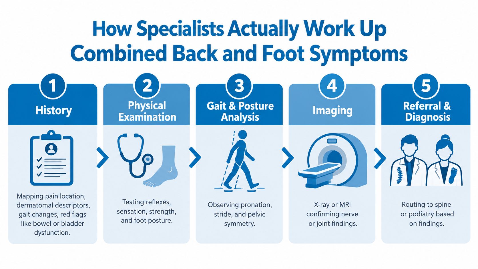
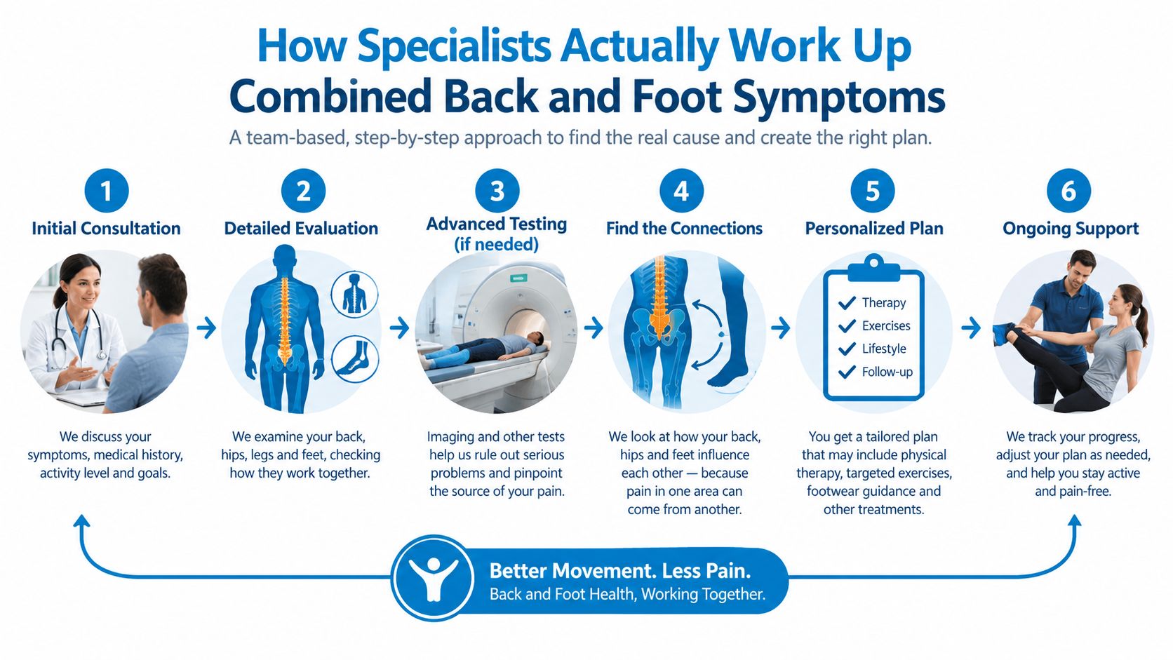
The most useful workup follows the symptom pathway instead of beginning with an isolated scan. Imaging can show structural changes that aren't causing the current pain, so the history and examination establish whether a finding is clinically relevant.
History comes first
The clinician maps the location and quality of pain, what triggers it, and how it travels. Burning or electric symptoms, numbness, weakness, changes in balance, and pain triggered by coughing or straining raise concern for nerve involvement. First-step heel pain, focal tendon tenderness, or pain linked to footwear suggests a different starting point.
The history also covers gait changes, work demands, previous injuries, systemic illness, and prior treatment. New bowel or bladder dysfunction, saddle numbness, or rapidly progressive weakness requires urgent medical assessment rather than routine foot treatment.
Examination links symptoms to anatomy
The physical examination may include straight-leg raise, femoral stretch, Slump testing, reflex assessment, strength testing, Babinski response, and clonus when clinically appropriate. Sensation is checked across relevant foot zones, sometimes with monofilament testing, while the clinician compares both sides.
The foot examination looks for focal tenderness, joint restriction, tendon pain, skin changes, pulses, arch behavior, and signs of nerve entrapment. Strength testing should include ankle dorsiflexion, plantarflexion, inversion, eversion, and great-toe movement when those actions correspond to the reported symptoms.
Posture and gait add the missing context
The Foot Posture Index six-criterion assessment, commonly called FPI-6, offers a structured way to classify foot posture. A study reported excellent inter-rater and test-retest reliability and concluded that FPI-6 is a valid clinical tool for assessing foot posture in people with low back pain. The FPI-6 research is relevant because reliable screening can identify a distal mechanical contributor without assuming that the foot is the sole cause.
Gait analysis may be observational or instrumented. It can reveal pronation, asymmetric stride, reduced push-off, pelvic drop, or a weight shift that isn't obvious when the patient is seated.
Nerve testing and imaging answer different questions
Electromyography and nerve-conduction studies are considered when the clinician needs to distinguish radiculopathy from peripheral neuropathy, assess persistent weakness, or clarify an atypical pattern. They complement the examination, but they don't replace it.
Weight-bearing foot radiographs can help evaluate alignment and structural mechanics when the foot appears to be a meaningful contributor. Lumbar MRI is considered when the history and examination suggest radiculopathy, significant stenosis, infection, tumor, or another condition requiring spinal visualization. Referral may involve pain medicine, spine care, neurology, orthopedics, podiatry, or physical therapy, depending on which findings dominate.
Foot Diagnoses That Are Commonly Mistaken for Spine Problems
Foot and spine conditions often share burning, aching, weakness, or walking intolerance. The distinguishing feature is usually not one symptom, but the combination of location, provocation, neurological findings, and response to local testing.
| Foot Diagnosis | Spine Mimic It Is Confused With | Distinguishing Clinical Clues |
|---|---|---|
| Plantar fasciitis | S1 radiculopathy | Focal heel insertion pain, local tenderness, and symptoms linked to foot loading favor a plantar source. A neurological deficit or radiating posterior-leg pattern raises spinal concern. |
| Achilles tendinitis | L5-S1 disc irritation | Posterior heel or tendon pain reproduced by resisted testing and palpation favors the Achilles. Radiating pain, sensory change, or reflex asymmetry points toward nerve involvement. |
| Morton neuroma | L4-L5 radiculopathy | Burning forefoot pain and web-space sensitivity, especially with local compression, support a neuroma. A broader dermatomal pattern or motor loss suggests the spine. |
| Metatarsalgia | Referred lumbar pain | Pain under the metatarsal region that follows forefoot loading suggests local overload. Diffuse symptoms with back movement or neurological findings need a spinal assessment. |
| Tarsal tunnel syndrome | Distal tibial nerve irritation from the spine | Symptoms along the sole or behind the medial ankle may be reproduced locally and may remain confined to the distal tibial nerve territory. |
| Peripheral neuropathy | Multilevel radiculopathy | Bilateral or length-dependent symptoms suggest a peripheral process, while asymmetric weakness, reflex change, and a root-consistent pattern increase concern for radiculopathy. |
The distinction isn't always clean. A patient can have plantar heel pain and lumbar nerve irritation at the same time. Local foot treatment may improve tenderness while the burning sensation persists because the nerve pathway remains active.
A foot-drop pattern deserves particular attention. Difficulty lifting the front of the foot can result from a lumbar nerve-root problem, a peripheral nerve lesion, or another neurological condition. The clinical discussion of foot drop reinforces why progressive weakness should not be managed as a routine shoe or arch-support issue.
The examination should also consider lumbar mimics of heel pain. A 2025 musculoskeletal review identified plantar fasciitis as a possible mimic of S1 radiculopathy and emphasized focused examination rather than assuming that every heel complaint is local. In patients with sciatica, altered plantar pressure and contact time can also reflect changes in lumbar-related loading, which makes a combined assessment more useful than a single-region diagnosis.
Integrated Treatment Options Across Spine and Podiatric Specialties
Treatment should follow the mechanism. The same intervention can be appropriate for one patient and ineffective for another because the symptoms may look similar while the source differs.
Conservative care addresses movement and capacity
For mechanical or referred back pain, treatment may include activity modification, graded exercise, manual therapy, hip and trunk strengthening, and progressive return to normal movement. For radicular symptoms, the plan may emphasize positions and activities that reduce nerve irritation while strength and mobility are restored.
Foot-directed care may include footwear changes, prefabricated or custom orthoses, calf mobility work, intrinsic foot strengthening, and a gradual increase in walking tolerance. Orthoses can reduce an excessive mechanical demand in selected patients, but they shouldn't be prescribed as a universal answer to back pain. A device that feels supportive can still fail if it doesn't match the patient's posture, activity, and local foot findings.
Medication and procedures have specific roles
Anti-inflammatory medication may help inflammatory or mechanical pain when medically appropriate. Neuropathic agents such as gabapentin or duloxetine may be considered for nerve-related symptoms, while a short oral steroid course may be appropriate in selected cases of acute radicular irritation. Medication decisions depend on medical history, kidney function, gastrointestinal risk, other prescriptions, and the examination.
Persistent radicular pain may lead to consideration of an epidural steroid injection or selective nerve-root block. These procedures can serve both therapeutic and diagnostic purposes, but they don't correct an unstable foot or replace rehabilitation. Radiofrequency ablation is directed at selected facet-mediated pain, not at a compressed nerve root or a local plantar fascia problem.
Surgical decompression or fusion is reserved for carefully selected situations such as progressive neurological deficit, refractory radiculopathy, or significant structural stenosis. Surgery shouldn't be chosen solely because an MRI shows degeneration.
Coordination prevents contradictory care
Peripheral neuropathy requires attention to its underlying contributors, including metabolic management when relevant, along with symptom-directed treatment. A podiatric clinician may address foot loading, while a spine specialist evaluates nerve irritation and a pain physician coordinates medication or image-guided procedures.
Interventional Pain Management evaluates back, nerve, joint, and foot-and-ankle conditions across pain management, orthopedic, and spine services, allowing treatment to be matched to the findings rather than selected by body region alone.
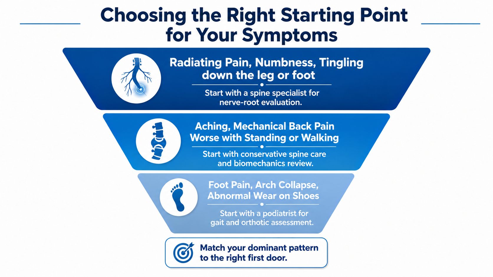
Choosing the Right Starting Point for Your Symptoms
The first appointment should match the dominant pattern, while keeping the other end of the chain in view.
Patients with radiating pain, numbness, tingling, or burning that follows a recognizable line from the back or buttock toward the foot should begin with a spine, pain, or neurological evaluation. The visit should include a symptom diary, previous imaging, a medication list, and a record of treatments that have already failed or helped.
Patients with diffuse back pain that worsens with standing or walking, visible arch collapse, asymmetric shoe wear, or a clear change after new footwear may begin with a podiatric or orthopedic foot-and-ankle assessment. The clinician should still screen lumbar movement, strength, sensation, and gait rather than treating the foot in isolation.
Mixed symptoms require a broader entry point. Bilateral foot burning with axial back pain may warrant neurological or pain-medicine assessment before assuming that a disc or arch is responsible. A local foot diagnosis becomes more convincing when local testing reproduces the pain and the neurological examination is normal.
Safety boundary: Saddle anesthesia, new bowel or bladder dysfunction, foot drop, or progressive weakness warrants urgent spine evaluation regardless of whether symptoms began in the back, calf, ankle, or foot.
Patients should bring comfortable walking shoes, a list of symptom triggers, and details about how far they can walk before symptoms begin. The most useful consultation is the one that compares the spine, nerve pathways, foot posture, and gait in the same decision pathway.
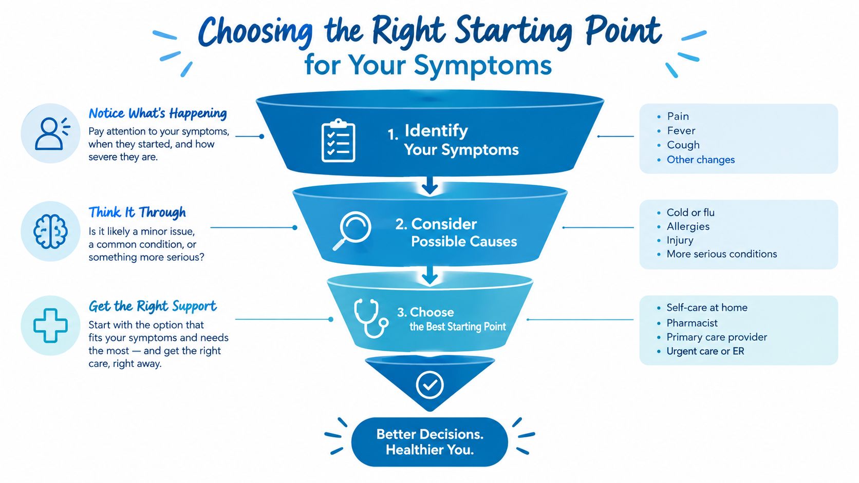
Interventional Pain Management can evaluate radiating back and foot symptoms with focused examination, imaging review, nerve-directed procedures, and coordination with orthopedic or foot-and-ankle care when needed. Patients in New Jersey and Staten Island can visit Interventional Pain Management to arrange an assessment that treats the back and foot as connected parts of the same clinical chain.