
For selected patients, minimally invasive spine procedures can reduce blood loss and hospital time without producing better long-term pain or function than open surgery. In a pooled MI-TLIF analysis, minimally invasive surgery was associated with 269.5 mL less blood loss and about 1.3 fewer hospital days, while surgery took about 21 minutes longer and involved more radiation exposure (meta-analysis).
A patient usually reaches this decision after months of trying to live around pain. Perhaps sitting through a workday triggers burning leg symptoms, walking now requires frequent rests, or another injection offers only temporary relief. The question becomes difficult because “minimally invasive” sounds like a promise: smaller access, less pain, faster recovery, and the same durable result.
That promise is too simple. Minimally invasive spine procedures are technical tools, not diagnoses and not guarantees. The sensible choice depends on the source of symptoms, spinal stability, bone quality, previous operations, medical health, surgeon experience, and the outcome that matters most in daily life.
Table of Contents
- Why This Decision Matters Before You Choose a Procedure
- What Minimally Invasive Spine Procedures Actually Mean
- How the Field Got Here
- The Four Main Types of Procedures
- Who Is a Good Candidate
- What Recovery Actually Looks Like
- The Misconceptions Worth Putting Aside
- Choosing the Option That Fits Your Life
Why This Decision Matters Before You Choose a Procedure
A patient with chronic sciatica may be deciding between another course of physical therapy, an injection, or surgery. Someone with spinal stenosis may be less concerned about the incision than about standing long enough to shop, sleeping without leg pain, or returning to a physically demanding job. Those priorities should shape the conversation before a procedure name does.
A consultation should answer three separate questions:
- What is causing the symptoms? An MRI finding matters only when it matches the pain pattern, examination, and functional limitations.
- What must the procedure accomplish? Removing pressure from a nerve is different from stabilizing an unstable segment or correcting deformity.
- Which trade-off matters most? A patient may value rapid early mobility, avoiding fusion, durable relief, a lower chance of revision, or a recovery compatible with caregiving responsibilities.
The phrase “minimally invasive” covers a wide range of interventions. An endoscopic decompression and a minimally invasive fusion may both use small access corridors, but they place very different demands on the spine and recovery. A smaller opening doesn't automatically make a complex operation appropriate.
Practical rule: The best procedure isn't the one with the smallest incision. It's the one that addresses the actual problem while fitting the patient's anatomy, goals, and realistic access to follow-up care.
Patients can prepare by bringing imaging reports, a medication list, records of prior injections or therapy, and a short description of activities they can no longer perform. The most useful question isn't “Which surgery is newest?” It is “What outcome should this operation improve, and what happens if that outcome isn't achieved?”
What Minimally Invasive Spine Procedures Actually Mean
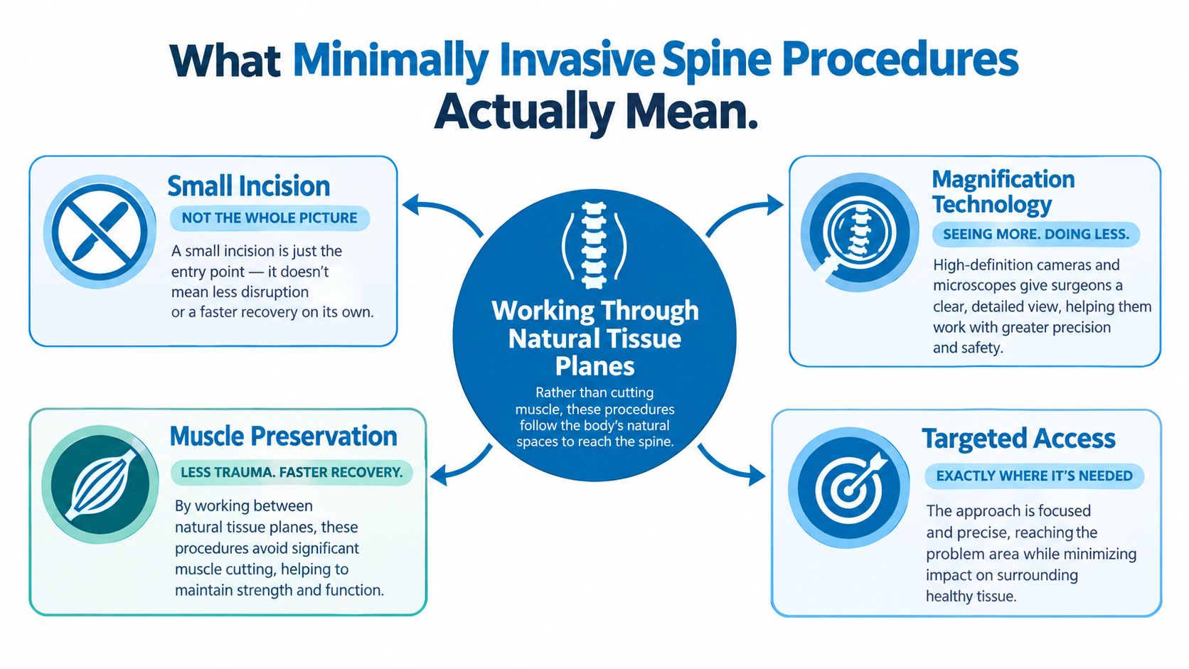
The simplest definition is accessing the spine while limiting unnecessary disruption to nearby muscle and soft tissue. That may involve a microscope, an endoscope, tubular retractors, fluoroscopy, or navigation. The surgeon still has to reach the affected nerve, disc, bone, or spinal segment, but the route is designed around a smaller working corridor and tissue-sparing planes.
A useful analogy is repairing a pipe through a carefully placed access hole instead of removing an entire section of wall. The hole may be smaller, but the repair still requires accurate diagnosis, the right tools, clear visualization, and enough room to complete the job safely. If the pipe is damaged across a broad area, a tiny opening may not provide the access needed.
This is why incision size is an incomplete measure. A surgeon may separate muscle fibers along their direction, use a tubular retractor to maintain a corridor, and magnify the operative field. An endoscope places a camera close to the target, while a microscope provides magnified viewing from outside the access tube. These choices affect visualization, instrument movement, tissue handling, imaging dependence, and the types of pathology that can be treated.
Why identical scans can lead to different plans
Two people can have similar MRI findings and receive different recommendations. One may have a single nerve compressed by a disc without instability. Another may have narrowing combined with vertebral slippage, deformity, weak bone, or extensive scar tissue from previous surgery. The first situation may suit focused decompression. The second may require stabilization or a wider approach.
Patients seeking a plain-language overview of endoscopic access can review endoscopic spine surgery, but the label alone shouldn't determine treatment. The meaningful question is whether the chosen corridor permits complete treatment of the specific pathology.
How the Field Got Here
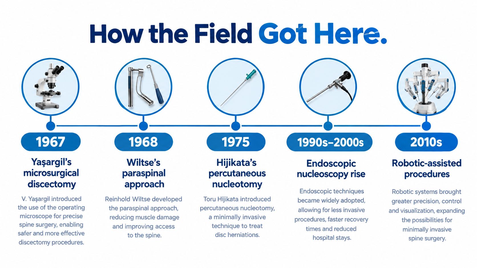
Modern minimally invasive spine procedures developed through successive efforts to preserve tissue while improving visualization, rather than through one sudden invention. In 1967, M. Gazi Yaşargil adapted the operating microscope for lumbar disc surgery. Magnification helped surgeons work through smaller exposures while seeing delicate structures more clearly (historical review).
In 1968, Leon L. Wiltse described a paraspinal, sacrospinalis-splitting approach. Instead of lifting muscle away from the spine through a more disruptive subperiosteal route, surgeons could follow intramuscular planes. The principle remains familiar: reach the target while respecting the body's existing anatomy.
A further milestone came in 1969, when chemonucleolysis was performed in humans. The same historical review identifies it as the first minimally invasive spine procedure. In 1975, Hijikata reported percutaneous nucleotomy, establishing a needle-based route for disc removal.
From narrow corridors to broader applications
During the 1980s, endoscopic development accelerated. Forst and Hausmann reported “nucleoscopy” in 1983, using a modified endoscope through a tubular working channel. By 1989, an endoscopic case series involving 109 patients reported a 72.5% success rate, including patients with spondylolisthesis and recurrent disc herniation, according to the same historical review.
Historical reviews describe the 1990s and early 2000s as the period when tubular retractors and percutaneous fixation spread widely. That history also explains the trade-off patients still face: a smaller access route may preserve more tissue, but it demands specialized training, equipment, and careful patient selection. Availability can differ between hospitals, so the procedure with the smallest incision may not be the option that best supports daily function.
The same 2024 historical review estimates that existing minimally invasive approaches could potentially accommodate as many as 900,000 cases annually. That figure describes possible scale, not a promise that every patient is suitable. Modern techniques are refined procedures with specific learning curves, not effortless substitutes for conventional surgery.
The Four Main Types of Procedures
A smaller incision does not automatically mean a better operation. The right choice depends on which problem is limiting daily life, such as leg pain, weakness, walking distance, instability, or loss of spinal support. “Minimally invasive spine surgery” describes an access strategy, not one operation.
Four categories patients commonly hear about
| Procedure type | Typical indication | Key advantage | Main trade-off |
|---|---|---|---|
| Endoscopic discectomy | Selected disc herniation with nerve compression | Focused access through a narrow corridor | Limited working space and demanding visualization |
| Tubular decompression | Foraminal or central stenosis affecting a nerve | Muscle-sparing access to remove pressure | Decompression alone may not address instability |
| Minimally invasive fusion, including MIS-TLIF | Spondylolisthesis, instability, or selected degenerative disease | Stabilization with less muscle disruption than some open approaches | Longer operation, radiation exposure, and fusion-related risks |
| Anterior lumbar interbody fusion variations | Conditions requiring anterior-column support, disc-height restoration, or selected alignment work | Access from the front can support disc-space reconstruction | Requires careful vascular, neurologic, and alignment assessment |
Endoscopic discectomy removes disc material pressing on a nerve through a narrow working corridor. It suits selected herniations, not every form of back pain. If instability or broad deformity drives the symptoms, removing disc material alone may leave the main problem untreated. This microdiscectomy overview explains the nerve-release concept, although the safest route still depends on anatomy and surgeon access.
Tubular decompression uses a working tube to reach a narrowed canal or foramen. The surgeon removes selected bone or ligament to give the nerve more room while preserving stabilizing tissue where possible. The trade-off is straightforward: if the segment is already unstable, or if adequate decompression would remove too much supporting bone, decompression alone may not restore useful function.
MIS-TLIF combines nerve decompression, disc-space work, interbody support, and fixation. A long-term systematic review of 16 studies and 1,321 patients found significant improvement in patient-reported outcomes after both MIS-TLIF and open TLIF, with no statistically significant difference in those measures at two years. Fusion rates were 80.5% for MIS-TLIF and 91.1% for open TLIF, a difference that was not statistically significant. Reoperation rates were 3.0% and 2.4%, respectively (systematic review). These figures describe study results, not a guarantee for an individual patient.
ALIF variations reach the lumbar disc space from the front. They may support disc-height restoration or anterior-column reconstruction, but they require careful assessment of vascular anatomy, nerves, and alignment. Access can be minimally invasive while recovery remains substantial, and equipment, specialist training, or hospital availability may limit which option a patient can realistically receive.
Who Is a Good Candidate
Candidacy isn't a single yes-or-no threshold. A surgeon layers evidence from symptoms, examination, imaging, spinal mechanics, prior treatment, and the patient's health before recommending a corridor.
The diagnostic match
The first test is whether the scan and symptoms tell the same story. Leg pain, numbness, weakness, walking limitation, or back pain should correspond to a plausible compressed nerve, narrowed passage, unstable segment, or disc problem. An incidental MRI abnormality shouldn't become the target only because it appears on the report.
Conservative care also matters. Depending on the diagnosis, the treatment history may include physical therapy, medication adjustments, activity changes, or an injection such as an epidural steroid injection. The purpose isn't to force every patient through endless treatment. It is to clarify whether symptoms persist despite reasonable nonoperative care and whether the proposed procedure addresses the remaining problem.
The anatomical and personal checklist
A surgeon then considers whether the selected corridor can provide complete treatment:
- Stability: Decompression alone may be unsuitable when the segment is unstable or when removing bone would create instability.
- Previous surgery: Scar tissue can alter the safest route and make a familiar corridor more difficult.
- Disease extent: Multilevel narrowing or deformity may require broader access than a single-level procedure.
- Bone quality: Fusion planning depends on whether the bone can support implants and healing.
- Body habitus and positioning: Physical anatomy can affect positioning, imaging, access, and wound management.
- Medical health: Cardiopulmonary conditions, diabetes, medications, and other risks influence anesthesia and recovery.
- Functional goals: The plan for a desk worker, caregiver, warehouse employee, or active older adult may differ even with similar imaging.
Patients should ask which finding makes the operation necessary, what anatomy could change the approach, and what nonoperative alternative remains reasonable.
What Recovery Actually Looks Like
A patient may leave the hospital feeling better while the spine still needs protection. Recovery runs on two clocks: the skin and muscle corridor can settle quickly, while nerve healing, conditioning, and fusion biology take much longer. A small incision does not make unrestricted lifting or strenuous work safe.
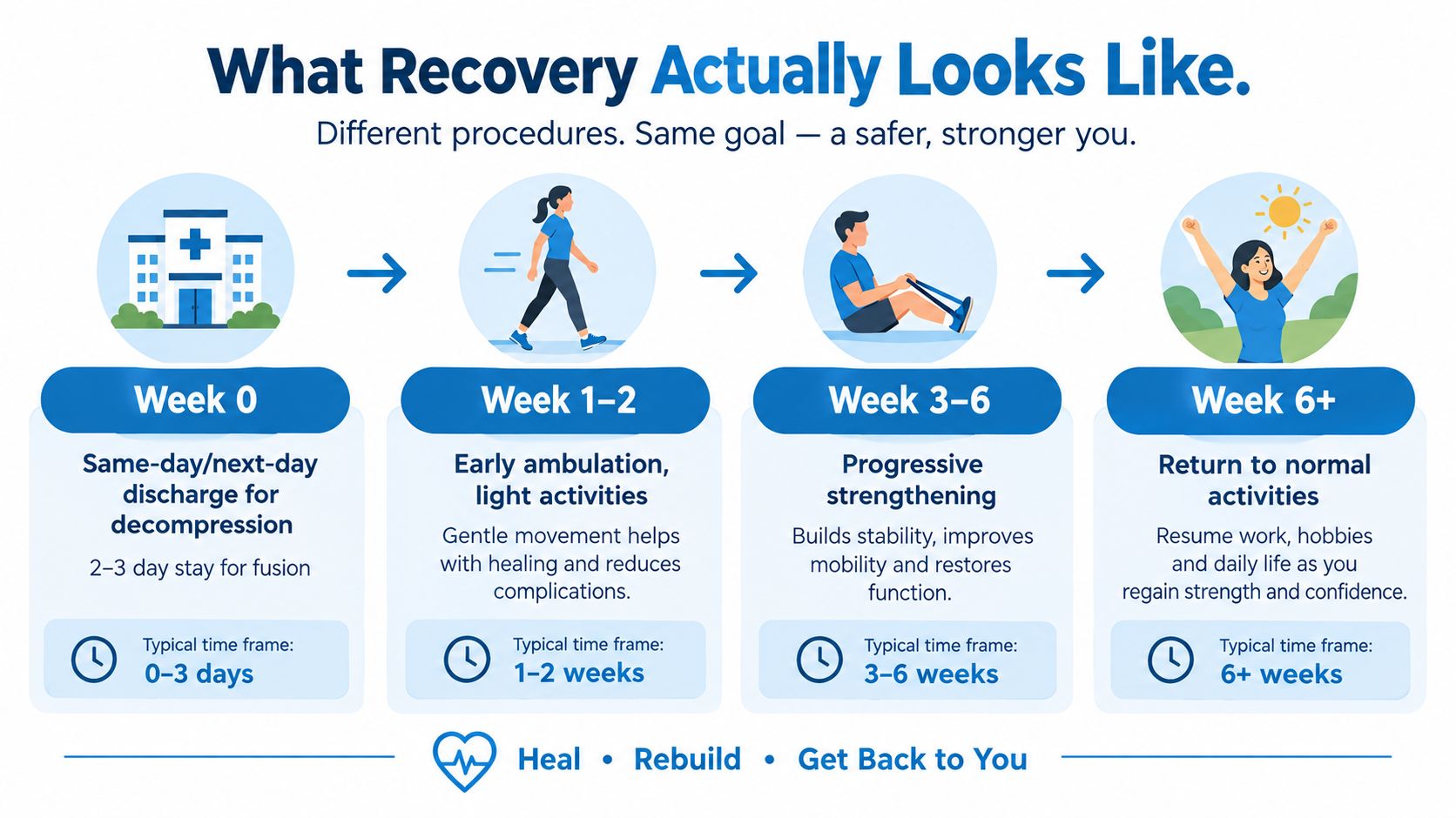
After decompression for nerve compression, discharge may occur the same day or the next day. Early recovery usually involves short walks, careful position changes, symptom control, and protection of the access area. During the first week, the original nerve pattern may begin to improve, but tingling or weakness can remain after the incision looks healed. Nerves recover on their own schedule.
Decompression and fusion follow different paths
Fusion usually requires a longer early recovery because the bone and implants need time to become stable. Hospitalization may last two to three days, compared with same-day or next-day discharge for many decompression procedures. Walking remains important in both cases, but lifting restrictions commonly continue for six to twelve weeks. The treating team sets the actual limits based on the operation and the patient's progress.
Pooled MI-TLIF evidence has associated the minimally invasive approach with shorter hospital stays and less blood loss than open fusion, but it also found longer operating times and greater radiation exposure. The review rated the evidence for the hospital-stay and blood-loss advantages as low quality (meta-analysis). Surgeon experience, anatomy, case selection, and the reason for fusion therefore remain important when estimating recovery.
Walking generally advances before heavier strengthening. Physical therapy may address posture, hip and core control, nerve mobility, and safe body mechanics. After fusion, activity depends more on healing and rehabilitation than on the incision's appearance.
Recovery planning should include practical demands, not only medical milestones. Arrange clear guidance about driving, work, lifting, exercise, wound care, and symptoms requiring urgent attention. Travel distance, available help at home, caregiving duties, and the physical demands of work can determine whether a proposed return-to-activity schedule is realistic.
The Misconceptions Worth Putting Aside
A smaller incision can sound like the obvious better choice, especially when you are worried about pain and recovery. The more useful question is what you need the operation to improve in daily life. Preserving muscle and other tissues may reduce some early surgical burdens, but it does not guarantee better long-term pain relief, function, durability, or freedom from revision.
The two-year equivalence noted in the procedure comparison above is the core fact behind this misconception: smaller access changed the short-term burden, not necessarily the long-term result. Both minimally invasive and open fusion can produce meaningful improvement, while the lasting patient-reported outcome may be similar. The incision is one part of the decision, not the outcome itself. A systematic review also illustrates why technique labels need context, because different operations and patient groups can produce different findings.
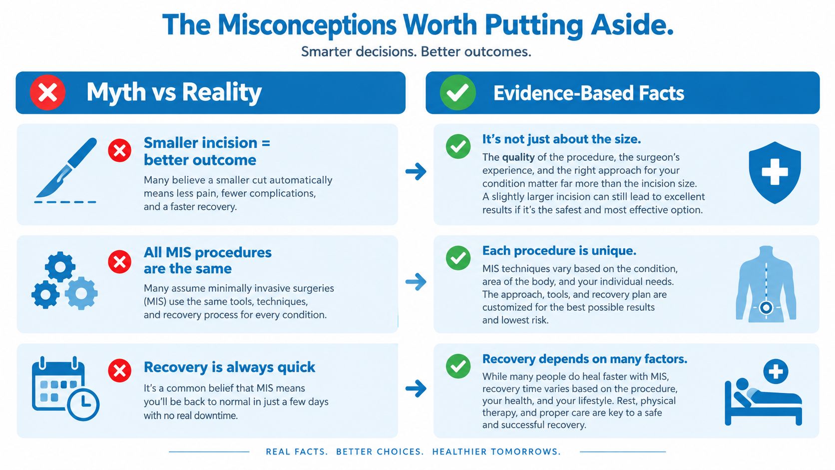
Different techniques create different risks
Endoscopic surgery changes the complication profile rather than removing risk. A review of 20,020 patients identified transient neurologic deficits, dural tears, and postoperative dysesthesia among important complications in endoscopic discectomy or decompression. Late mechanical implant failure appeared more characteristic of endoscopic interbody fusion (endoscopic complication review).
The operation determines what must be watched closely. Decompression involves working carefully around the nerves through a confined channel. Fusion adds questions about cage position, fixation integrity, alignment, and delayed construct failure. Because the review combined varied techniques and spinal regions, its overall rates should not be treated as an individual prediction.
Access is part of safety
A technically appealing procedure can still be a poor practical fit if authorization is delayed, the trained surgeon is far away, transportation is unreliable, therapy is unavailable, or urgent and revision care would be difficult to reach. Surgical access can break down at four points: reaching a provider, detecting a surgical indication, progressing to surgery, and receiving optimal care (surgical-access review).
Technique differences also may not change the result that matters most at home or work. A comparison of unilateral endoscopic and unilateral biportal endoscopic procedures for lumbar stenosis included 17 studies and 1,457 patients. Through 12 months, it found no significant differences in postoperative pain, disability, blood loss, hospital stay, or complication rates. Biportal surgery had shorter operative time and greater dural-sac expansion. Ask which expected benefit will change your daily life, then weigh whether the required care is realistically available.
Choosing the Option That Fits Your Life
A useful consultation starts with one priority stated plainly. Is the main goal faster early mobility, avoiding fusion, durable leg-pain relief, preserving the ability to lift at work, reducing revision risk, or returning to caregiving? The answer helps the surgeon explain which trade-offs matter instead of presenting incision size as the whole decision.
Patients should ask:
- How does the imaging match the symptoms?
- What exactly will the procedure change?
- Why is this approach preferable to decompression, fusion, or continued nonoperative care?
- How often does the surgeon perform this specific operation?
- What complications are most relevant to this patient?
- What is the realistic return-to-work and lifting timeline?
- What is the plan if pain persists or the construct doesn't heal?
- How will insurance, travel, therapy, and follow-up affect the treatment burden?
Minimally invasive spine procedures can be powerful when deliberately matched to the pathology and the person. They shouldn't be selected because the name sounds gentler. A sound decision leaves the patient understanding both the likely benefit and the inconvenient parts of recovery.
Interventional Pain Management evaluates back, neck, joint, and nerve-related pain through diagnostic review, interventional treatments, and spine care options matched to clinical needs. Patients in New Jersey and Staten Island can visit Interventional Pain Management to request an evaluation and discuss whether continued nonsurgical care or a minimally invasive spine procedure fits the symptoms, anatomy, and daily goals.