
A patient may hear “you may need a cervical fusion” after months of arm pain, tingling fingers, hand weakness, or a neck that no longer responds to therapy and medication. The phrase can sound final, but it doesn't describe the whole decision. It describes one possible step in a treatment pathway that may also include observation, rehabilitation, injections, decompression without fusion, or a motion-preserving implant.
Cervical discectomy and fusion, usually called ACDF, is designed to remove pressure from a nerve root or the spinal cord and stabilize the affected level. It can be highly useful for carefully selected patients, but it permanently changes motion at the treated segment and requires a recovery plan that extends beyond the operating room.
Table of Contents
- When a Surgeon First Mentions Cervical Discectomy and Fusion
- What the Procedure Actually Does to Your Neck
- Who Actually Needs Cervical Discectomy and Fusion
- Inside the Operating Room Step by Step
- Risks and Complications Worth Understanding
- Recovery Timeline and What Each Week Actually Looks Like
- Comparing ACDF to Motion-Preserving and Posterior Alternatives
- Long-Term Outcomes and Questions to Bring to Your Surgeon
When a Surgeon First Mentions Cervical Discectomy and Fusion
The conversation often begins with a familiar story. A patient has tried physical therapy, changed activities, taken medication, and perhaps received an injection. The arm pain still interrupts sleep, numbness makes buttoning a shirt difficult, or weakness makes a hand feel unreliable. An MRI then shows a disc herniation, bone spur, or narrowed canal that matches the symptoms.
The important question isn't whether an MRI looks abnormal. Many adults have age-related changes without needing surgery. The question is whether the scan, examination, and daily limitations point to the same pain-generating problem.
Why nonoperative care comes first
For nerve-root symptoms, a clinician may begin with activity modification, targeted exercises, medication review, and injections when appropriate. This approach gives irritated tissue an opportunity to settle and helps determine whether symptoms are improving without an operation.
Surgery becomes more persuasive when symptoms remain disabling despite reasonable conservative care, weakness progresses, or the spinal cord is compressed. Myelopathy, meaning spinal cord dysfunction, can affect balance, hand coordination, walking, and dexterity. Instability or deformity may create a separate reason to consider stabilization even when pain isn't the only concern.
Practical rule: The strongest surgical recommendation connects three findings, the patient's symptoms, the neurological examination, and imaging at the same level.
ACDF shouldn't be viewed as an automatic response to neck pain or a disc finding. It's a planned decision between alternatives, with the choice shaped by the location of compression, the number of affected levels, spinal alignment, facet-joint condition, bone quality, medical history, and the patient's functional goals.
The operation may solve the current nerve-compression problem, but it doesn't freeze the rest of the neck in time. After treatment, some patients need surveillance and rehabilitation rather than another operation. Others may later develop symptoms from a different level, although imaging changes alone don't prove that a new surgery is needed.
What the Procedure Actually Does to Your Neck
The cervical disc sits between two vertebrae and helps the neck move while distributing load. With degeneration or injury, the disc can bulge or herniate. Bone spurs and thickened tissue can also reduce the space used by a nerve root or the spinal cord.
A compressed nerve can produce pain that travels from the neck into the shoulder, arm, or hand. It may also cause tingling, numbness, or weakness. Spinal cord compression creates a different concern, because it can interfere with coordination, balance, and fine hand function.
Decompression followed by stabilization
During ACDF, the surgeon reaches the spine from the front of the neck and removes the damaged disc. The surgeon also clears away compressive disc material, bone spurs, or other tissue that is pressing on the spinal cord or exiting nerve root.
That is the discectomy. It creates room for the nerve or cord, but it also leaves an empty disc space. The surgeon restores that space with an interbody graft or cage, often supplemented with an anterior plate and screws. The spacer helps maintain foraminal height and alignment while bone grows through or around the implant.
The intended final result is an arthrodesis, or solid fusion, in which the two neighboring vertebrae become one stable unit. The technical goals are neural decompression, preservation of the restored disc height, and reliable bone healing, as described in this clinical review of ACDF technique and fusion considerations.
A useful analogy is replacing a worn-out spacer between two blocks. The surgeon removes the damaged material, opens the narrowed passage, inserts a precisely sized support, and allows the two blocks to heal together. The analogy has limits, because bone healing is biological and can be affected by smoking, osteoporosis, diabetes, medications, and the number of treated levels.
Patients seeking broader background on stabilization can also review this explanation of spinal fusion and its treatment role. The key distinction is simple: ACDF doesn't merely remove a disc. It removes the source of compression and then sacrifices motion at that level to create stability.
Who Actually Needs Cervical Discectomy and Fusion
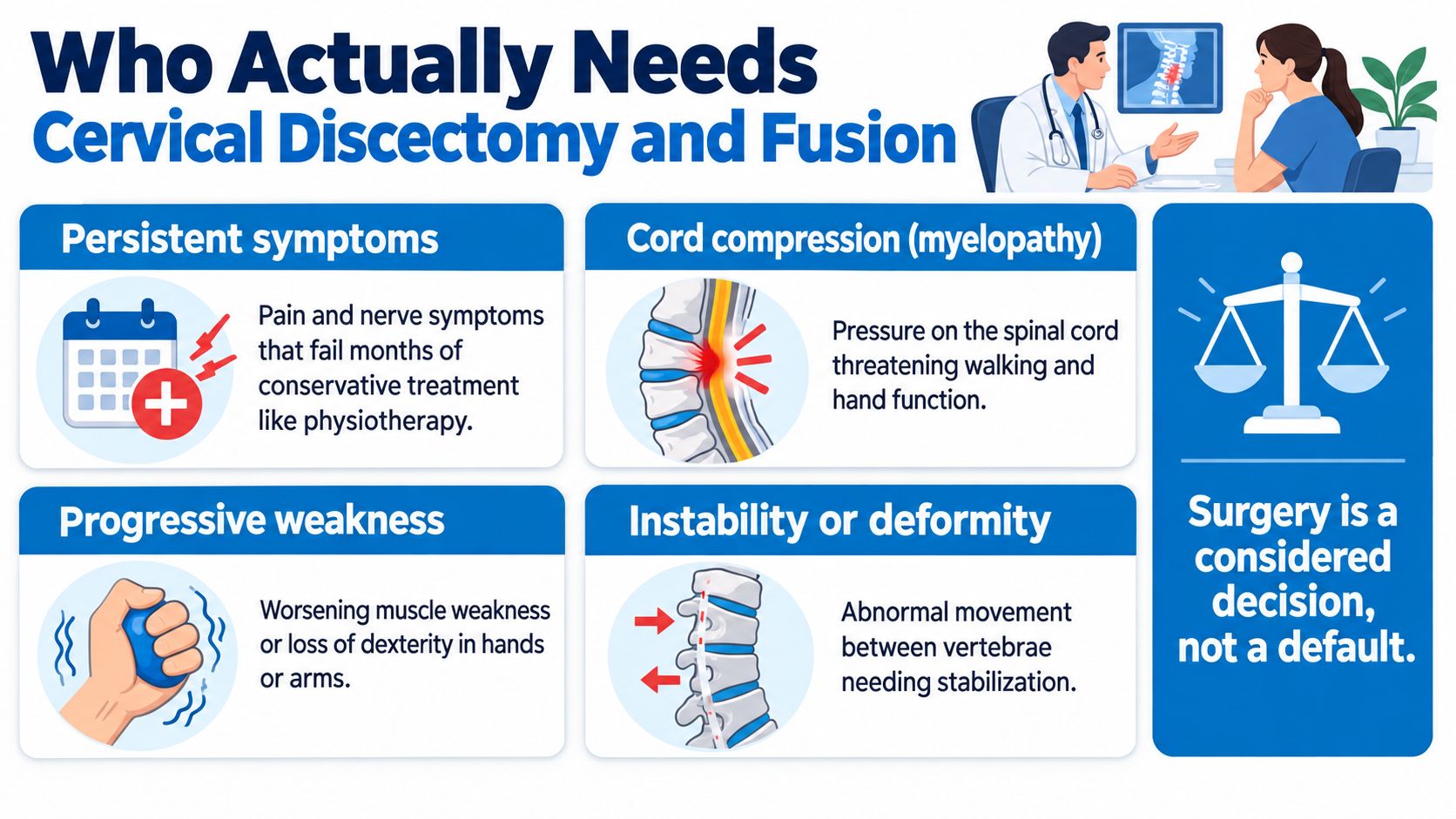
A painful neck does not automatically require ACDF. The operation is usually considered when the clinical problem has a structural explanation and nonoperative treatment hasn't provided adequate relief, or when neurological function is at risk.
The common decision patterns
Persistent radiculopathy involves pain, numbness, tingling, or weakness caused by a compressed cervical nerve root. Surgery becomes more reasonable when symptoms remain functionally limiting, the examination confirms nerve involvement, and imaging shows matching compression.
Myelopathy involves the spinal cord rather than only an exiting nerve. Worsening hand clumsiness, balance problems, changes in walking, or progressive weakness deserve prompt assessment. The urgency depends on the examination and the degree of cord compromise.
Instability or deformity can make decompression alone insufficient. If vertebrae move abnormally or alignment needs correction, fusion may provide the structural support that a motion-preserving decompression cannot.
Selected trauma and multilevel disease require individualized planning. The surgeon must determine whether the main compression comes from the front or back, whether the spinal cord is involved, and whether one level or several levels account for the symptoms.
A patient with a single disc herniation and preserved stability may receive a different recommendation from a patient with multilevel stenosis, severe facet arthritis, deformity, or previous cervical surgery. Similar MRI findings don't guarantee similar operations.
Where ACDF fits in practice
A national United States database analysis covering approximately 1.2 million cervical spine operations from 2010 through 2022 found that ACDF represented 61.6% of procedures. Laminoplasty represented 22.1%, posterior cervical fusion 14.0%, and cervical disc arthroplasty 3.4%, according to the national analysis of cervical spine surgery trends.
ACDF, laminoplasty, and posterior fusion were most common among adults aged 55 to 74, while disc arthroplasty was more frequently performed in younger patients. Across the period studied, total cervical-spine surgery volume increased from 86,272 to 96,976 procedures, while annual ACDF volume declined from 60,435 to 51,012 in the same source.
Those figures show ACDF's historical role, but they don't decide an individual case. The operation remains a common benchmark while alternatives gain importance for selected anatomy and goals. A patient should ask which specific structure is being treated, why fusion is necessary, and what non-fusion option has been considered.
For readers trying to understand the underlying disc problem before discussing surgery, this guide to a herniated or bulging disc provides useful context.
Inside the Operating Room Step by Step
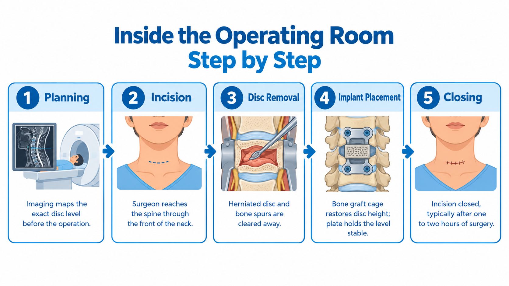
The operation starts before the incision. The surgical team reviews MRI or CT images, confirms the symptomatic level, evaluates alignment, and considers medical factors that can affect anesthesia or bone healing. Smoking, diabetes, osteoporosis risk, medications, and previous cervical operations should be part of that preparation.
Positioning and access
After general anesthesia, the patient lies on the back with the neck positioned carefully. The surgeon makes an incision at the front of the neck, usually within a natural skin crease when possible. The tissues are gently moved aside to reach the cervical spine without cutting through the large posterior neck muscles.
The team confirms the correct level with imaging. Surgeons also work around structures such as the esophagus, trachea, blood vessels, and nerves involved in voice function. Intraoperative imaging and, in selected cases, neuromonitoring help the team verify anatomy and protect neurological function.
Removing the source of pressure
The surgeon opens the disc space and removes the damaged disc material. Small instruments and magnification help clear the central and foraminal areas. Bone spurs or thickened ligament may also be removed when they contribute to compression.
This is the moment when decompression occurs. The patient won't feel the removal because of anesthesia, but the operation's purpose is direct: create room for the spinal cord or nerve root and restore the passageway through which the nerve exits.
The vertebral endplates are then prepared. This step matters because the implant must sit securely and allow bone to grow across the space. The surgeon selects an appropriately sized graft or cage, places it between the vertebrae, and may add an anterior plate and screws for immediate construct stability.
Closure and the first sensations
The incision is closed in layers. After awakening, a patient may notice a sore throat, discomfort with swallowing, hoarseness, shoulder soreness, or neck tightness. These symptoms can result from the anterior approach and tissue retraction, and they don't automatically mean that the nerve or esophagus has been injured.
The surgeon's instructions determine collar use, wound care, lifting limits, and discharge timing. Before leaving, the patient should know which symptoms require urgent contact, how medications will be reduced, and when follow-up imaging or rehabilitation will be considered.
Risks and Complications Worth Understanding
ACDF has a substantial clinical evidence base, but no operation is risk-free. A pooled analysis of 50,584 patients from 267 cohorts reported an overall postoperative complication rate of 16.0%, with 13.8% experiencing nonhome discharge, 3.7% readmission, and 0.1% mortality, as reported in this meta-analysis of ACDF complications and outcomes.
The same analysis reported a 20.5% complication rate for one-level ACDF. That figure should be interpreted carefully because studies may define complications differently and may use different follow-up periods. It doesn't mean that every patient faces the same risk, nor does it imply that all complications are severe or permanent.
What patients may encounter
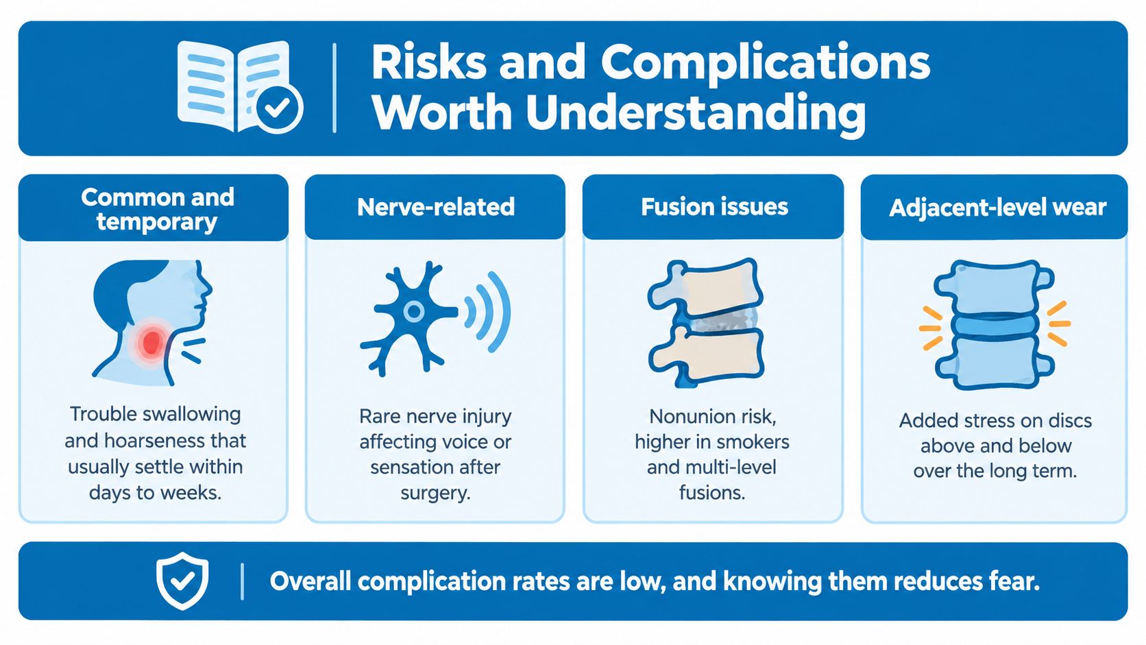
Early swallowing difficulty and voice changes are among the complications patients notice most. Other possibilities include postoperative hematoma, respiratory problems, infection, recurrent laryngeal-nerve symptoms, instrumentation failure, and esophageal injury. A rapidly expanding neck swelling or new breathing difficulty requires urgent medical attention.
Nonhome discharge means a patient doesn't go directly home after the hospital stay. It may reflect medical complexity, mobility needs, pain control, or the need for additional support. Readmission can occur for wound issues, swallowing or breathing concerns, infection, pain, or other postoperative problems.
Fusion-specific risks deserve separate discussion. A radiographic nonunion means the bones haven't fully joined on imaging. Some nonunions cause no symptoms, while a clinically meaningful pseudarthrosis can produce persistent pain, instability, or a need for revision surgery.
Factors that change the risk profile
The surgeon should assess smoking status, bone density, diabetes control, operative levels, previous cervical surgery, and overall health. These factors can influence fusion biology, implant loading, wound healing, and the ability to recover safely at home.
A solid fusion is an imaging goal, not a guarantee that every future neck symptom has been eliminated.
Informed consent should therefore address more than a generic list. Patients need to understand the likely swallowing and voice symptoms, the possibility of additional support after discharge, the chance of readmission, and the specific risks created by an anterior approach. A careful discussion also explains what the surgeon will do if the implant settles, the bones fail to unite, or symptoms persist.
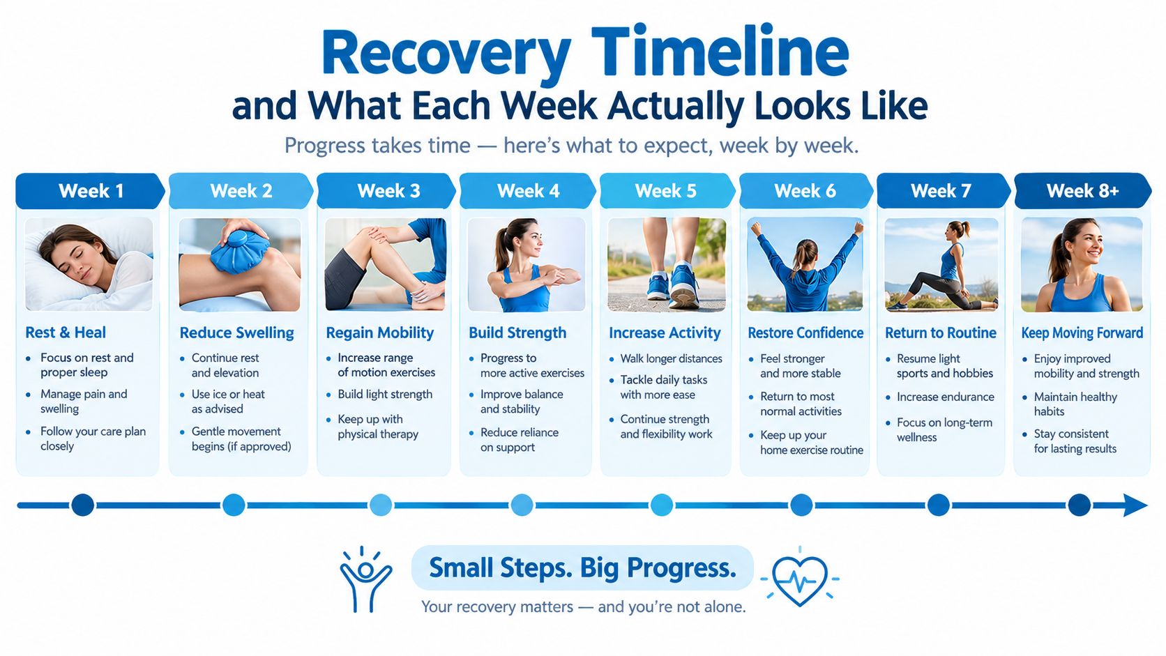
Recovery Timeline and What Each Week Actually Looks Like
Recovery after ACDF has two tracks. The nerve may feel better before the neck incision does, while the bone continues healing after the patient has resumed many ordinary activities. That mismatch can be confusing, especially when early symptom relief creates the impression that the entire recovery is complete.
The early days
During the first several days, patients usually focus on incision care, medication schedules, short walks, hydration, and safe swallowing. A collar may be prescribed, but collar use varies by surgeon, construct, and patient factors. Lifting and driving restrictions also depend on the operation and the treating team.
Arm pain may improve quickly after decompression, while numbness or weakness can take longer because an irritated nerve needs time to recover. Neck soreness, fatigue, and swallowing discomfort can be more noticeable than expected.
During the first few weeks, walking is generally increased gradually. Desk work may resume when concentration, medication effects, pain, and surgeon restrictions allow. Driving should wait until the patient can turn safely, react normally, and is no longer taking medications that impair alertness.
Rehabilitation and the fusion process
Therapy isn't just a way to stretch a stiff neck. A rehabilitation plan may address posture, shoulder-blade control, endurance, movement patterns, and the gradual return to work or sport. The timing must respect the surgeon's restrictions and the stability of the construct.
A 2025 Veterans Health Administration longitudinal study found that median opioid use fell from 25.0 morphine-milligram equivalents before surgery to 0 at one year, yet more than one-third of patients still used opioids at one year. High preoperative consumption was the strongest predictor of persistent use, with an odds ratio of 17.1, according to the longitudinal study of opioid use and ACDF recovery.
That finding makes medication planning part of surgical preparation, not an afterthought. A patient already taking opioids should receive a coordinated review of the indication, expected tapering process, alternative pain strategies, and signs that ongoing pain needs reassessment rather than additional medication.
The same study associated therapy begun within six weeks with greater 12-month improvements in physical-health and myelopathy scores than therapy started later. Timing must remain individualized, but prolonged inactivity isn't automatically safer.
Later recovery
Over subsequent months, the graft continues to incorporate and the patient gradually resumes heavier activity if imaging, symptoms, and examination support progression. Follow-up may assess alignment, implant position, and evidence of fusion, not just whether pain has improved.
Recovery planning: The most useful timeline is individualized around the patient's job, medication exposure, neurological findings, bone health, and rehabilitation access.
Persistent or returning symptoms deserve evaluation. They may reflect slow nerve recovery, incomplete decompression, pseudarthrosis, hardware concerns, or a different cervical level. They shouldn't be labeled surgical failure without examining the cause.
Comparing ACDF to Motion-Preserving and Posterior Alternatives
ACDF is common because it combines direct anterior decompression with stabilization. It isn't automatically the best operation for every source of cervical compression. The meaningful comparison depends on anatomy, alignment, instability, facet joints, bone quality, number of levels, and whether the spinal cord is involved.
| Approach | Motion at Treated Level | Best-Suited Indications | Adjacent-Segment Risk Profile |
|---|---|---|---|
| ACDF | Motion is intentionally eliminated at the fused level | Anterior disc or bone compression, instability, selected deformity, and conditions requiring stabilization | Fusion changes load and motion patterns. Future adjacent-level symptoms remain possible |
| Cervical disc arthroplasty | Motion is intended to be preserved | Carefully selected disc disease without major facet disease, instability, deformity, osteoporosis, or unsuitable multilevel degeneration | May reduce adjacent-segment degeneration and reoperation in selected patients, but doesn't eliminate risk |
| Laminoplasty | Motion is generally preserved across treated posterior levels | Selected multilevel spinal cord compression with suitable alignment and anatomy | Avoids fusion at treated levels, but future disease can still occur |
| Posterior cervical fusion | Motion is reduced across fused posterior levels | Posterior compression, instability, deformity, or cases where anterior access isn't the safest route | Shares the broader trade-off of stabilization with reduced motion |
A 2025 systematic review of 17 randomized controlled trials with at least four years of follow-up found higher neurological success, better range of motion, lower neck-disability and pain scores, and significantly lower adjacent-segment degeneration and reoperation rates with arthroplasty than ACDF. The review found no significant differences in operative time or adverse events, but candidacy remains decisive, as discussed in this systematic review comparing arthroplasty and ACDF.
Motion preservation shouldn't be presented as a guarantee. Significant facet-joint disease, instability, deformity, osteoporosis, or multilevel degeneration may make an artificial disc unsuitable. Patients exploring that option can review an explanation of artificial disc replacement surgery, then ask the surgeon which exclusion factors apply to the individual case.
A posterior procedure may be preferable when compression is mainly behind the cord or when anatomy makes an anterior route less appropriate. The central question is not which operation sounds most modern. It's which operation addresses the actual pathology while preserving as much healthy function as the situation allows.
Long-Term Outcomes and Questions to Bring to Your Surgeon
A fused level becomes stable but loses its segmental motion. In a 10-year randomized comparison, motion at the operated level in the fusion group decreased from 45° at baseline to 36° at 10 years, while the arthroplasty group increased from 43° to 50°. Patient-reported outcomes were similar, and preserved motion didn't eliminate MRI-defined adjacent-segment pathology, according to this long-term comparison of cervical arthroplasty and fusion.
Before consenting, patients should ask:
- Which finding explains the symptoms?
- Why is fusion preferred over decompression alone or arthroplasty?
- What graft, cage, plate, or screw construct is planned?
- How will smoking, bone health, diabetes, and medications affect healing?
- When should rehabilitation begin?
- What imaging will confirm alignment and fusion?
- Which returning symptoms require prompt review?
Interventional Pain Management offers diagnostic evaluation, image-guided injections, rehabilitation coordination, and spine-surgery assessment across nonoperative and operative treatment pathways. Patients with persistent neck or arm symptoms can bring their imaging, medication history, and treatment goals to a focused consultation with Interventional Pain Management to discuss whether continued conservative care, an intervention, or surgical planning is appropriate.