
A lumbar MRI can look frightening. The report may describe “severe” narrowing, multiple levels, thickened ligaments, enlarged joints, or a slipped vertebra. Yet the person holding that report may be dealing with a very specific problem, such as leg pain after walking, numbness in one foot, or back pain that has little to do with the tightest-looking level.
That mismatch creates the central problem with spinal stenosis surgery options. A procedure shouldn't be selected because its name sounds modern, aggressive, or minimally invasive. It should be selected because it treats the structure compressing the nerve and preserves stability whenever possible. A careful decision separates ligament thickening from facet overgrowth, disc-related compression from bony narrowing, and isolated stenosis from a mechanically unstable spine.
Table of Contents
- Why the Right Procedure Starts With the Cause, Not the Name
- How Stenosis Develops in the Spine
- Decompression, Fusion, and Spacers Compared
- Minimally Invasive and Image-Guided Techniques
- When Fusion Is the Right Call and When It Is Overkill
- What Pre-Surgical Planning Should Look Like
- Matching the Procedure to Your Stenosis Profile
- Risks, Reoperations, and What Recovery Looks Like
Why the Right Procedure Starts With the Cause, Not the Name
A typical consultation begins with a stack of terms the patient has heard from different sources: laminectomy, fusion, decompression, endoscopic surgery, spacer. The MRI may look dramatic, but the actual complaint is often simpler. Walking causes burning pain in both legs, leaning forward over a shopping cart helps, and sitting brings relief.
That patient may not need a fusion. If the symptoms come from nerves crowded in the central canal and the vertebrae remain stable, decompression can create room without permanently joining spinal segments. The fact that the scan calls the stenosis severe doesn't, by itself, change that logic.
Practical rule: Name the compressing structure first. Choose the operation second.
Four anatomical causes appear repeatedly:
- Ligamentum flavum hypertrophy: Thickened ligament can occupy the back of the canal and contribute to central narrowing.
- Facet hypertrophy: Arthritic facet joints can narrow the lateral recess or contribute to foraminal compression.
- Disc bulge or collapse: A disc can press on a nerve directly or reduce foraminal height.
- Bony overgrowth: Spurs and enlarged joints can create hard compression that soft-tissue removal alone won't address.
The same patient can have more than one cause. That's why a small-incision procedure may be entirely appropriate for one MRI and inadequate for another. A focused decompression can fit isolated ligament or bone compression, while a fusion may become necessary when removing the compressive anatomy would destabilize the segment.
The useful question isn't “Which surgery is best?” It's “What is narrowing the space, which nerve is affected, and will removing that structure leave the spine stable?”
How Stenosis Develops in the Spine
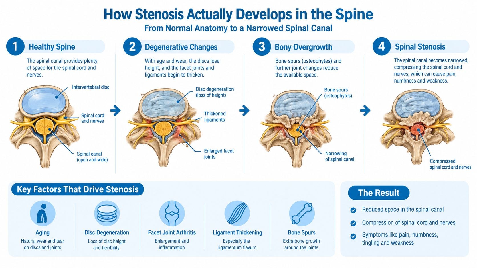
Lumbar stenosis develops when the passages for spinal nerves become too narrow. The narrowing may affect the central canal, lateral recess, or neural foramen. That location matters because it often determines which nerve is compressed and which operation can relieve it without removing more stabilizing anatomy than necessary.
The central canal runs behind the vertebral bodies. Narrowing there can crowd several nerve roots and cause neurogenic claudication, including aching, heaviness, numbness, or burning in the legs during standing or walking. Sitting or leaning forward often helps because flexion gives the nerves more room. Extension reduces that space and can worsen symptoms.
The lateral recess is the channel an individual nerve root follows before exiting the spine. Enlarged facet joints, disc material, or bone can narrow it and produce symptoms in one leg. Foraminal stenosis occurs farther to the side, where the exiting nerve passes through its opening. It commonly causes dermatomal pain, tingling, or weakness that follows a classic pinched-nerve pattern.
The structures that take up space
Several parts of the motion segment can narrow the nerve passage at the same time:
- Ligamentum flavum: Thickening and buckling can push into the canal, particularly during extension.
- Facet joints: Arthritis can enlarge the joints and narrow the lateral recess or foramen.
- Intervertebral discs: Bulging, herniation, or loss of disc height can crowd nerve roots.
- Bone: Osteophytes and other overgrowth can create fixed compression that soft-tissue removal alone may not resolve.
A report from lumbar spinal stenosis specialists may list several abnormalities, but the MRI does not identify the pain generator by itself. The examination, pain distribution, walking tolerance, weakness, reflexes, and response to position must match the images. That match is what should guide surgery.
A cyclist may tolerate activity better than a pedestrian with the same MRI. Cycling keeps the lumbar spine relatively flexed, while prolonged standing and walking often extend the back and increase nerve crowding. That positional pattern is useful evidence, not a diagnosis by itself. The cause of compression, more than the label “minimally invasive,” should determine the procedure.
Decompression, Fusion, and Spacers Compared
A patient with leg pain while standing may need only a focused decompression. A patient with vertebral instability or deformity may need decompression with fusion. The anatomy decides. Decompression removes what is pressing on the nerve. Fusion adds stabilization when the segment has a mechanical problem or when adequate nerve release would otherwise remove too much supporting bone. An interspinous spacer limits extension and fits a much narrower group.
For leg-dominant symptoms caused by lumbar stenosis, decompression alone generally performs as well as decompression with instrumented fusion when there is no meaningful instability, deformity, or major mechanical back pain. A randomized trial reported two-year disability scores of 27 versus 24, with no significant difference. Six-minute walking distances were 397 meters versus 405 meters, also without a significant difference, in the randomized comparison of decompression and fusion.
The procedure families
Decompression alone includes laminectomy, laminotomy, foraminotomy, and targeted bilateral approaches. It suits central, lateral recess, or foraminal compression when the surgeon can remove the ligament, bone, disc material, or enlarged joint tissue causing the problem while preserving enough facet joint and other stabilizing anatomy. Its main failure modes are residual compression, recurrent narrowing, or instability after removal of too much supporting structure.
Decompression plus instrumented fusion combines nerve release with screws, rods, and a fusion construct, such as an interbody or posterolateral fusion. Choose it when instability, deformity, symptomatic spondylolisthesis, or substantial mechanical back pain changes the mechanical problem. Fusion adds surgical burden, hardware-related risks, and a longer recovery than a limited decompression, so it should have a specific mechanical rationale.
Interspinous spacers sit between the posterior spinal elements and limit extension. They may help a carefully selected patient with position-dependent symptoms and anatomy that accommodates the device. They fit poorly with significant foraminal compression, major facet or bony overgrowth, instability, deformity, or symptoms that fail to improve with flexion.
| Criterion | Decompression alone | Decompression plus fusion | Interspinous spacer |
|---|---|---|---|
| Primary target | Nerve compression from bone, ligament, disc, or joint enlargement | Nerve compression plus instability or deformity | Extension-related narrowing in selected anatomy |
| Best fit | Leg-dominant symptoms with a stable segment | Stenosis with instability, deformity, or substantial mechanical back pain | Carefully selected positional claudication |
| Main advantage | Relieves pressure while preserving motion | Decompresses and stabilizes the segment | Less extensive structural removal |
| Main concern | Residual compression or instability if too much bone is removed | Greater morbidity and hardware-related issues | Limited indications and uncertain durability in complex disease |
| Wrong reason to choose it | Assuming every severe MRI needs fusion | Adding stability without evidence of a mechanical problem | Choosing it only because it sounds less invasive |
The practical test is anatomical fit. Identify whether ligamentum flavum thickening, facet enlargement, instability, or deformity drives the narrowing, then choose the operation that reaches that cause without adding unnecessary reconstruction. A smaller operation is valuable only when it adequately treats the compression.
Minimally Invasive and Image-Guided Techniques
“Minimally invasive” describes the surgical corridor, not the diagnosis. Tubular and endoscopic decompression may use smaller access routes, but they still perform the same fundamental job as decompression through a larger exposure. If the patient has central or foraminal narrowing from bone and facet enlargement, the surgeon must still remove enough of that anatomy to free the nerve.
Tubular techniques can suit patients whose compression is focal and whose anatomy can be reached through a limited corridor. Endoscopic approaches may also address selected lateral recess or foraminal problems. The smaller access route can reduce muscle disruption, but it doesn't make an unsuitable operation suitable.
More detail about the technique and its indications appears in endoscopic spine surgery options.
Where mild or PILD fits
Percutaneous image-guided lumbar decompression, commonly called mild or PILD, is different. It uses a small working channel to remove part of a thickened ligamentum flavum. That makes it a structure-specific treatment, not a general method for every form of stenosis.
It isn't designed to remove substantial facet hypertrophy or hard bony overgrowth. It also won't correct deformity or stabilize an unstable segment. A patient considering it should ask one direct question: Does the MRI show ligamentum flavum hypertrophy as the dominant source of narrowing?
A 2025 systematic review identified 334 records, then included 12 studies involving 500 patients. The review found significant improvements in pain and disability and low adverse-event rates, while emphasizing the need for larger, better-designed randomized trials in the systematic review of percutaneous image-guided lumbar decompression.
The evidence supports a measured position. These procedures can be useful when the anatomy matches, but the available research doesn't justify presenting them as universal replacements for conventional decompression. Less tissue disruption is attractive. Undertreating the actual compression is not.
When Fusion Is the Right Call and When It Is Overkill
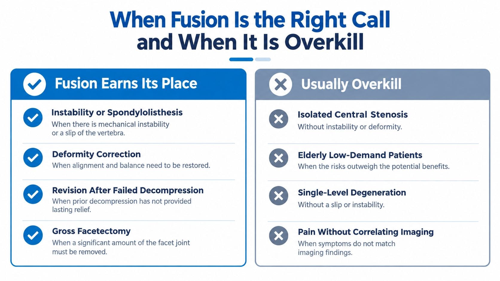
Fusion earns its place when the spine has a mechanical problem that decompression alone won't solve. It becomes technically more defensible when the segment moves abnormally, a deformity changes the load pattern, or the planned decompression would remove stabilizing anatomy.
Relevant situations include:
- Dynamic instability: Flexion-extension radiographs show abnormal translation or angular motion.
- Symptomatic spondylolisthesis: A degenerative or isthmic slip contributes to the patient's symptoms and mechanical dysfunction.
- Deformity: Adult scoliosis or kyphosis changes alignment and load distribution.
- Revision disease: Recurrent stenosis at a previously decompressed level or stenosis beside an existing fusion may require a different mechanical strategy.
- Necessary bone removal: Extensive bilateral facet removal or major pars resection could create iatrogenic instability.
A review discussing these indications cites planning thresholds of more than 5 millimeters of translation or approximately 10 to 15 degrees of angular rotation, particularly when a slip exceeds 25%. Those figures are planning benchmarks, not a universal law, and the review is available in the discussion of lumbar instability and fusion indications.
Questions that expose weak reasoning
Randomized evidence hasn't established one universally accepted definition of instability. That means a patient should ask the surgeon to show the actual finding rather than accept the word “unstable” as a complete explanation.
Useful questions include:
- What structure is causing the stenosis?
- Is there motion on flexion-extension imaging?
- How much facet or pars anatomy must be removed?
- Is the slip stable, or does it move with position?
- Is the main complaint leg pain, mechanical back pain, or both?
- What would happen if decompression alone were performed?
Fusion should be added because the mechanical problem requires it, not because the MRI looks severe.
A stable slip may still be treated with decompression alone in selected patients. Conversely, a seemingly modest narrowing may warrant stabilization if the segment is mechanically compromised. The decision belongs to the anatomy and symptom generator, not to the most alarming phrase in the radiology report.
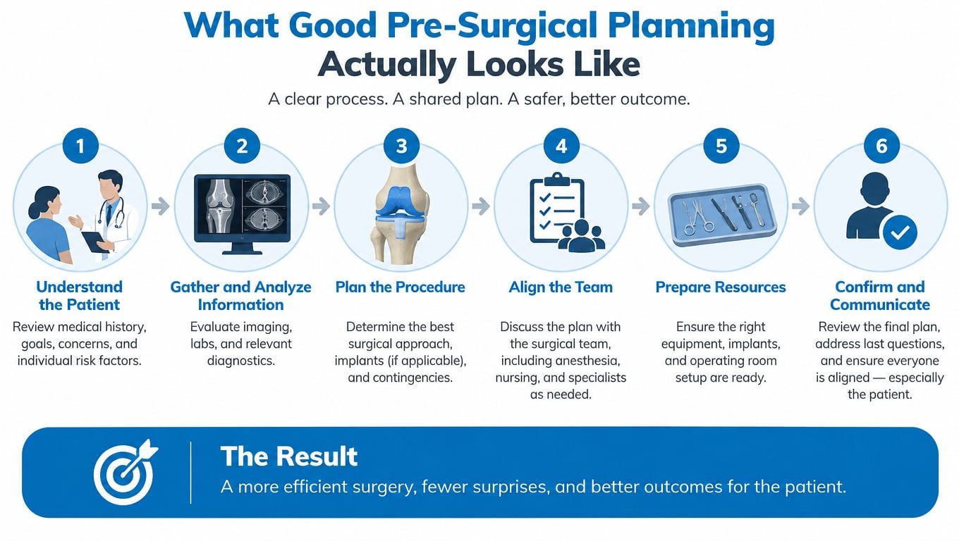
What Pre-Surgical Planning Should Look Like
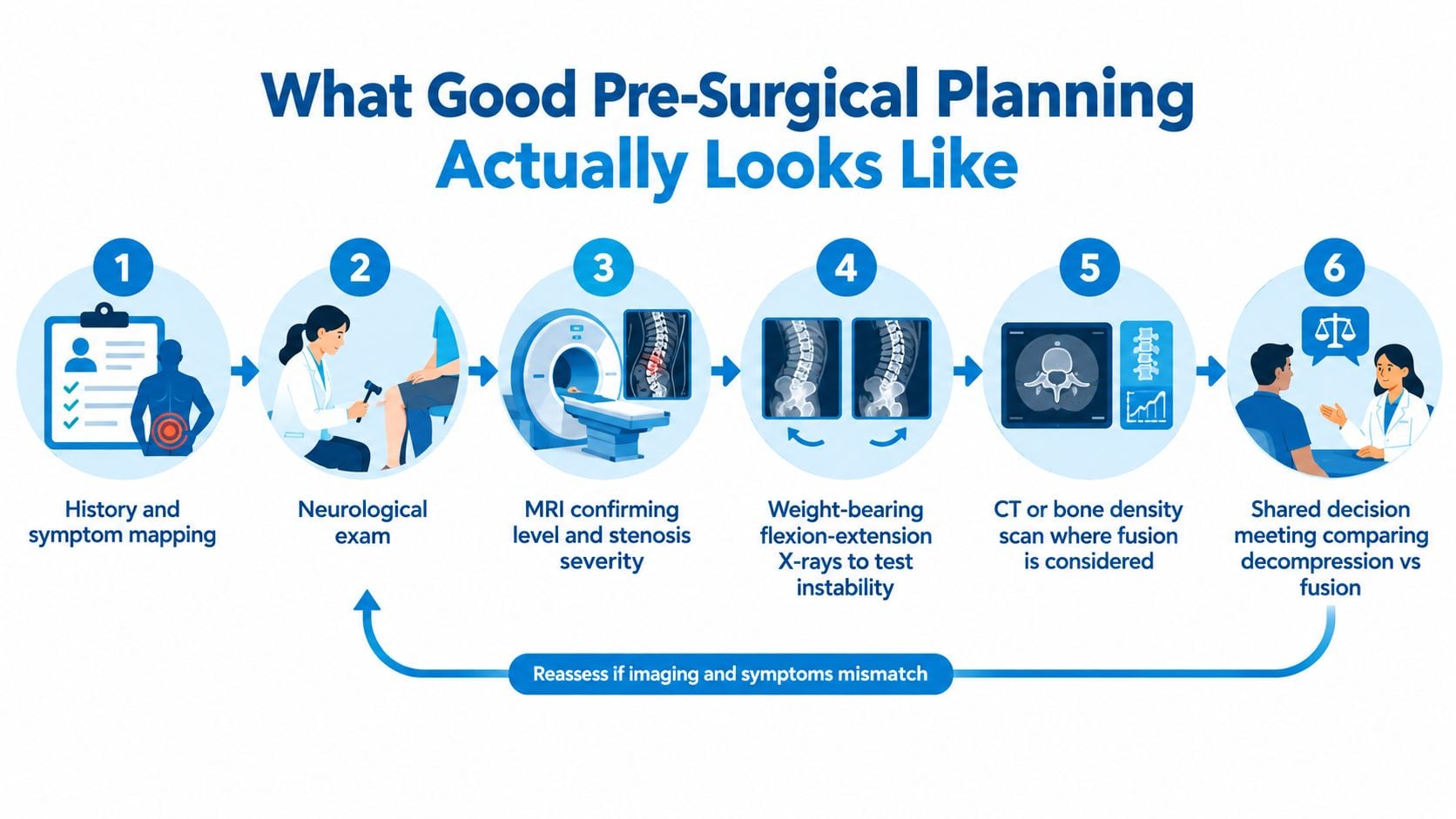
Good planning begins with the symptom pattern and the anatomical cause of narrowing. Map where the pain travels, what triggers it, whether standing or walking is limited, and whether bending forward provides relief. Leg-dominant pain usually points toward nerve compression. Isolated axial back pain demands a different explanation before surgery is considered.
The neurological examination must confirm that the scan matters. Strength, sensation, reflexes, gait, and functional weakness show whether compression is affecting the patient. MRI often reveals narrowing that is incidental, so the surgeon must match the image to the disability.
Diagnostic workup before surgery
- History and symptom map: Identify the likely nerve or nerves involved and the activities that reproduce symptoms.
- Neurological examination: Check weakness, sensory change, reflex asymmetry, gait, and functional loss.
- Standing radiographs: Weight-bearing images assess alignment and can reveal a slip that is less apparent on a supine MRI.
- MRI review: Define the exact level and determine whether ligament, facet, disc, or another structure is compressing the nerve.
- CT when needed: CT clarifies hard-bone anatomy, particularly when prior surgery or complex facet and pars anatomy makes MRI incomplete.
- Bone-density assessment: This matters if the planned operation may require instrumentation or fusion.
If several levels appear abnormal, a diagnostic injection can help identify the symptomatic level. It cannot replace the history, examination, and imaging review, but it can clarify the target when those findings do not agree.
The consultation should produce a precise operative rationale. The surgeon should identify the compressed nerve, name the structure causing the stenosis, explain what will be removed, and state which stabilizing anatomy will be preserved. The proposed operation should follow that anatomy, not the label “minimally invasive” or the most dramatic phrase in the radiology report. If the target and trade-offs remain unclear, consent should wait.
Matching the Procedure to Your Stenosis Profile
The most useful recommendation is profile-based. The following examples show how the cause, symptoms, and stability findings work together.
Isolated ligamentum flavum hypertrophy with claudication may fit percutaneous image-guided decompression when the ligament is the dominant compressive structure and no other major narrowing requires treatment. If bone or facet enlargement also occupies the canal, a conventional decompression may be more complete.
Multilevel degenerative stenosis with leg-dominant symptoms usually points toward decompression at the levels that correlate with the clinical pattern. Fusion shouldn't be added merely because several levels look worn. The surgeon must first determine whether the spine is stable and whether the planned bone removal can preserve stability.
Single-level foraminal stenosis with radiculopathy often requires a targeted foraminotomy, disc treatment, or another approach that reaches the exiting nerve. If restoring foraminal height or removing the facet would destabilize the segment, fusion may be considered. The correct choice depends on whether the compression is soft, hard, or mechanically driven.
Degenerative spondylolisthesis with substantial mechanical back pain deserves a stability-focused discussion. A stable slip with primarily leg symptoms may not automatically require fusion, while motion, deformity, or disabling mechanical pain can make stabilization more reasonable.
Recurrent stenosis beside a prior fusion requires revision planning, not a generic minimally invasive label. Existing hardware, adjacent-segment narrowing, alignment, bone quality, and the amount of prior facet removal all affect the approach. If symptoms don't correlate with the images, continued non-operative care may be more appropriate than another operation.
Risks, Reoperations, and What Recovery Looks Like
A smaller incision does not guarantee a more durable result. Recurrence depends on the degenerative process, whether the nerve was fully decompressed, segment stability, and disease at other levels. A minimally invasive approach can limit muscle and tissue disruption, but it cannot correct the wrong anatomical target or stop degeneration elsewhere.
Reoperation deserves a direct discussion. In the randomized trial discussed earlier, additional lumbar surgery during a mean 6.5-year follow-up occurred in 22% of fusion patients and 21% of decompression-only patients. Those figures do not predict your personal outcome, but they challenge the belief that fusion automatically prevents future surgery. The anatomy still controls the decision: ligamentum flavum thickening, facet overgrowth, instability, and deformity carry different risks and require different operations.
Recovery follows the operation
Recovery reflects your health, age, neurological status, number of treated levels, and procedure. The operation should match the cause of compression, because a smaller approach is not automatically the better approach.
- Limited decompression: Many patients walk soon after surgery, while leg pain often improves before numbness or weakness. Incisional soreness may settle first, with nerve recovery continuing over time.
- Endoscopic or tubular decompression: The smaller access route may reduce early muscle discomfort. It does not shorten the nerve's biological recovery.
- Fusion: Recovery usually takes longer because the procedure treats nerve compression and spinal stabilization. Activity must progress while the fusion and instrumentation heal.
- Image-guided ligament decompression: This limited approach may reduce anesthesia and tissue burden in carefully selected patients. Its evidence base is less mature than that of established decompression procedures, so it should not be presented as a universal substitute.
Your surgeon should explain infection, bleeding, dural injury, nerve injury, persistent symptoms, recurrent stenosis, adjacent-level disease, hardware problems, nonunion after fusion, and the possibility that the operated level was not the primary pain source. The lack of one accepted definition of instability makes the surgeon's reasoning especially important. Ask what finding on your imaging justifies each part of the proposed operation.
The most reassuring recommendation explains what the operation will not fix.
Before consenting, ask which symptom is most likely to improve, what could remain, how long recovery should take, and what non-operative option remains available. A second opinion makes sense when fusion is recommended without documented instability, deformity, or a clear mechanical reason.
Interventional Pain Management evaluates spinal stenosis through history, examination, imaging review, image-guided interventions, and surgical consultation when indicated. Patients in New Jersey and Staten Island can discuss decompression, fusion, endoscopic procedures, and non-operative alternatives by visiting Interventional Pain Management and requesting an evaluation matched to their anatomy and symptoms.