
A person with scoliosis may wake with a familiar ache, sit through a workday, and feel the pain sharpen while standing, walking, or climbing stairs. After an MRI, the report may highlight a curve, a worn disc, narrowed nerve opening, or several findings at once. The difficult question isn't just how large the curve is. It's which structure is producing the pain today, and which treatment can address that structure without creating unnecessary risk.
Scoliosis affects an estimated 2% to 3% of the population, representing about six to nine million people in the United States. Back pain is among the most common complaints associated with the condition, and reported back-pain prevalence in adults with scoliosis ranges from 40% to 90% (American Association of Neurological Surgeons overview of scoliosis). Effective back pain relief for scoliosis starts with a careful diagnosis, not an automatic decision to straighten the spine.
Table of Contents
- Why Scoliosis Back Pain Is Harder to Treat Than It Looks
- Identifying the True Pain Generator in Scoliosis
- Conservative Care That Actually Moves the Needle
- Interventional Procedures for Targeted Relief
- When Surgery Becomes the Right Next Step
- Building Your Individualized Treatment Plan
Why Scoliosis Back Pain Is Harder to Treat Than It Looks
A 42-year-old patient had been told that worsening pain meant the scoliosis was progressing. The explanation seemed plausible because the curve was obvious on X-rays. Yet the symptoms had changed: a deep ache became sharp with sitting, coughing, and bending, then began traveling into the leg. New imaging showed a disc herniation near the curve's apex.
The curve was real, but it was not the pain generator. Treating the Cobb angle alone would have overlooked the herniated disc and irritated nerve. This pattern is common in practice. A visible deformity draws attention, while a disc, facet, nerve, or muscle imbalance may be responsible for the symptoms that need treatment.
One curve can create several mechanical problems
Scoliosis changes how forces pass through the spine. The pelvis, shoulders, and nearby spinal segments may compensate to keep the body upright. Several problems can then overlap:
- Asymmetric loading: Muscles on one side work harder to maintain balance, leading to fatigue and localized aching.
- Disc and facet wear: Uneven pressure may accelerate degeneration at selected levels. Imaging changes alone do not prove that a disc or facet is painful.
- Muscle imbalance: Shortened, overactive muscles may coexist with weak stabilizers, provoking pain during standing, lifting, or prolonged sitting.
- Compensatory curves: A secondary curve may preserve upright posture while increasing stress in another region.
A review of exercise and nonoperative care in scoliosis notes that exercise can reduce pain and disability, although the source of pain may remain uncertain. It may arise from the curvature, degeneration, muscle imbalance, or nerve irritation. That distinction determines the treatment. Exercise may improve endurance and control, while a nerve-directed procedure may be more appropriate for radiating symptoms. Neither approach automatically corrects the other problem.
Adults and adolescents need different expectations
Adolescent idiopathic scoliosis and adult degenerative scoliosis require different clinical priorities. Adolescents can have pain with limited degeneration. Adults more often have a combination of an established curve, disc collapse, facet arthritis, stenosis, and altered sagittal balance.
Back pain is common in adolescents with idiopathic scoliosis. A 2024 study of back pain in adolescent idiopathic scoliosis found pain in 48% of newly diagnosed patients. Earlier studies reported 23% to 68%, compared with about 33% in the general adolescent population. For adolescents, care may involve monitoring growth, controlling symptoms, maintaining activity, and assessing progression risk. For adults, identifying the painful structure and restoring function usually matter more than the curve measurement alone.
Clinical rule: A Cobb angle describes spinal geometry. It does not identify the painful tissue by itself.
The practical sequence is clear: identify the likely pain generator, then match treatment to it. Exercise and therapy may address mechanical strain or deconditioning. Medication or an injection may target a specific painful structure. Surgery is reserved for selected cases involving neurologic compromise, disabling symptoms, or deformity that cannot be managed safely by less invasive care.
Identifying the True Pain Generator in Scoliosis
A useful assessment combines the symptom pattern, physical examination, and imaging. MRI findings matter, but an abnormal disc or facet joint can be painless. The most persuasive diagnosis is the one in which the symptoms, examination, and imaging point in the same direction.
Four patterns clinicians look for
Curve-related mechanical pain is often a broad ache or fatigue sensation along the convex side of the curve. Standing, sitting, or repetitive activity may aggravate it, while changing position or resting may help. Examination may show asymmetric muscle endurance, altered pelvic or scapular control, and pain without a clear dermatomal leg pattern. Imaging confirms the curve but may not reveal a single damaged structure responsible for symptoms.
Disc-related pain tends to feel deep and central or paraspinal. Sitting, bending, vibration, and flexed postures may be more provocative than walking. MRI may show disc dehydration, collapse, or signal changes, but those findings need to match the clinical pattern. A disc herniation becomes more convincing when it also explains radiating leg pain or a neurologic deficit.
Facet-mediated pain is usually more localized beside the spine. Extension and rotation may reproduce it, and examination may reveal focal paramedian tenderness. Hypertrophic or arthritic facet joints on imaging support the diagnosis, but a diagnostic medial branch block may be needed before considering longer-lasting facet treatment.
Nerve compression produces a different quality of pain. Burning, electric, shooting, or aching symptoms may travel through the buttock and leg in a recognizable distribution. Numbness, weakness, reflex changes, or positive nerve-tension signs increase concern. MRI should show a matching foraminal or central narrowing, disc herniation, or other source of nerve irritation.
A practical comparison
| Pain Generator | Hallmark Pain Pattern | Provocative Exam | Key Imaging Finding |
|---|---|---|---|
| Curve-related mechanics | Broad aching, fatigue, activity-related discomfort | Asymmetric endurance or trunk control, without a consistent nerve pattern | Structural scoliosis and compensatory alignment |
| Degenerative disc | Deep midline or paraspinal ache, often worse with sitting or flexion | Pain with loaded flexion or sustained sitting | Disc degeneration, collapse, or compatible MRI signal change |
| Facet joint | Localized paramedian pain, often worse with extension and rotation | Focal tenderness and extension-rotation pain | Facet hypertrophy or arthritic change |
| Nerve compression | Radiating, burning, electric, or dermatomal leg pain | Tension signs, sensory change, weakness, or reflex difference | Foraminal or central stenosis, or a matching disc herniation |
The sacroiliac joint can also imitate lower lumbar pain, particularly when symptoms sit near the buttock and vary with transfers, stairs, or single-leg loading. A focused evaluation of sacroiliac joint dysfunction can prevent every lower-back symptom from being attributed to the scoliosis.
The central diagnostic mistake is treating the most impressive image rather than the most convincing pain pattern. A modest curve with severe foraminal narrowing may require nerve-directed care, while a larger stable curve may mainly need conditioning and load management.
Conservative Care That Actually Moves the Needle
Conservative treatment works best as a sequence, not as a random collection of stretches, medications, and passive appointments. The first step is to match the program to the suspected pain generator while preserving general strength and confidence in movement.
Start with controlled, scoliosis-specific movement
A scoliosis-trained physical therapist can use rotational breathing, active curve correction, and methods influenced by Schroth or SEAS to improve trunk awareness and control. The aim isn't to promise that exercise will straighten an adult spine. The more realistic aim is improved endurance, better pelvic and scapular stabilization, reduced asymmetric strain, and greater tolerance for daily tasks.
General conditioning still matters. Walking, water-based exercise, cycling, and progressive strengthening can maintain capacity without repeatedly provoking the painful direction. Exercises should be adjusted when they produce sharp, radiating, or progressively worsening symptoms. Mild muscular fatigue is different from nerve pain.
The treatment emphasis changes with the generator:
- Disc-sensitive pain: Reduce repeated loaded flexion during the irritable phase, then rebuild tolerance through graded trunk and hip strengthening.
- Facet-sensitive pain: Limit repeated end-range extension and rotation initially, while improving hip mobility and trunk control.
- Muscle imbalance: Use endurance work, breathing mechanics, and progressive loading rather than relying only on massage or stretching.
- Radicular symptoms: Add carefully selected neural mobility work and monitor strength, sensation, and walking tolerance.
Make the daily environment less provocative
Ergonomics won't remodel a curve, but it can reduce repeated irritation. A sit-stand workstation, supported lumbar lordosis, a screen at a comfortable height, and regular position changes can help someone who hurts after prolonged desk work. The key is variation. Holding a rigid “perfect posture” all day can become another source of fatigue.
Sleep position, lifting technique, and recovery habits also deserve attention. Loads should stay close to the body, twisting under load should be minimized, and flare-ups should prompt a temporary reduction in intensity rather than complete inactivity.
Use bracing and medication selectively
Adult bracing may provide temporary support for selected patients with chronic pain, but it shouldn't be presented as a reliable method for correcting an established adult curve. In a 75-patient adult scoliosis bracing pilot study, 65% reported improved back pain and 75% reported improved worst or leg pain, yet only 25% reached the minimal clinically important difference for back pain (adult scoliosis bracing pilot study). The benefit was primarily symptomatic, and leg-pain improvement was greater than back-pain improvement.
Medication should support rehabilitation rather than replace it. Clinicians may consider anti-inflammatory medication when medically appropriate, short courses of oral steroids for selected flares, or neuropathic-pain medicines such as gabapentinoids or duloxetine when nerve features dominate. Kidney disease, gastrointestinal risk, cardiovascular conditions, anticoagulants, pregnancy, and other medications must be reviewed before regular anti-inflammatory use. Long-term opioids can create dependence, tolerance, sedation, and reduced function, so they rarely solve the underlying mechanical problem.
Interventional Procedures for Targeted Relief
A patient may have a visible curve on imaging but pain from a different structure. Before recommending an injection, identify whether the likely generator is a nerve root, disc, facet joint, sacroiliac joint, or muscle imbalance. The procedure should answer a specific diagnostic or treatment question, then guide rehabilitation or the next clinical decision.
Match the procedure to the suspected source
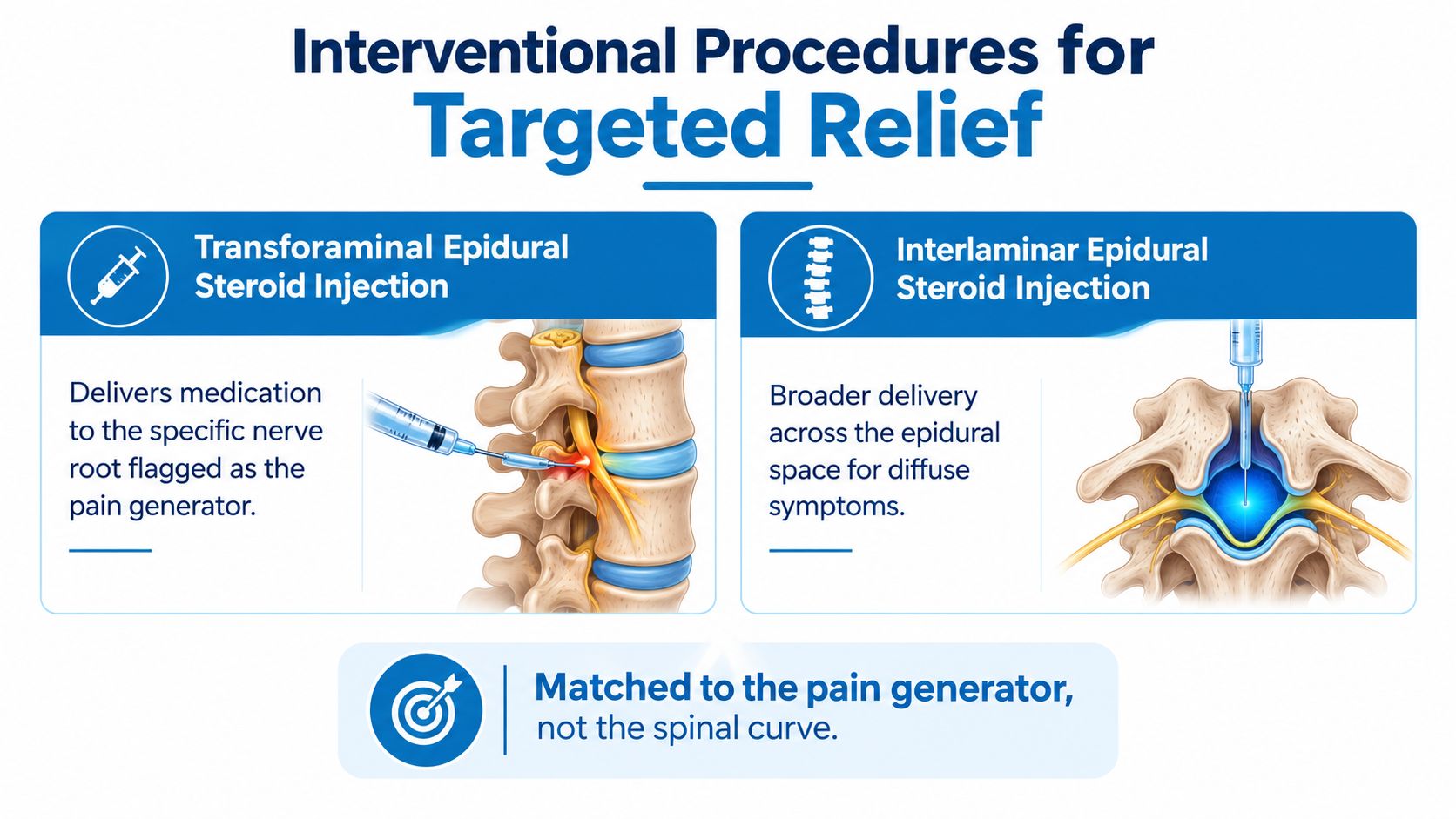
A transforaminal epidural steroid injection places medication beside a selected nerve root. It may fit a patient with leg pain or other radicular symptoms that match disc herniation or foraminal stenosis on imaging. An interlaminar epidural injection spreads medication more broadly through the epidural space and may suit less focal or multilevel nerve irritation. Both approaches generally aim to reduce inflammation long enough to improve walking, therapy participation, and daily function. Neither corrects the spinal curve.
Facet-mediated pain requires a different strategy. Medial branch blocks can test whether facet joints contribute to localized pain, particularly when extension reproduces symptoms. Patients who obtain meaningful, temporary relief from diagnostic blocks may be considered for radiofrequency ablation. A sacroiliac injection is more appropriate when buttock-centered pain, examination findings, and symptom location point toward that joint.
| Procedure | Main target | Best clinical fit | Practical limitation |
|---|---|---|---|
| Transforaminal epidural injection | Specific irritated nerve root | Focal radicular pain with matching imaging | Relief may be temporary and does not remove structural stenosis |
| Interlaminar epidural injection | Broader epidural inflammation | Diffuse or multilevel nerve irritation | Less selective for one nerve root |
| Medial branch block | Facet joint nerve supply | Localized extension-related pain | Diagnostic value depends on careful interpretation |
| Radiofrequency ablation | Medial branch nerves | Reproducible facet-mediated pain after diagnostic testing | Does not treat disc, curve, or nerve compression |
| Sacroiliac injection | Sacroiliac joint | Buttock-centered pain with supportive examination | The joint may be only one contributor |
In adult spinal deformity, injections can help selected patients, but improvement may fade. One reported injection study found 37.2% success at one year and 27.3% at two years. Those results support realistic counseling about durability rather than promises of permanent relief.
Image-guided epidural steroid injections carry procedural risks, including infection, bleeding, dural puncture, temporary blood-sugar elevation, mood changes, and a short-term pain flare. Spinal cord stimulation may suit selected patients with persistent neuropathic or postsurgical pain when structural surgery is not the appropriate next option. Intrathecal pumps are reserved for carefully evaluated refractory cases because device management and medication complications can be substantial.
When Surgery Becomes the Right Next Step
Surgery becomes reasonable when the problem is structural, symptoms remain disabling, and the expected benefit outweighs the loss of motion, recovery burden, and complication risk. Persistent pain alone doesn't automatically justify a long fusion. The decision depends on whether the painful structure can be addressed with a smaller operation or whether deformity and imbalance require broader reconstruction.
Urgent assessment is warranted for progressive neurologic weakness, signs of cauda equina compression, or rapidly worsening deformity in a growing child. In adults, documented curve progression beyond 50 to 55 degrees may influence surgical planning, but the measurement is only one part of the decision. Pain, disability, sagittal balance, bone quality, health status, and functional goals often matter more than the Cobb angle in isolation.
Choosing the scale of the operation
- Decompression alone may suit selected patients whose main problem is nerve compression and whose alignment remains stable.
- Short-segment fusion can address a focal painful or unstable level while preserving more motion, provided the surrounding spine can tolerate the altered load.
- Long-construct fusion may be needed when multiple levels, major imbalance, or progressive deformity drive the symptoms, but it involves greater surgical exposure, longer recovery, and more stress on adjacent segments.
Comparative adult scoliosis data found that surgically treated patients improved mean back-pain scores from 6.3 to 2.6 and disability scores from 35 to 20 at two years, while nonoperative patients had no significant change in those measures. The result shows that surgery can provide substantial improvement for selected patients, not that every painful curve should be fused.
A careful surgical consultation should explain the pain generator, the proposed levels, the expected functional trade-offs, possible revision needs, and alternatives. Information about scoliosis correction is most useful when it is discussed alongside the patient's neurologic examination, imaging, bone health, and daily limitations.
Surgery should be planned around the symptoms and alignment that need correction, not around an X-ray number alone.
Building Your Individualized Treatment Plan
A workable plan begins with organized information. Patients should bring the actual imaging, reports from prior studies, a medication list, and a short symptom diary. The diary should record where the pain starts, whether it travels, what positions trigger it, sleep disruption, walking tolerance, medication use, and the activities that matter most.
Function deserves priority over a single pain score. “Stand long enough to cook,” “walk through a grocery store,” or “return to recreational activity” gives a clinical team a measurable target. A reassessment every eight to twelve weeks can examine pain, function, medication reliance, strength, and tolerance for exercise rather than waiting until a crisis forces a change.
Assemble the right clinical team
A physiatrist can coordinate diagnosis and rehabilitation. A scoliosis-trained physical therapist can build movement strategies around the curve and the dominant pain generator. An interventional pain clinician can use diagnostic blocks or injections when appropriate. An orthotist may help with a selected brace, while a spine surgeon can evaluate deformity, stenosis, instability, and neurologic risk.
A second opinion is reasonable when a major fusion is proposed without a clear pain generator, when symptoms and imaging don't match, or when nonoperative options haven't been explained. It is also sensible when the recommended operation seems much larger than the functional problem being treated.

Troubleshooting common setbacks
- Exercise soreness: Mild, short-lived muscle soreness may mean the load needs to be progressed more gradually. Sharp, radiating, or neurologic symptoms require prompt reassessment.
- Pain rebound after a procedure: A temporary flare can occur, but fever, increasing weakness, severe headache, or worsening neurologic symptoms need urgent medical attention.
- Recovery stalls: Recheck the diagnosis, sleep, medication effects, conditioning level, and work demands. A plateau may mean the original pain generator was incomplete or another source has emerged.
The strongest plan remains flexible. It measures meaningful function, responds to new neurologic findings, and avoids escalating treatment just because an image looks dramatic.
Interventional Pain Management offers diagnostic evaluation, image-guided injections, nerve blocks, rehabilitation coordination, spinal cord stimulation, and spine surgery assessment for selected patients with scoliosis-related pain. To match treatment to the actual pain generator rather than the curve alone, visit Interventional Pain Management.