
You notice it during a run, a long walk, or a gym session. The top of the foot starts as a small, annoying spot, then it nags more with each impact, and later it settles down once the shoe comes off and the foot rests. That pattern is easy to dismiss as “just overuse,” but when the pain is pinpoint, repeatable, and tied to loading, it deserves a more careful explanation.
A stress fracture is usually a load problem, not a single dramatic injury. The bone takes repetitive force faster than it can repair itself, so tiny cracks begin to form. On the top of the foot, that often shows up as pain that worsens with walking, running, jumping, or even daily steps, then eases with rest, which is one reason it can be confused with tendon irritation early on.
Table of Contents
- What a Stress Fracture on Top of the Foot Actually Is
- Stress Fracture vs Extensor Tendonitis on Top of the Foot
- How Clinicians Diagnose Top of Foot Stress Fractures
- Treatment Options From Rest to Surgical Repair
- Realistic Recovery Timelines and Return to Activity
- Prevention and Load Management That Actually Works
- When to Seek Specialty Care and What to Expect
What a Stress Fracture on Top of the Foot Actually Is
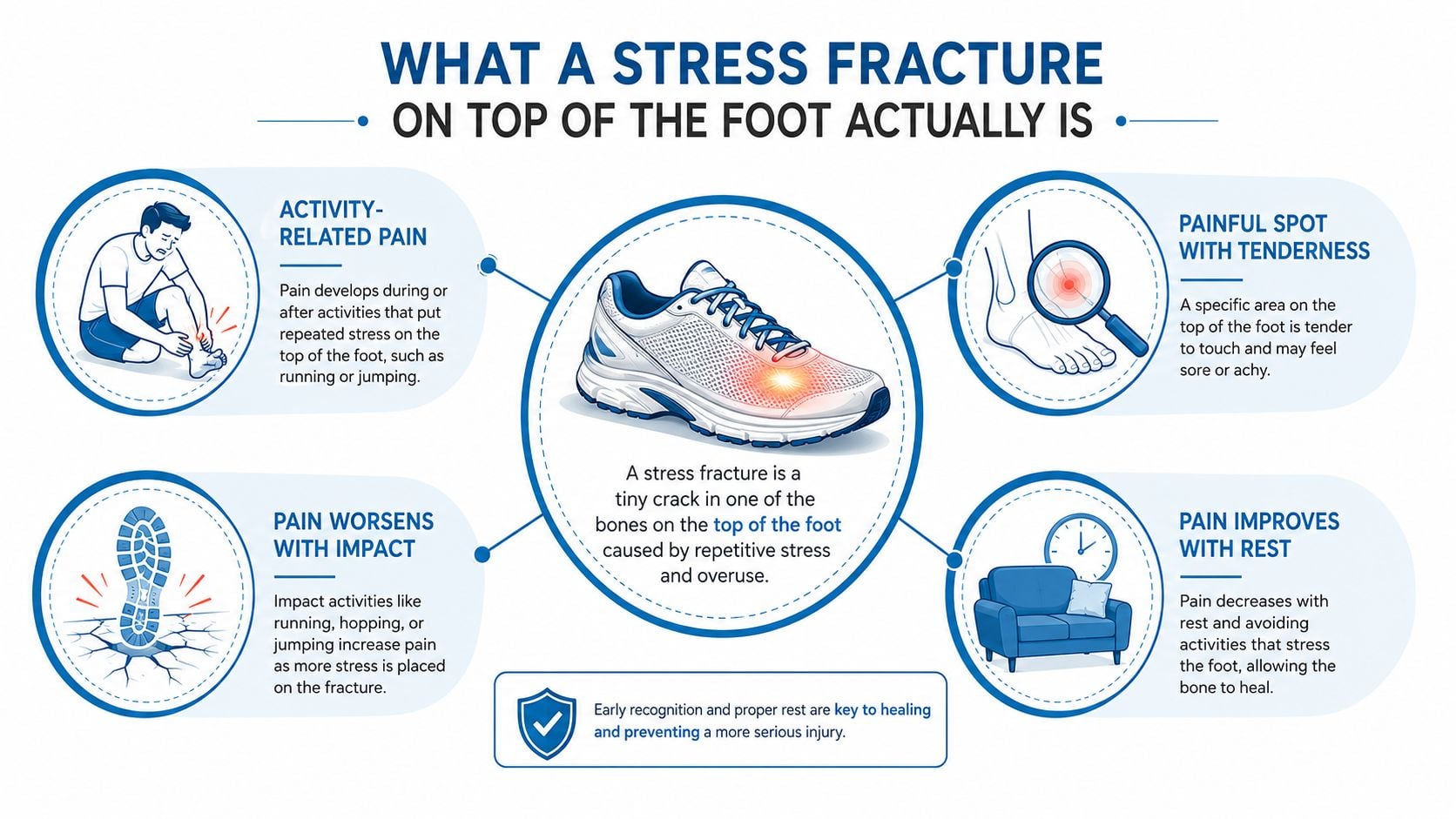
A runner often notices this first as a small, tender spot on the top of the foot that hurts more near the end of a workout than at the start. The confusing part is that the pain can seem mild enough to ignore, then it flares with the next walk, jog, or stair climb. That stop-start behavior is one reason people keep trying to “work through it” longer than they should.
The bone is failing at repair, not breaking all at once
A stress fracture is a small crack from too much stress on a bone, not a single traumatic break. Cleveland Clinic describes stress fractures that way, and CHOP's metatarsal guide notes that the main symptom is pain on the top of the foot that can progress to pain with daily activities such as walking, with a classic pattern of worsening during impact and improving with rest (Cleveland Clinic). That pattern is the clue that separates a bone problem from a random ache.
Practical rule: if one exact spot hurts every time the foot loads, and the pain quiets down when activity stops, bone injury moves higher on the list.
The top of the foot can involve several bones, especially the metatarsals, and sometimes the navicular, cuneiforms, or talus. Location matters because each bone behaves a little differently under load, and some heal more predictably than others. A bone that takes force in a high-stress zone needs more protection than one in a lower-risk area.
An easy analogy is a credit card. Bend it once and it's fine, but bend the same spot again and again, and a line of weakness starts to appear. Bone works differently from a card, of course, but the idea is the same, repetitive force creates damage faster than repair can keep up.
Training through that pain usually makes the crack more stubborn, not less. The bone gets more irritated, surrounding tissue tightens up, and the person ends up with a longer shutdown instead of a quicker fix. That's why early recognition matters more than toughness here.
Stress Fracture vs Extensor Tendonitis on Top of the Foot
Many people get stuck here, because pain on the top of the foot can come from either bone or tendon. The distinction matters because stress fracture and extensor tendonitis can start with the same complaint, but the clues on exam are different. A clinician looks for whether the pain is focal and bony or diffuse and tendon-line based.
The most useful clue is where the pain lives
Stress fracture pain usually has one clear spot that can be reproduced by pressing directly on the bone. Extensor tendonitis more often feels like soreness along the tendon path, and it can be aggravated by moving the toes upward against resistance. The article on top-of-foot pain from Baptist Health makes the point that top-of-foot pain can be either a stress fracture or extensor tendonitis, but the better discriminator is the symptom pattern, not the label alone (Baptist Health).
| Feature | Stress Fracture | Extensor Tendonitis |
|---|---|---|
| Pain location | One pinpoint bony spot on the top of the foot | More spread out along the tendon line |
| What reproduces pain | Direct pressure over bone, impact loading, hopping | Resisted toe extension, repeated toe lifting |
| Pain during activity | Often worsens the longer the foot is loaded | Can warm up or change with tendon use |
| Pain after rest | May settle, then return quickly with impact | May feel less sharp after loosening up |
| Touch test | One exact spot is often very tender | Tenderness is usually more linear or diffuse |
A simple decision rule helps
If the pain is reproducible with one fingertip on one bone, and it returns the moment running or jumping restarts, fracture rises on the list. If the pain spreads along the top of the foot and shows up more with toe motion than with bone pressure, tendon irritation becomes more likely. That's not a final diagnosis, but it's a useful self-screen.
The line between the two can blur early, which is why normal-looking movement doesn't rule out a stress fracture. The foot can still function while a microcrack is forming, especially if the person is fit and determined. That's also why persistent pain after a few days of unloading deserves a closer look rather than another week of guesswork.
This orthopedic overview of tendon irritation and related pain patterns can be useful if the pain feels more tendon-like, but a clinician still needs to match symptoms to exam findings.
If pressing on one exact spot causes a sharp, familiar pain, that's more concerning for bone than for soft tissue.
How Clinicians Diagnose Top of Foot Stress Fractures
A specialist visit usually starts with your story, not with imaging. The clinician asks when the pain began, whether training volume rose, whether shoes changed, and whether walking, running, or jumping increased in a way that overloaded the foot. Bone health risk factors matter too. In female athletes, menstrual history can provide an important clue, because missed or irregular periods can point to a broader problem with bone loading.
The first question is often whether this behaves like a bone injury or like extensor tendon irritation. A stress fracture usually gives one very focal, easy-to-find spot of pain over a bone, while tendon pain tends to spread along the top of the foot and change more with toe movement. That difference matters because both can hurt on the dorsal foot, but they do not behave the same way under exam.
The exam looks for a very specific pain signature
The physical exam focuses on direct palpation, the hop test, and other maneuvers that load the bone in a controlled way. A clinician may also use a tuning fork over the painful area, check the forefoot with a squeeze test, and compare both feet for swelling or a change in gait. The goal is to reproduce the exact pattern that suggests osseous injury rather than generalized soreness.
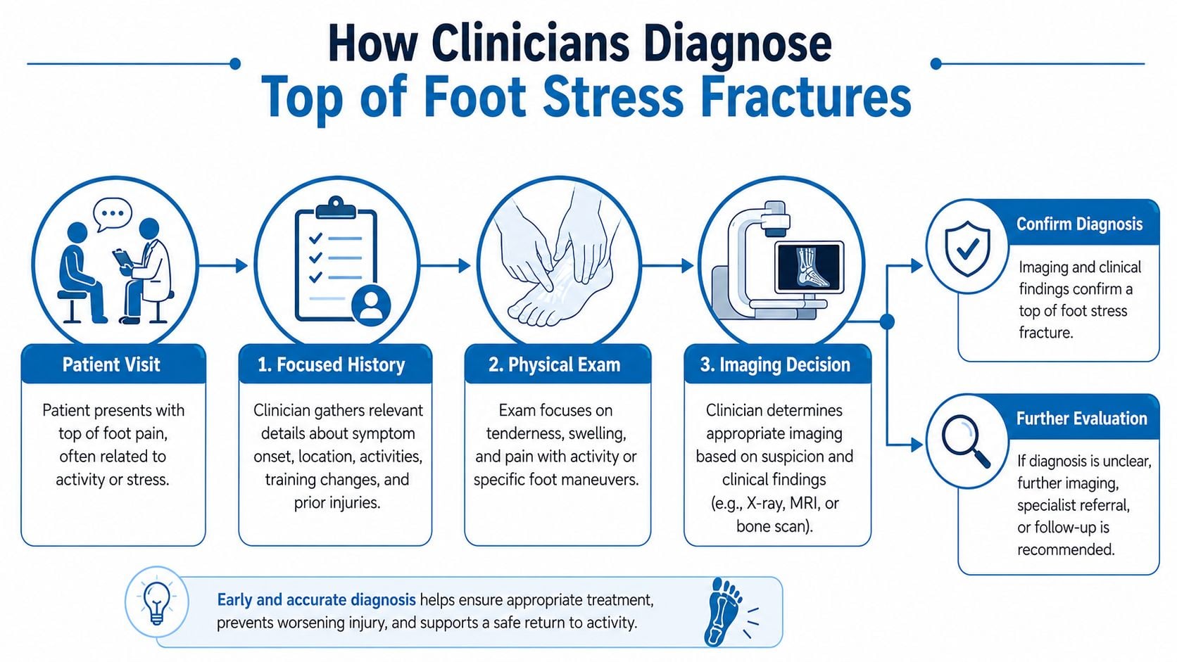
A useful way to think about the exam is simple. If one fingertip on one bone recreates the familiar pain sharply, that raises concern for a stress fracture. If the discomfort is more spread out and tied to toe motion or tendon use, soft-tissue irritation moves higher on the list. That is not a final diagnosis, but it helps explain why a careful exam can separate the two early on.
Imaging follows a ladder, not a guess
X-rays are often the first test, but early stress fractures can look normal. A normal film does not end the workup if the pain pattern is convincing. MRI is usually the best early test when the goal is to see a stress reaction before a fracture line becomes obvious, while CT is used more selectively when the clinician needs a clearer look at a fracture line in higher-risk bones such as the navicular or talus. Bone scan is used less often now, since MRI usually gives more useful detail.
A normal early X-ray does not mean nothing is wrong. It often means the bone injury is still too early to show clearly on plain film.
Clinical guides from StatPearls and similar orthopedic references describe stress-fracture care as starting with relative rest or non-weight-bearing when needed, and more protection for high-risk bones such as the navicular. That is one reason persistent pain on the top of the foot deserves attention after a training increase, especially when the tenderness stays sharply localized.
When the exam and the story do not line up cleanly, imaging helps sort out the next step. A clinician may decide the foot needs more imaging, more protection, or both, based on the exact bone involved and how focal the pain is. For a broader look at fracture patterns and bone-injury care, see this overview of foot and ankle fractures.
Treatment Options From Rest to Surgical Repair
Treatment depends on where the crack is and how risky that bone is. A low-risk metatarsal stress fracture does not get the same plan as a navicular or talus injury, because the healing behavior is different. The treatment plan has to match the bone involved, not just the label “stress fracture.”
The first step is unloading the bone
For many foot stress fractures, treatment begins with relative rest, which means cutting back activity enough that the bone stops taking repeated impact. A stiff-soled shoe or walking boot can reduce load through the injured area, and crutches make sense when every step hurts. In many foot and ankle stress fractures, healing often falls in the 4 to 6 week range, and some cases take up to 12 weeks, especially when the bone is higher risk.
Protective care can escalate when the bone needs more help
If pain keeps showing up with everyday walking, clinicians may move to non-weight-bearing, especially for navicular injuries. The navicular is a classic high-risk site, so it gets more caution than a routine metatarsal crack. In practice, the plan is based on pain, bone location, and imaging findings together, the way a car mechanic does not ignore where the noise is coming from before deciding what needs to be opened up.
A bone stimulator sometimes enters the plan for slow healing or stubborn cases, especially when the goal is to support remodeling while the bone stays protected. If symptoms still do not settle, the clinician may look for other reasons the injury is not calming down, such as mechanical overload, nutrition issues, or a repeated injury pattern.
Interventional pain care can also help when pain is blocking sleep or rehabilitation. In selected patients, image-guided nerve blocks or targeted injections may calm pain enough for safer function while the fracture is protected. At Interventional Pain Management, that kind of evaluation can be coordinated with orthopedic review when symptoms are complex, which matters when a foot injury is part of a larger musculoskeletal picture.
Surgery is reserved for specific situations
Surgery is considered when the fracture line is displaced, when a navicular nonunion or talar injury is present, or when a high-demand athlete needs a more stable solution after the risks and benefits are reviewed. Surgical repair is not the usual path for a simple metatarsal stress fracture, but it becomes more relevant when the bone is high risk or healing has stalled. The decision is about stability and return of function, not about being aggressive for its own sake.
This overview of foot and ankle fractures fits naturally here because the same visit may involve both fracture management and pain-control planning.
Realistic Recovery Timelines and Return to Activity
Recovery works best when it follows symptoms, not wishful thinking. The foot needs a period of unloading first, then a gradual rebuild of tolerance, then a return to impact only when the bone is quiet under everyday stress. Pushing that sequence too fast often brings the pain back.
Early recovery means protecting the bone, not testing it
The first phase usually centers on pain control and unloading. That may mean a boot, a stiff shoe, or temporary crutches, depending on how much pain shows up with walking. If daily steps still reproduce a focal ache, the bone is not ready for impact work yet.
Middle recovery is where cross-training matters. Swimming, cycling, and rowing can help preserve fitness without hammering the injured area, as long as they don't trigger foot pain. The transition back to activity should feel boringly gradual, because that's what bone likes.
A useful milestone checklist
- Walking feels quiet: ordinary steps don't recreate the same pinpoint pain.
- No rebound pain: symptoms don't come roaring back after light activity.
- Swelling is settling: the foot looks and feels less reactive, not more.
- Impact is added last: jogging comes after pain-free walking and low-impact cardio.
Low-risk metatarsal injuries often settle faster than navicular or talus injuries, which are more likely to need longer protection. The 2 to 12 week range from the earlier section matters here because the same diagnosis can hide very different healing paths. A longer timeline is not a failure, it's often the safer plan for the bone involved.
If pain returns with the first few steps of a return-to-run attempt, that's a sign to step back, not push harder.
Red flags deserve earlier follow-up. Pain at rest, night pain, swelling that increases instead of fades, or a sudden sharp change in symptoms can mean the bone is under more stress than expected. When that happens, the return plan needs to slow down before a small crack becomes a bigger one.
Prevention and Load Management That Actually Works
Prevention starts with load management, not shoe hype. A new pair of shoes can help, but it won't fix a training plan that jumps too fast or a body that's already behind on recovery. The bone responds to total stress, not just what's on the feet.
Training load is the main variable
A steady progression is safer than abrupt changes in mileage, hills, speed work, or surface. The familiar 10 percent rule is a simple way to keep weekly mileage changes from spiking too sharply, even though real-world training still needs common sense. Sudden changes in intensity matter too, because intervals and hills can overload the top of the foot even when overall mileage looks modest.
The best prevention checklist is practical:
- Build gradually: keep increases modest and consistent.
- Track intensity: hard sessions count, not just distance.
- Strengthen the chain: foot and hip strength both matter.
- Replace worn shoes: dead cushioning doesn't help load distribution.
- Screen repeat injuries: recurrent stress fractures deserve a deeper look.
Nutrition and bone health can't be ignored
Bone remodeling needs adequate building blocks. Calcium and vitamin D support that process, and a helpful overview of those nutrients is available in this guide to calcium, vitamin D, and magnesium. For athletes with repeated injuries or menstrual irregularities, bone density screening may be part of a more complete evaluation because the issue may not be the foot alone.
Footwear still matters, but it should stay in proportion. A shoe should fit well, match the activity, and be replaced before it's completely worn out. Rotating shoes can also help spread load patterns a bit, though it won't undo a bad training block.
When to Seek Specialty Care and What to Expect
Specialty care makes sense when the pain pattern stops behaving like simple soreness. Focal pain lasting more than two weeks, pain at rest or at night, swelling that lingers, or a history of stress fracture should move the person beyond home care. Those clues suggest that the foot needs a focused exam and possibly imaging rather than another round of guessing.
A multidisciplinary visit should feel organized
A good orthopedic or pain visit starts with the history, then moves to the exam, then to imaging coordination if the symptoms warrant it. The clinician may also build a plan that combines unloading, rehabilitation, and, when needed, interventional pain procedures or surgical referral. That kind of coordinated care is especially helpful when the pain is interfering with sleep, work, or safe walking.
The orthopedic service page is relevant here because persistent foot pain often needs that same type of structured evaluation, not just advice to rest.
Bring a few things to the first visit so the appointment stays efficient:
- Training log: recent mileage changes, speed work, hills, and impact volume.
- Shoe history: what's been worn, what changed, and how old the pair is.
- Prior imaging: old X-rays or MRIs, if any exist.
- Symptom notes: where the pain is, when it worsens, and what eases it.
The practice model matters too. Interventional Pain Management serves New Jersey and Staten Island with seven-days-a-week availability, multiple locations, and support services that can help with transportation and insurance navigation. For someone trying to get a timely answer about foot pain, that access can shorten the gap between “this hurts” and “this is what to do next.”
If the top of the foot has been hurting with walking, running, or daily steps, Interventional Pain Management can help sort out whether the problem looks more like a stress fracture, tendon irritation, or another load-related injury. Visit Interventional Pain Management to arrange an evaluation and get a plan that matches the bone involved, the pain pattern, and the pace of recovery.