
A patient may wake after surgery expecting a few sleepy hours, only to find that an arm, hand, or foot still feels heavy, numb, or strangely weak. That moment can be frightening, especially when the block was supposed to improve comfort rather than create a new problem. The important question isn't whether symptoms are present. It's how they change, where they occur, and how long they last.
Nerve block damage is uncommon, but temporary numbness, tingling, or weakness can overlap with normal anesthetic recovery. The first 24 to 48 hours are usually an observation window, not a final diagnosis. Clear information helps patients recognize expected recovery, contact the anesthesia or surgical team at the right time, and seek urgent help when the pattern suggests compression or another treatable cause.
Table of Contents
- When Numbness After a Nerve Block Isn't Just the Anesthetic
- What Nerve Block Damage Actually Means in Clinical Terms
- How Nerve Blocks Injure Nerves and Why Severe Symptoms Need Urgent Attention
- Patient Risk Factors That Raise the Chance of Nerve Injury
- Symptoms to Watch For and When to Call Your Physician
- How Doctors Diagnose and Stage Nerve Block Damage
- Treatment Options and Realistic Recovery Timelines
- Prevention, Specialist Care, and Where to Go From Here
When Numbness After a Nerve Block Isn't Just the Anesthetic
A 54-year-old office worker had shoulder surgery with an interscalene block. His arm was expected to feel “asleep” for several hours. The next morning, he still could not lift his wrist, and the numbness felt unlike the softer heaviness he noticed immediately after surgery.
Lingering numbness, tingling, heaviness, or weakness does not automatically indicate permanent nerve injury. The timing and pattern still deserve attention. Local anesthetic may be fading slowly, while surgical irritation, swelling, or positioning can also affect nerve function.
A peripheral nerve block temporarily interrupts pain signals between a body region and the brain. As the medication wears off, feeling and movement usually return gradually. Pins and needles can occur during this transition, and patchy sensation may make recovery seem uneven.
The pattern matters: a symptom that is steadily improving deserves different attention from one that is dense, worsening, or accompanied by severe pain. A useful comparison is a dimmer switch. Normal recovery often brings signals back gradually, while a nerve problem may leave sensation or strength unusually absent, distorted, or painful.
Serious injury caused specifically by a peripheral nerve block is uncommon. A review found postoperative neurologic symptoms at about 1% during the first 2 weeks, falling to roughly 3 per 10,000 by 1 year. That pattern indicates that many early symptoms resolve, while a much smaller group persists long term (the review of neurologic complications after peripheral nerve blocks). The Royal College of Anaesthetists advises patients to contact the hospital if symptoms continue beyond 48 hours (its patient guidance on nerve damage after a block).
A clear overview of how nerve blocks provide pain relief can help explain why numbness occurs in the first place. The practical question is whether the anesthetic effect is fading and function is returning in a reassuring direction. Dense or worsening weakness, severe pain, or symptoms that do not begin improving should prompt a call to the treating team.
What Nerve Block Damage Actually Means in Clinical Terms
Clinically, nerve block damage means an unintended change in sensation, movement, or pain within the area supplied by a nerve involved in regional anesthesia. The key question is not only whether a symptom exists. Its duration, severity, and direction matter. Symptoms that steadily recede fit expected recovery more closely than dense numbness, new weakness, or worsening pain.
Anesthetic block temporarily silences nerve conduction. Structural injury disrupts the axon itself, so recovery may take weeks to months. That difference explains why temporary numbness can be expected while persistent or changing symptoms deserve medical review.
Three broad clinical patterns
Doctors describe nerve effects as a spectrum:
- Transient neuropraxia is a temporary conduction problem. Sensation or strength may change for days or weeks as the nerve recovers.
- Prolonged deficit continues beyond the early recovery period. It may require examination, observation, and possibly electrodiagnostic testing.
- Permanent injury is a lasting deficit after the nerve has had a reasonable opportunity to recover. This outcome is rare.
A large review reported temporary postoperative neurologic symptoms ranging from 0 to 2.2% at 3 months, 0 to 0.8% at 6 months, and 0 to 0.2% at 1 year. In the same review, permanent peripheral nerve injury ranged from roughly 0.014% to 0.04% (the review of peripheral nerve injury after regional anesthesia). These figures support an important distinction: early nerve symptoms can occur, while confirmed persistent injury is much less common.
The practical clinical task is to track the pattern over time. A mild, shrinking area of altered sensation suggests recovery is underway. A deficit that stays dense, spreads, or affects strength needs assessment, because examination findings help distinguish delayed anesthetic recovery from nerve dysfunction.
Patients may hear different names for procedures involving injections. A plain-language guide to common pain-management injection types can help distinguish a nerve block from other treatments.
How Nerve Blocks Injure Nerves and Why Severe Symptoms Need Urgent Attention
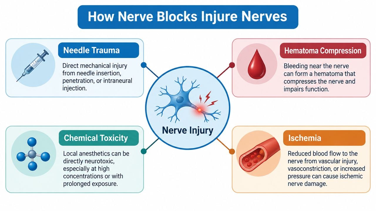
A nerve can be affected by the injection, by pressure around it, by the medication, or by events connected to surgery. The cause isn't always obvious because several stresses can occur during the same procedure.
Four pathways doctors consider
Direct needle trauma occurs when the needle contacts or enters nerve tissue. A particularly serious concern is injection within the fascicle, the bundle containing nerve fibers. Mechanical disruption can affect the axons and the protective connective layers around them.
Raised intrafascicular pressure can occur when fluid is injected inside the nerve's protective compartment. Pressure may reduce small-vessel blood flow and create ischemic injury. This is one reason injection should stop when the clinician encounters concerning resistance or unusual patient feedback.
Local anesthetic toxicity refers to chemical injury from exposure to the anesthetic. Laboratory and clinical guidance identify toxicity as a possible contributor, especially when medication reaches sensitive internal nerve structures.
Compression from a secondary cause may follow a hematoma, infection, abscess, swelling, or surgical positioning. In these cases, the block may not be the sole cause. A nerve can also be stretched or compressed during surgery, particularly when the limb remains in a difficult position.
Why urgent assessment matters
A mild, gradually improving tingling pattern is different from a dense motor deficit that appears suddenly or worsens. Severe weakness, escalating pain, tense swelling, or pain that sharply increases when a joint is gently moved can indicate compression or a circulation-threatening process.
Hematoma and infection are especially important because early treatment may reverse the pressure on the nerve. Expert guidance recommends urgent evaluation for severe deficits and specialist nerve-surgery assessment when a lesion remains persistent beyond 3 to 5 months (guidance on neurologic complications after regional anesthesia). Patients shouldn't wait for a routine follow-up if weakness is rapidly progressing, the limb is becoming cold or discolored, or swelling is becoming firm and painful.
Patient Risk Factors That Raise the Chance of Nerve Injury
Risk doesn't belong to the injection alone. A nerve may already have reduced reserve before surgery, and the operation may add pressure, stretch, inflammation, or reduced blood flow. That's why the consent discussion should include the patient's health, the planned operation, the block location, and positioning.
Medical conditions and medication history
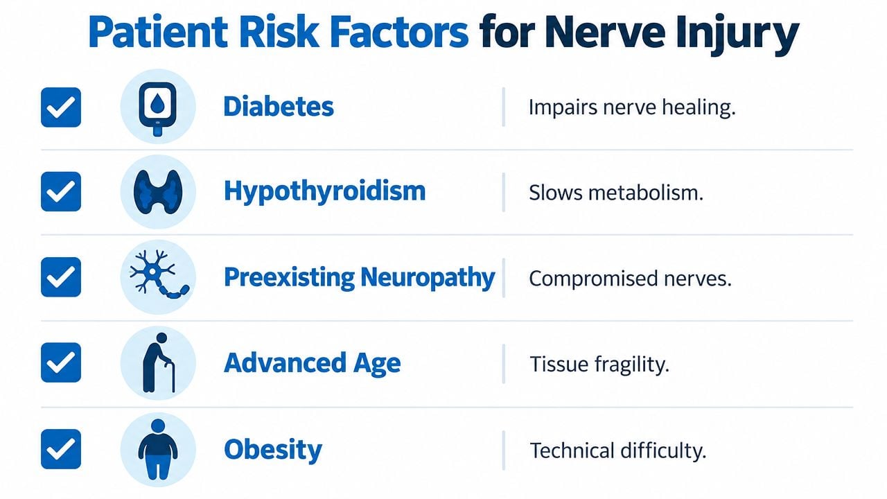
Diabetes can affect nerve health and healing, even when a patient has no obvious symptoms. Preexisting neuropathy, whether related to diabetes, prior chemotherapy, or another cause, can make it harder to identify a new deficit and may leave the nerve less tolerant of additional stress.
Hypothyroidism and advanced age may also influence tissue resilience and recovery. These factors don't mean a patient can't receive a block. They mean the anesthesiologist should document baseline sensation and strength carefully so postoperative changes can be recognized.
Anticoagulants matter because they can increase bleeding risk, while steroid exposure and other medical treatments may influence infection risk, tissue quality, or healing. Medication decisions must come from the treating team. Patients should provide a complete list rather than stopping anything independently.
Procedure and positioning factors
Body habitus can affect access and positioning, but it isn't a judgment about a patient's responsibility. Surgical factors may include tourniquet use, pressure against a vulnerable area, prolonged stretching, or implant placement near a nerve. These events can create symptoms that appear after surgery even when the block itself was performed appropriately.
A useful preprocedure checklist includes:
- Baseline symptoms: Report numbness, burning, weakness, balance problems, or reduced grip before the procedure.
- Metabolic conditions: Disclose diabetes, thyroid disease, and other conditions affecting nerves or circulation.
- Bleeding medicines: Provide exact information about anticoagulants, antiplatelet drugs, and supplements.
- Previous reactions: Mention any prior block that caused unusually prolonged numbness, severe pain, or weakness.
- Intraoperative concerns: Ask how the team will protect the limb during positioning and whether postoperative checks are planned.
Risk factors change the level of attention required. They don't predict an individual outcome, and they don't turn normal postoperative numbness into proof of nerve block damage.
Symptoms to Watch For and When to Call Your Physician
The most useful triage tool combines symptom type, timing, and direction of change. Numbness alone isn't enough to diagnose injury. The same sensation can be expected while anesthetic fades or concerning when it becomes more intense after function should be returning.
A practical symptom pattern
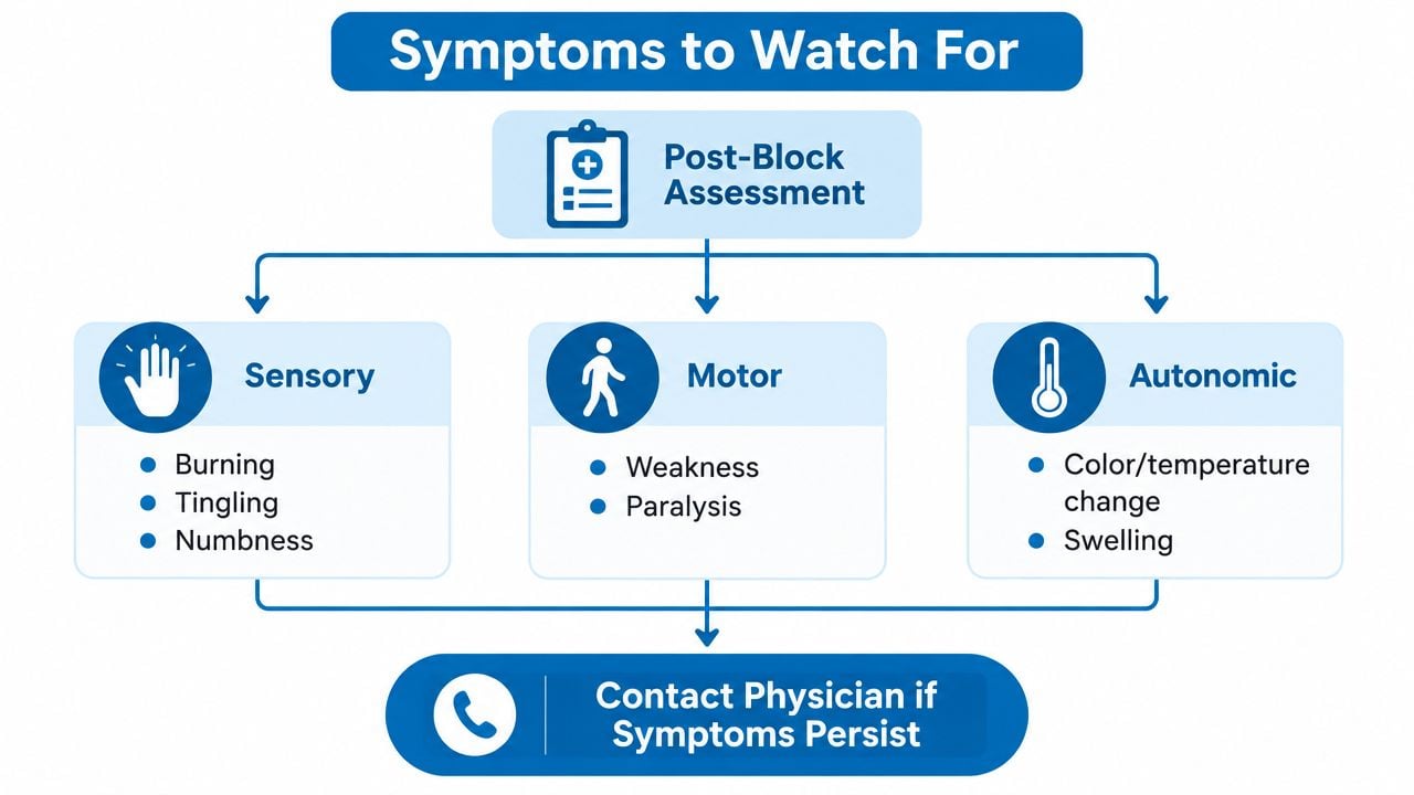
Sensory changes include burning, electric shocks, tingling, hypersensitivity, or a patch of numb skin. Mild tingling during recovery can be reassuring if sensation is returning. Persistent or expanding numbness, especially with increasing burning pain, deserves a call to the anesthesia team or surgeon.
Motor changes include difficulty lifting the wrist, moving the foot, bending the fingers, or bearing weight. Weakness that remains after the expected block effect should be reported. Complete paralysis, rapidly worsening weakness, or a limb that cannot be moved requires urgent assessment rather than a routine appointment.
Autonomic or circulation changes include unusual color, temperature difference, marked swelling, sweating changes, or a cold limb. These symptoms can point toward pressure or blood-flow problems and should be assessed promptly.
A patient can use the following action guide:
- During the first recovery period: Protect the numb limb from heat, pressure, and accidental injury. Follow the surgical team's instructions and check whether sensation and movement are gradually returning.
- After 48 hours: Contact the hospital or treating team if numbness or weakness continues, as recommended in patient guidance from the Royal College of Anaesthetists.
- After several days: Report worsening pain, new burning or shooting pain, weakness that isn't improving, or symptoms that spread beyond the expected nerve territory.
- At any time: Seek urgent care for sudden severe pain, tense swelling, rapidly progressive weakness, significant color or temperature change, or new bladder or bowel problems.
Patients with weakness affecting walking or ankle control should take fall precautions. Information about foot drop and related weakness may help explain why a motor deficit needs prompt clinical attention, but it doesn't replace an examination.
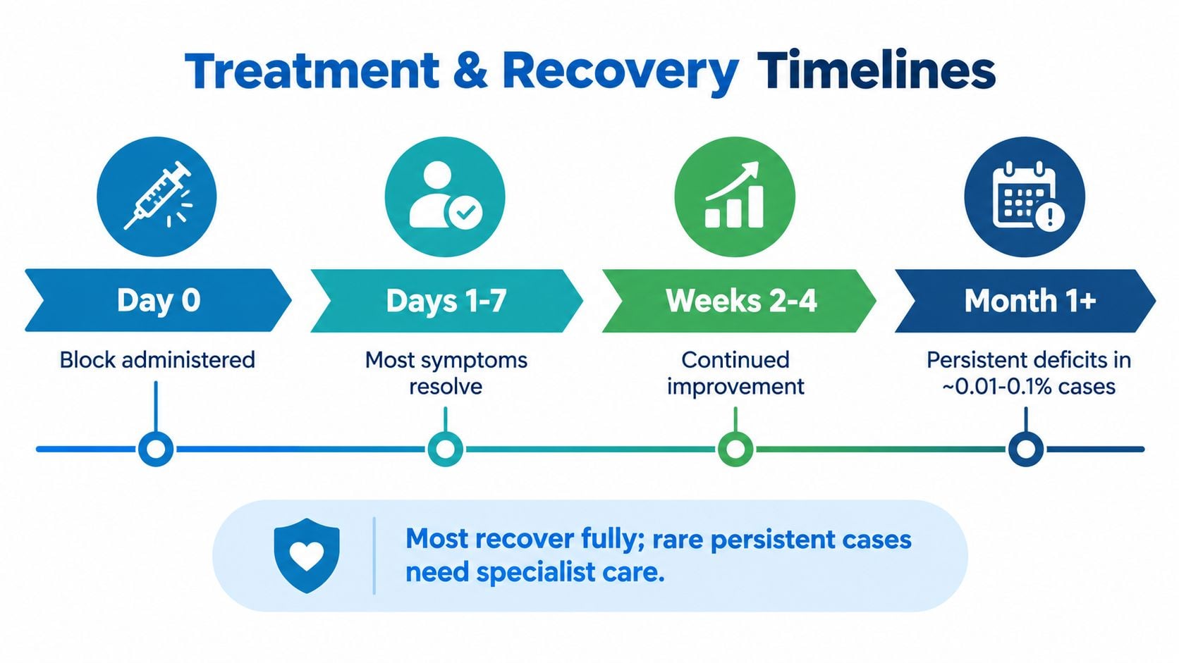
The key distinction is improvement versus deterioration. A patient who can feel small changes each day is following a different course from someone whose weakness or pain is intensifying.
How Doctors Diagnose and Stage Nerve Block Damage
Diagnosis starts with a careful history, not a single scan. The clinician records the block type and location, the timing of symptoms, the operation, positioning, tourniquet use, medications, and the patient's preoperative neurologic status.
The examination then maps sensation and strength. Comparing the affected side with the opposite side helps identify whether the problem follows one nerve, a plexus, a nerve root, or a less specific postoperative pattern. The clinician also checks reflexes, skin condition, circulation, swelling, and signs of pressure.
Tests answer different questions
Nerve-conduction studies measure how electrical signals travel along a nerve. EMG examines electrical activity in muscles and can show whether denervation has occurred. Testing too early may miss changes that need time to develop, so the treating specialist chooses timing based on the examination and suspected injury.
High-resolution ultrasound can show nerve shape, fascicles, swelling, continuity, or a nearby hematoma. MRI or MR neurography can provide additional information about soft tissues, inflammation, compression, or deeper anatomy.
| Test | What it detects | Typical timing |
|---|---|---|
| Focused neurologic examination | Distribution of sensory loss, weakness, reflex change, swelling, and circulation findings | Immediately and during follow-up |
| Nerve-conduction study | Signal transmission along sensory and motor nerves | Scheduled according to symptom timing and clinical question |
| EMG | Muscle denervation and patterns of nerve dysfunction | Often more informative after changes have had time to develop |
| High-resolution ultrasound | Nerve structure, swelling, discontinuity, or nearby hematoma | When a superficial nerve or compressive cause needs evaluation |
| MRI or MR neurography | Deep soft-tissue change, inflammation, and structural compression | When anatomy is unclear or a deeper lesion is suspected |
Doctors may describe severity using systems such as Seddon or Sunderland. These classifications help distinguish a temporary conduction problem from axonal disruption or more severe structural damage. A nerve surgeon, neurologist, or pain specialist becomes especially important when weakness is profound, symptoms persist, or testing suggests a lesion requiring targeted treatment.
Treatment Options and Realistic Recovery Timelines
Treatment depends on the symptom pattern, its direction, and the suspected cause. Improving sensation without new weakness often calls for protecting the area, observing changes, and beginning rehabilitation. Severe pain, a collection pressing on the nerve, or worsening motor function requires faster specialist assessment.
The recovery ladder
Conservative care is usually considered when the nerve appears intact and symptoms are improving. Physical or occupational therapy can preserve movement, build strength safely, reduce fall risk, and protect a numb limb. Range-of-motion exercises, desensitization, and skin protection may be included. For burning or electric pain that disrupts sleep or function, clinicians may consider medicines used for neuropathic pain, such as gabapentin or duloxetine.
Procedural treatment may help when neuropathic pain continues without progressive weakness. Depending on the suspected mechanism, a specialist might discuss sympathetic blocks, peripheral nerve stimulation, or pulsed radiofrequency. The treatment should match the pain pattern and examination, rather than follow a fixed sequence.
Surgical care is reserved for a documented structural problem or a deficit that is not recovering as expected. Depending on the lesion, a peripheral nerve surgeon may consider decompression, neurolysis, repair, grafting, or another reconstructive approach. The decision rests on the examination, electrodiagnostic findings, imaging, and evidence of recovery.
The practical question is often duration and pattern, not whether numbness is present. When symptoms persist beyond the initial observation window, clinicians shift from observation to staged rehabilitation and, when indicated, electrodiagnostic testing to guide the next step. Persistent weakness should have a documented follow-up plan, even when pain is manageable.
What recovery can look like
Recovery is rarely perfectly even. Tingling may appear as sensation returns, strength may trail behind feeling, and pain can vary with activity. A single unusual sensation does not establish permanent injury. Lack of improvement, spreading symptoms, or new weakness matters more than one isolated change.
Escalation matters: Symptoms that persist beyond the timeframe recommended by the evaluating team should prompt specialty review rather than indefinite observation.
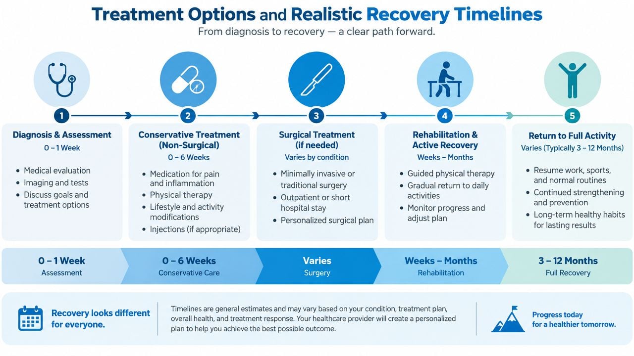
Rehabilitation protects function while the nerve heals. A therapist may maintain joint flexibility, support a weak limb, reduce falls, and teach skin checks when sensation is reduced. Keep follow-up appointments, record whether symptoms are improving or worsening, and contact the treating team promptly if the pattern changes.
Prevention, Specialist Care, and Where to Go From Here
The assumption that any numbness after a block equals permanent damage is incorrect. The opposite assumption, that every persistent deficit will resolve without assessment, is also unsafe. Prevention reduces risk, but it can't guarantee a symptom-free recovery because surgery, positioning, patient factors, and the block may all contribute.
Experienced regional anesthesia teams use layered safeguards. These can include ultrasound guidance, nerve stimulation when appropriate, attention to injection pressure, avoidance of intraneural or intrafascicular injection, and the lowest effective anesthetic concentration and dose. The patient can support that process by reporting baseline neuropathy, diabetes, thyroid disease, anticoagulant use, prior block problems, and unusual electric or burning pain during injection.
After surgery, positioning instructions matter. A numb limb is less able to signal pressure or heat injury, so patients should protect it, check the skin, and avoid relying on strength that hasn't returned. Any concern should be documented with the time of onset, affected area, degree of weakness, and whether symptoms are improving or worsening.
A patient with persistent or significant symptoms should contact the anesthesia team and surgeon first. If a deficit continues beyond four to six weeks, or if weakness is substantial earlier, referral to a neurologist, peripheral nerve surgeon, or chronic pain specialist with relevant experience is reasonable. An EMG and nerve-conduction assessment may help before that visit, depending on the clinical examination and timing.
Interventional Pain Management can evaluate neuropathic and post-surgical pain, perform selected interventional procedures, and coordinate orthopedic or spine care when the clinical findings call for a broader assessment. The practical goal is not to assume the block caused the problem. It is to identify the cause, protect function, and match treatment to the recovery pattern.
Patients with persistent numbness, weakness, or burning pain after a nerve block can seek a focused evaluation rather than waiting without guidance. Interventional Pain Management offers pain, orthopedic, anesthesiology, and spine services that can help assess symptoms and coordinate an appropriate next step.