
A patient with L4-L5 pain often reaches the same uncomfortable crossroads: the leg pain has lasted long enough to disrupt sleep, work, exercise, or basic movement, yet surgery still feels like a major step. An MRI may show a disc extrusion, narrowing around a nerve, or a slipping vertebra, but an image alone can't decide whether a small decompression, an endoscopic procedure, or a fusion is appropriate.
That's why minimally invasive spine surgery at L4-L5 isn't one operation. It's a group of techniques that use smaller, more focused corridors to reach the affected nerve or spinal segment. The useful question isn't just whether minimally invasive surgery is “better.” It's which approach matches the anatomy, the stability of the segment, the symptoms, and the patient's recovery priorities.
Table of Contents
- When L4-L5 Pain Stops Responding and Surgery Enters the Conversation
- What Minimally Invasive Spine Surgery Actually Means at L4-L5
- Who Qualifies for L4-L5 Minimally Invasive Surgery
- Comparing the Main L4-L5 Minimally Invasive Techniques
- Inside an L4-L5 Minimally Invasive Procedure and the First Weeks of Recovery
- Why Less Invasive Is Not Always the Right Answer
- Benefits, Risks, and What the 2024–2026 Evidence Shows
- Your Next Step After Reading This Guide
When L4-L5 Pain Stops Responding and Surgery Enters the Conversation
Consider a 52-year-old physical therapist who develops sharp pain from the lower back into the leg. After six weeks of structured care, including oral steroids, McKenzie-style directional-preference exercises, and one epidural steroid injection, the pain has plateaued. A straight-leg raise remains positive, and MRI shows a paracentral L4-L5 disc extrusion that corresponds with the symptoms.
The next appointment may begin with a practical question: should another injection be tried? But the more useful discussion is broader. Does the compressed nerve need direct decompression? Is the disc problem isolated, or is the L4-L5 segment also unstable? Does the patient need rapid relief to return to clinical work, or is continued non-operative care still reasonable?
An epidural injection can be part of that decision process, particularly when the diagnosis and symptom pattern remain clear but surgery isn't yet necessary. Information about the role of this treatment is available through lumbar epidural steroid injection care.
Three reasons a surgical consultation becomes important
Progressive weakness: New or worsening weakness in ankle lifting, toe walking, heel walking, or another muscle group deserves prompt evaluation. A surgeon needs to determine whether ongoing nerve compression threatens recovery.
Cauda equina warning signs: New bowel or bladder changes, saddle numbness, or rapidly developing weakness require urgent medical assessment rather than routine scheduling.
Functional collapse: Surgery becomes a more reasonable conversation when appropriately delivered medication, therapy, activity modification, and injections haven't restored essential activities, and imaging shows a matching structural cause.
A consultation doesn't automatically lead to an operation. It translates symptoms, examination findings, imaging, work demands, and expectations into a decision tree. For a disc extrusion, that may lead toward microdiscectomy or endoscopic discectomy. For stenosis with instability, decompression alone may be insufficient. For a stable, isolated disc problem, fusion may add unnecessary complexity.
Practical rule: The MRI should explain the symptoms, but the examination and functional impact should help determine whether treatment needs to escalate.
What Minimally Invasive Spine Surgery Actually Means at L4-L5
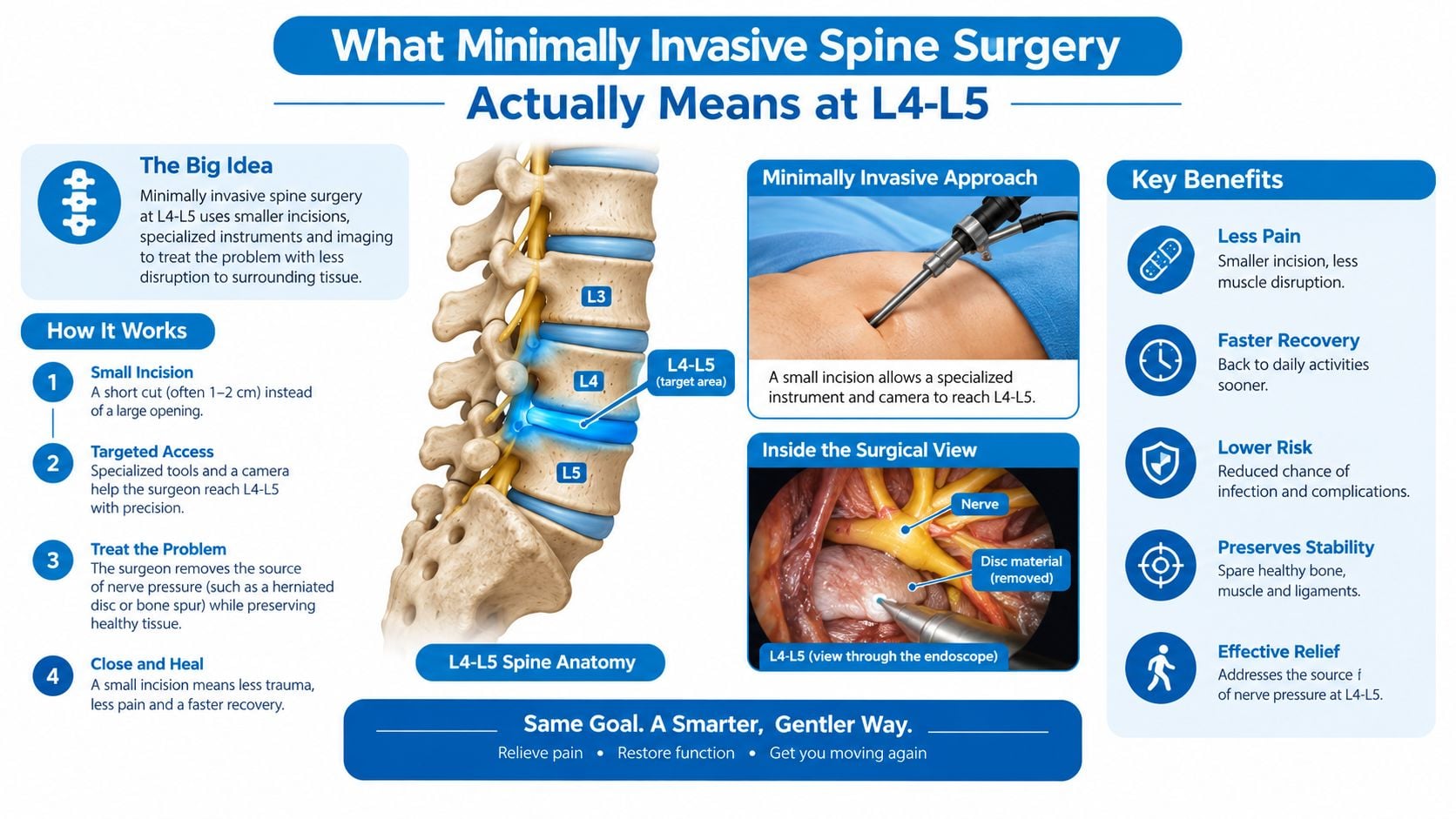
At L4-L5, minimally invasive surgery can be understood as keyhole carpentry. Instead of opening a broad midline corridor and stripping muscle away from the spine, the surgeon creates a focused path through the muscles with tubular retractors, a microscope, or an endoscope. The instruments pass through that corridor to remove pressure from a nerve or stabilize the segment.
The target doesn't change. A decompression still removes the disc fragment, bone, or thickened tissue that crowds a nerve. A fusion still prepares the disc space, places an implant, and stabilizes the vertebrae when movement or collapse makes decompression alone unreliable. What changes is the route and the amount of surrounding tissue disturbed.
How the field reached modern L4-L5 techniques
The history is longer than the term “modern MISS” suggests. Early lumbar disc treatment began with chemonucleolysis in 1969, widely cited as the first minimally invasive spine procedure. The field then accelerated with the Williams microdiscectomy in 1978, which shifted lumbar disc removal from a roughly 6-inch open incision toward a microsurgical approach. Endoscopic lumbar surgery emerged in the early 1980s, and the first reported laparoscopic lumbar discectomy followed in 1991. By the early 2000s, minimally invasive methods had expanded into several procedures, including fusion, as described in this historical review of minimally invasive spine surgery.
The preservation goal is straightforward:
- Muscle attachment: Tubular dilation separates fibers rather than requiring broad muscle stripping.
- Posterior support structures: A focused corridor can limit disruption to the multifidus and posterior tension band.
- Soft-tissue trauma: Less exposure can reduce the area affected by dissection, although it doesn't eliminate surgical pain or risk.
- Precision: Magnification and image guidance help the surgeon work around the nerve root and bony landmarks.
Patients should also understand what MISS doesn't guarantee. A smaller incision doesn't make a fusion biologically automatic, and it doesn't allow a surgeon to decompress a nerve without removing whatever structure is causing compression. The approach is less invasive in access, not necessarily less significant in purpose. Surgeons may use endoscopic spine surgery techniques when the anatomy and pathology provide a safe working corridor.
Who Qualifies for L4-L5 Minimally Invasive Surgery
Candidacy depends on the problem at L4-L5, not just the presence of back pain or an abnormal MRI. A disc herniation, spinal stenosis, and degenerative spondylolisthesis place different demands on the operation.
Disc herniation
A favorable pattern includes a contained or modestly extruded fragment that matches radicular symptoms and the neurological examination. A patient who has completed a structured period of non-operative care, often 6 to 12 weeks, may be considered when leg pain or weakness remains disabling and the imaging shows a surgically accessible fragment.
A surgeon becomes more cautious when the main complaint is axial back pain without convincing nerve compression, when the disc has substantial collapse, or when the fragment has migrated into a corridor that makes a limited approach difficult. The question isn't whether the MRI looks dramatic. It's whether removing the specific fragment is likely to address the dominant problem.
Stenosis
Tubular decompression can fit single-level or limited multilevel narrowing in the central canal or lateral recess when the segment remains stable. The operation may involve a laminotomy, undercutting, or focused removal of thickened tissue and bone.
Severe deformity, broad instability, or narrowing that requires extensive facet removal can change the plan. Removing too much stabilizing anatomy through a small incision can create a larger problem, which is why access size shouldn't drive the decision.
Spondylolisthesis
For selected degenerative slips, MIS-TLIF may be considered when the slip is lower grade, mobile on dynamic radiographs, and associated with foraminal narrowing or instability. Facet effusion and Modic changes can add context, but they don't independently dictate fusion.
High-grade translation, major deformity, active infection, and severe osteoporosis can make a limited fusion corridor inappropriate. Previous open surgery may also leave scarred planes that reduce the advantages of a minimally invasive route.
| Pathology at L4-L5 | Strong MISS Candidate | Poor or Excluded Candidate |
|---|---|---|
| Disc herniation | Matching radiculopathy, accessible fragment, persistent symptoms after structured care | Pain without matching compression, severe collapse, inaccessible migration |
| Spinal stenosis | Focal central or lateral recess narrowing without major instability | Extensive facet destruction required, marked deformity, unstable segment |
| Degenerative spondylolisthesis | Lower-grade mobile slip with instability or foraminal compromise | High-grade slip, major deformity, active infection, severe osteoporosis |
| Recurrent or multilevel disease | Carefully selected focal recurrence with a clear target | Broad multilevel deformity or heavily scarred operative planes |
| General medical status | Able to tolerate anesthesia and follow rehabilitation | Uncontrolled conditions that make anesthesia or healing unsafe |
The final decision requires a physical examination, imaging review, medical assessment, and a discussion of what the operation can and can't change. A technically feasible procedure isn't automatically the right procedure.
Comparing the Main L4-L5 Minimally Invasive Techniques
The main techniques differ by what needs to be fixed. Microdiscectomy removes a nerve-compressing disc fragment. Tubular decompression enlarges a narrowed passage. Fusion-based procedures address instability, disc-space collapse, or recurrent disease where decompression alone may leave the segment mechanically unsound.
A microscope-assisted tubular microdiscectomy is often used for a posterior disc herniation that can be reached through a focused corridor. Full endoscopic discectomy can approach selected foraminal, extraforaminal, or recurrent fragments through a working channel. At L4-L5, the choice between transforaminal and interlaminar endoscopy depends heavily on the iliac crest, the traversing nerve root, and the direction of fragment migration. A 2026 meta-analysis found no significant differences between these approaches in overall complications, reoperation, operative time, hospital stay, or patient-reported outcomes, including ODI and leg-pain scores, as reported in this comparative analysis of L4-L5 endoscopic discectomy approaches.
Tubular decompression is a different operation. It addresses stenosis by removing enough bone and ligament to free the nerve while trying to preserve stability. MIS-TLIF, OLIF, and Endo-TLIF add interbody support and fixation when the segment needs stabilization or restoration of foraminal height.
What each approach is trying to accomplish
| Technique | Corridor / Approach | Best-Fit L4-L5 Pathology | Typical Incision | Key Limitation |
|---|---|---|---|---|
| Tubular microdiscectomy | Posterior tubular corridor with microscope | Contained or extruded posterior disc herniation | Focused small incision | Doesn't correct instability or major disc-space collapse |
| Full endoscopic discectomy | Transforaminal or interlaminar working channel | Selected foraminal, lateral, or recurrent herniation | Very small working-channel access | Corridor can be limited by bone, iliac crest, or migration |
| Tubular decompression | Posterior laminotomy and undercutting | Focal central or lateral recess stenosis | Focused posterior incision | May be inadequate when decompression would destabilize the segment |
| MIS-TLIF | Posterior transforaminal corridor with fixation | Instability, spondylolisthesis, recurrent same-level disease | Several small access points | More extensive than decompression and still depends on fusion biology |
| OLIF or LLIF | Lateral corridor through the disc space | Foraminal collapse or selected indirect decompression cases | Lateral access incision | Indirect decompression may not address every type of severe nerve compression |
| Endo-TLIF | Endoscopic posterior fusion corridor | Selected fusion cases requiring a smaller working channel | Focused posterior access | Technique remains dependent on anatomy and surgeon experience |
The table isn't a ranking. It's a matching exercise. A patient with leg-dominant pain from a free disc fragment may need nerve decompression, not hardware. A patient with foraminal collapse and a mobile slip may need restoration of disc height and stabilization, even if the incision is small.
Inside an L4-L5 Minimally Invasive Procedure and the First Weeks of Recovery
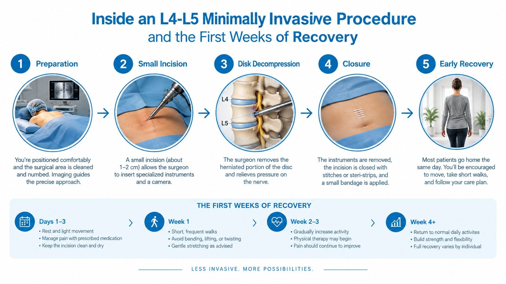
The day usually starts before the operating room. The surgical team confirms the symptoms, reviews the MRI and radiographs, marks the operative level, and checks medications, allergies, and anesthesia risks. The patient is then positioned face down on a Wilson frame or Jackson table, depending on the operation and the team's preference.
Fluoroscopy or navigation confirms the L4-L5 level before the incision. Sequential dilators create the working corridor, and a tubular retractor holds the path open. Through that channel, the surgeon uses a microscope or endoscope to identify the lamina, facet, disc fragment, and nerve root. The goal is controlled decompression, not just removal of as much tissue as possible.
After the nerve is free and bleeding is controlled, the instruments are removed and the incision is closed. A decompression patient may walk in the recovery area the same day. Fusion patients generally need more observation because implants, anesthesia, mobility, and pain control require a different postoperative plan.
A practical four-week recovery view
Week one: Walking is encouraged within the surgeon's restrictions. Decompression patients often focus on wound care, short walks, position changes, and controlling muscle soreness. Leg pain may improve quickly, while numbness or weakness can take longer.
Week two: Light daily activity can increase if symptoms are settling. A desk-based patient may begin discussing a return to light duty, but the plan depends on pain, medication use, neurological status, and job demands.
Weeks three and four: After microdiscectomy, some patients move toward light-duty work during the broader 2-to-6-week range. Fusion patients remain under stricter lifting and bending limits, with heavier lifting commonly deferred for 6 to 12 weeks, depending on healing and the surgeon's protocol.
Rehabilitation: Physical therapy timing varies. Walking may begin immediately, while formal strengthening waits until the wound and nerve irritation have settled. A brace is generally reserved for situations in which the surgeon believes additional support is useful, rather than being required for every MISS procedure.
A recovery plan should be individualized. Persistent weakness, worsening leg pain, fever, drainage, new bowel or bladder symptoms, or severe swelling warrants prompt contact with the surgical team.
Why Less Invasive Is Not Always the Right Answer
A smaller incision reduces access-related tissue disruption, but it doesn't change the mechanical problem at L4-L5. Severe facet overgrowth may require a broader decompression. A deformity may need correction rather than a limited nerve release. A mobile degenerative slip may become less stable if the operation removes the structures that were holding it in place.
That's the central trade-off. MISS can reduce muscle disruption and support a smoother early recovery, but it still has to accomplish a complete decompression or a solid fusion. A minimally invasive operation that leaves instability untreated isn't automatically the safer choice.
What the fusion data actually show
A 2024 Scientific Reports study compared MIS-TLIF, OLIF, and Endo-TLIF for L4-L5 degenerative spondylolisthesis. At 12 months, complete fusion occurred in 84 of 98 MIS-TLIF patients, or 85.7%, 101 of 107 OLIF patients, or 94.4%, and 96 of 114 Endo-TLIF patients, or 84.2%. ODI scores also improved from 38.2 to 16.1 with MIS-TLIF, 31.4 to 14.4 with OLIF, and 32.9 to 15.7 with Endo-TLIF, based on the study's reported group averages in this L4-L5 fusion comparison.
Those findings support meaningful improvement, but they don't prove that every patient should receive a minimally invasive fusion. The cohort involved a particular diagnosis, selected techniques, and a defined follow-up period. Fusion biology still depends on bone quality, smoking status, medical conditions, implant positioning, and the reason fusion was needed.
An open or hybrid procedure may be more appropriate for extensive deformity, severe osteoporosis, high-grade instability, or scarred anatomy from prior operations. A formal fusion discussion should explain what problem the hardware addresses, what happens if decompression alone is chosen, and how the surgeon will confirm healing. Patients can review broader spinal fusion treatment information, but the operative recommendation must remain individualized.
The least invasive operation is only conservative if it treats the actual source of symptoms without creating a need for revision.
Benefits, Risks, and What the 2024–2026 Evidence Shows
The evidence supports a measured conclusion. Minimally invasive approaches can produce meaningful relief and may reduce early tissue trauma, but comparative research doesn't establish one universal winner for every L4-L5 diagnosis.
The strongest recent data are indication-specific. The 2024 fusion comparison showed substantial ODI improvement and complete fusion rates in the mid-80% to mid-90% range at one year for the three studied approaches. The 2026 endoscopic discectomy meta-analysis found comparable outcomes between transforaminal and interlaminar routes, suggesting that anatomy often matters more than a general claim that one corridor is superior.
A 2025 retrospective study of 180 lumbar stenosis patients found similar complication and reoperation rates between endoscopic and tubular MISS. The endoscopic group had less immediate postoperative pain, no infections compared with a 5.0% infection rate in the tubular group, and shorter in-room-to-incision and closure times, according to the comparative stenosis study.
Benefits that matter early
- Focused tissue access: The surgeon can reach the target while limiting broad muscle exposure.
- Recovery priorities: Some patients value early walking, reduced immediate pain, or a faster return to light activity.
- Approach flexibility: Endoscopic, tubular, biportal, and fusion-based methods can be selected according to the pathology rather than a single MISS label.
Questions the literature hasn't settled
Recent reviews note that longer-term pain and disability outcomes can be similar across several approaches, while endoscopic techniques may offer short-term recovery advantages in selected comparisons. A 2026 scoping review also emphasizes that the field still relies heavily on lower-quality studies and has limited randomized evidence, as discussed in this review of minimally invasive lumbar surgery evidence.
That uncertainty matters. Risks include nerve irritation, infection, spinal fluid leakage, incomplete decompression, recurrent symptoms, and the possibility of additional surgery. The correct consent conversation should cover both the benefits expected from the selected operation and the consequences of choosing a technique that doesn't match the anatomy.
Your Next Step After Reading This Guide
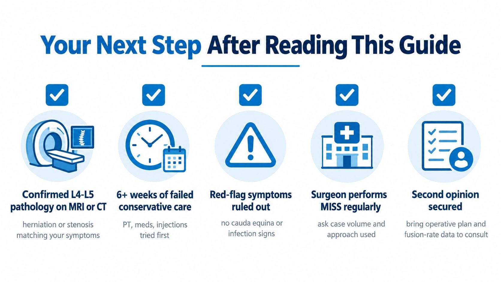
A patient in New Jersey or Staten Island can prepare for a spine consultation by treating the appointment as a matching problem, not a search for the smallest incision. The key question is whether the proposed approach fits the L4-L5 anatomy, the neurological findings, the stability pattern, and the recovery goal.
A consultation checklist
Bring the complete record. Imaging should include the actual MRI or CT images when available, not only the written report. Prior injection notes, therapy records, medication trials, and radiographs can show what has already been attempted and whether the segment moves abnormally.
Write a symptom timeline. The record should distinguish back pain from leg pain, identify numbness or weakness, and describe the activities that have become difficult. A surgeon needs to know whether the main limitation is walking, sitting, lifting, sleeping, working, or exercising.
Ask for the specific operation. “Minimally invasive” is too broad by itself. The consultation should identify whether the plan is tubular microdiscectomy, endoscopic discectomy, decompression, MIS-TLIF, OLIF, Endo-TLIF, or another approach, and explain why that corridor fits the MRI.
Clarify the stability question. Patients should ask whether dynamic radiographs show movement, whether decompression alone could destabilize the segment, and what findings would make fusion necessary.
Seek another opinion when uncertainty remains. A second consultation is especially reasonable when fusion is proposed, symptoms and imaging don't match, or the recommended operation seems more extensive than the functional problem.
Urgent evaluation is needed for progressive weakness, bowel or bladder changes, or saddle anesthesia. Those symptoms shouldn't wait for a routine second opinion or an elective appointment.
The most useful decision isn't “surgery or no surgery” in the abstract. It's whether a clearly identified L4-L5 problem requires decompression, stabilization, continued non-operative treatment, or urgent assessment.

Interventional Pain Management offers diagnostic review, epidural injections, nerve blocks, orthopedic evaluation, endoscopic procedures, minimally invasive spine surgery, and fusion care aligned with the clinical findings. Patients with persistent L4-L5 symptoms can bring their imaging and treatment history to a multidisciplinary consultation by visiting Interventional Pain Management.