
You're doing the grocery store shuffle, one hand on the cart, because your legs feel heavy before you've even made it to the dairy aisle. Then the MRI report arrives and says mild spinal stenosis, which sounds reassuring until the walking pain, numbness, or cramping still shows up every time you stand too long. That disconnect is common, and it's exactly why the word mild deserves a closer look.
What matters most is not the label alone, but how much space is left for the nerves, which nerves are irritated, and what happens when the spine is loaded during standing or walking. Mild spinal stenosis is a real anatomical finding, but it can still be functionally important. The safest way to read it is alongside symptoms, walking tolerance, and a clinician's exam, not as a stand-alone verdict.
Table of Contents
- When the MRI Says Mild but the Legs Feel Heavy
- What Mild Spinal Stenosis Means
- Why the Spine Narrows With Age
- Recognizing the Symptoms Behind the Label
- How Doctors Confirm the Diagnosis
- Conservative Care That Actually Works
- When Interventional and Surgical Care Make Sense
- Red Flags and When to See a Specialist
When the MRI Says Mild but the Legs Feel Heavy
A patient in the exam room often sounds frustrated in the same way. The scan says mild, yet the person can't get through a shopping trip, a walk around the block, or a shift at work without stopping to lean forward and rest. That mismatch is not a contradiction, it's a clue.
The term mild describes anatomy, not how much a person hurts or how far they can walk. In lumbar spinal stenosis, the canal can be only partially narrowed, yet the space for the nerves may still be tight enough to cause leg heaviness, cramping, tingling, or numbness when the spine is loaded. The result is a diagnosis that can look small on paper and feel big in daily life.
Practical rule: if standing and walking make symptoms worse, and sitting or bending forward makes them better, the label on the MRI is only part of the story.
That's why the next question is never just “What did the report say?” It's “What does the body do during the day?” A person who can sit comfortably but struggles to stand through dinner, wait in line, or finish a short walk may have a functional problem that deserves attention even if the scan sounds modest. The care pathway usually starts with conservative treatment, then moves stepwise toward procedures or surgery only when symptoms, function, and neurologic findings justify it.
What Mild Spinal Stenosis Means
In research and diagnosis, mild spinal stenosis is usually defined as a central spinal canal narrowing of one-third or less. Moderate stenosis means the canal is narrowed by one-third to two-thirds, and severe stenosis means more than two-thirds narrowing. That definition matters because the word mild describes a measurement, not a person's symptoms.
A hallway analogy helps here. If a hallway has lost about a third of its width, traffic may still move, but there is less reserve when demand goes up. Once the space shrinks more, small changes in posture, swelling, or nerve sensitivity can create much more trouble than the MRI label suggests.
The grading system helps radiologists describe anatomy, but it does not tell the whole story by itself. That matters because lumbar spinal stenosis is common in everyday practice. A systematic review found the mean prevalence of lumbar spinal stenosis was 11% in the general population, 25% in primary care, and 29% in secondary care when diagnosed clinically, while radiologic prevalence estimates ranged from 11% to 38% depending on the population studied. Seeing stenosis on a report is not unusual, so the key question is whether the narrowing fits the symptoms and exam findings (Pain Medicine review).
| Severity | Canal Narrowing | Typical Symptoms | Initial Direction |
|---|---|---|---|
| Mild | One-third or less | May be minimal, or may still cause walking discomfort if nerves are sensitive | Conservative care first |
| Moderate | One-third to two-thirds | More likely to produce activity-related leg symptoms | Conservative care, with closer follow-up |
| Severe | More than two-thirds | Higher risk of persistent pain, walking limits, or neurologic findings | Specialist evaluation and possible intervention |
That is why a person with “mild” stenosis can still feel stuck. The label does not measure walking tolerance, leg heaviness, or how quickly symptoms appear when standing. In clinic, an interventional pain specialist and an ortho-spine surgeon usually look at the same question from different angles, then step treatment up only when the functional picture justifies it.
Why the Spine Narrows With Age
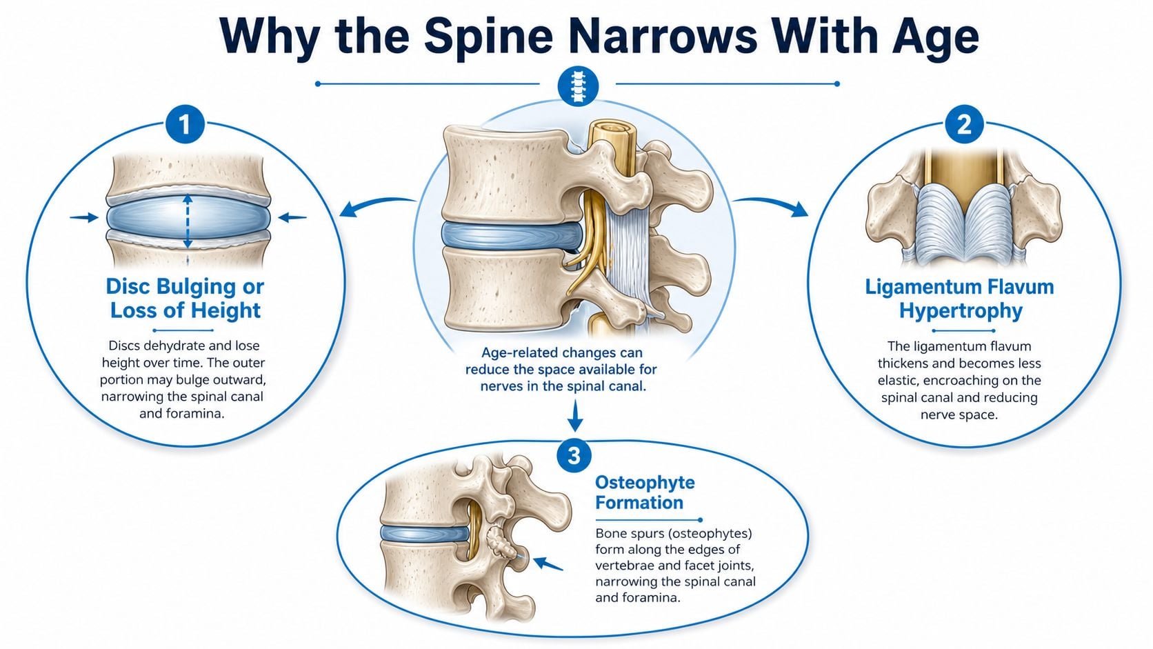
Most adult lumbar stenosis develops slowly, not from one sudden injury. As the spine ages, the disc can lose height or bulge, the ligamentum flavum can thicken, and the facet joints can enlarge from arthritis. Each of those changes takes a little more room away from the canal and the nerve roots.
Several small changes can also create a bottleneck effect in the spine. One issue by itself may not cause trouble, but together they leave less reserve space for the nerves, especially when someone stands, walks, or extends the back.
That pattern is why acquired stenosis is so often seen in older adults. Reports and reviews have described lumbar spinal stenosis as common in later life, and the age-related rise fits the way these structures wear over time (PubMed review).
Reports may use acquired stenosis or congenital stenosis, and the distinction matters. Congenital narrowing means the canal started out smaller from birth, so age-related change can become symptomatic sooner. Acquired narrowing is the more typical adult pattern, and it tends to worsen with repeated loading, posture, and arthritis over time.
Degenerative disc disease and disc height loss often sit in the same neighborhood as stenosis, so it is common for a report to mention both. The key point is not which label appears first. It is how much room remains for nerve movement when the spine is upright and active.
Recognizing the Symptoms Behind the Label
The classic pattern is neurogenic claudication. That means the legs may feel heavy, weak, crampy, tingly, or numb when a person stands or walks, then ease when sitting, bending forward, or leaning on something like a shopping cart. The relief with flexion is a major clue, because bending forward often opens a little more room in the canal.
Symptoms can vary widely because different nerve roots are compressed in different people. One person may notice foot numbness, another may feel both calves tighten on hills, and another may mainly describe balance changes or a sense that the legs won't “go.” The MRI label can be the same while the day-to-day experience is very different.
The scan doesn't walk down the street. The patient does. Function still matters more than the report title.
This is also where confusion with vascular leg pain can arise. Both can limit walking, but spinal stenosis often improves with sitting or leaning forward, while other circulation-related problems behave differently. A careful history helps separate those patterns before anyone jumps to treatment.
A clinician also listens for details that patients often mention casually but that matter a lot in stenosis care: night-time cramping, numb feet, slower walking pace, needing to stop after a short distance, or a posture that feels safer than standing upright. Those details often point to dynamic nerve irritation, not just a static picture on MRI. Once that pattern is clear, the next step is to match it with a focused exam and, when appropriate, imaging.
How Doctors Confirm the Diagnosis
The diagnosis starts with the story. A clinician asks what makes the pain or heaviness appear, whether sitting helps, how far the person can walk, and whether the symptoms spread into one leg or both. That history often tells more than the report title does.
A focused physical exam then checks strength, reflexes, sensation, balance, and walking mechanics. The goal is to see whether the nerves are only irritated during activity or whether they are showing persistent signs of dysfunction. If the exam suggests stenosis, imaging helps define the anatomy, but it doesn't replace the history.
MRI is the main study because it can show canal narrowing, ligament thickening, disc bulging, and facet enlargement. X-rays may be added when alignment or instability is a concern. In some cases, CT or EMG can help sort out anatomy or nerve function when the picture is not straightforward.
Clinical principle: imaging confirms structure, but symptoms and function decide whether that structure is actually the problem.
That's why the same scan can mean different things in different people. One person may have a mild narrowing that never matters, while another has a modest-looking scan but very real walking limits. Diagnosis is strongest when the image, the history, and the exam all point in the same direction.
Conservative Care That Actually Works
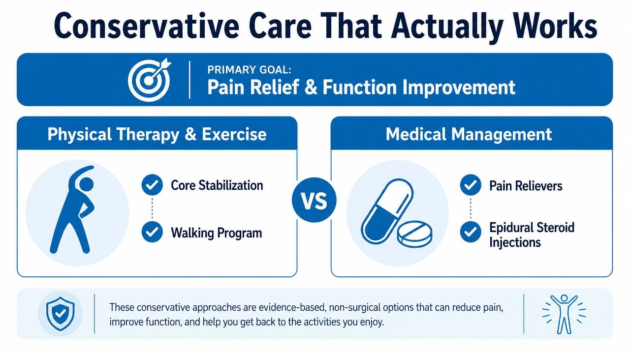
For mild spinal stenosis, the first treatment is usually conservative care, not surgery. Major references list activity modification, physical therapy, NSAIDs or oral analgesics, and sometimes bracing as initial management, with epidural steroid injections added when symptoms persist (NCBI Bookshelf). That stepwise approach makes sense because the problem is often mechanical and activity-related.
Physical therapy usually focuses on flexion-based exercise, core stabilization, and walking tolerance. These measures help people stay active without repeatedly provoking the same nerve compression pattern. A walking aid can also help some patients because leaning slightly forward may reduce symptoms during longer outings.
Epidural steroid injections can be part of the plan, but the expected benefit is limited. In the historical review cited earlier, epidural steroid injections were described as offering only modest relief lasting less than 3 weeks (PubMed review). That makes injections useful for symptom control in selected patients, but not a cure for the underlying narrowing.
| Conservative Option | What It Does | Evidence Strength | Typical Duration of Benefit |
|---|---|---|---|
| Physical therapy | Builds trunk support and improves tolerance to flexed or supported movement | Common first-line care | Often ongoing, depends on adherence |
| NSAIDs or oral analgesics | Reduces pain and inflammation symptoms | Standard initial care | Short-term symptom relief |
| Activity modification | Avoids positions that trigger nerve compression | Practical foundation of care | Works as long as habits change |
| Walking aids or bracing | Helps reduce painful loading during walking | Selected patients benefit | Variable, tied to use |
| Epidural steroid injections | Calms inflammation around irritated nerves | Helpful for selected patients | Often short-lived |
Lifestyle changes are not a backup plan. They are the base layer of care at this stage. A structured plan that respects posture, pacing, sleep, and daily movement often gives better day-to-day control than hoping the scan label will stay quiet on its own.
When Interventional and Surgical Care Make Sense
Escalation becomes reasonable when symptoms keep limiting life despite a structured conservative trial, when walking distance keeps shrinking, or when a neurologic deficit appears. Progressive weakness, new foot drop, bowel or bladder changes, or rapidly worsening numbness move the situation into a different category. Those findings need urgent assessment, not more waiting.
For selected patients with hypertrophic ligamentum flavum, a minimally invasive option called MILD can be appropriate after conservative care fails. Expert guidance describes ideal candidates as having neurogenic claudication for more than 3 months, ligamentum flavum hypertrophy greater than 2.5 mm, and central canal area less than 100 mm² on MRI, while excluding grade II or higher spondylolisthesis or major instability (Johns Hopkins MILD procedure). The logic is direct. Removing excess ligament can restore space and reduce dynamic compression during standing and walking.
Surgical decompression is usually reserved for more severe disability, progressive neurologic loss, or structural instability. Fusion enters the picture when the spine needs stabilization, not just more room. That's why a multidisciplinary spine team looks at the whole picture, not only whether a person wants to avoid surgery.
A practical resource on symptom control during chronic pain care is Thermo Recovery Wear for arthritis, especially for people who need comfort strategies while a spine plan is being adjusted. It's one piece of the larger toolkit, not a substitute for diagnosis or treatment.
Image-guided epidural steroid injections may be part of that escalation pathway when inflammation and nerve irritation are contributing to the pain. The right move depends on symptoms, exam findings, and stability, not on forcing every patient into the same branch of care.
Red Flags and When to See a Specialist
Some symptoms need urgent medical attention. Sudden weakness, foot drop, loss of bowel or bladder control, saddle anesthesia, or rapidly progressive numbness should not wait for a routine appointment. Those signs can point to serious nerve compromise.
Other signs are not emergencies, but they do justify a specialist visit. Declining walking distance, persistent leg pain despite conservative care, sleep disruption, or symptoms that interfere with work or daily chores are all good reasons to book a spine evaluation. A multidisciplinary interventional pain and ortho-spine visit can review the imaging, match it to the exam, and decide whether continued conservative care, an injection, a minimally invasive procedure, or surgery fits best.
The most useful next step is often a clear plan, not more guessing. If the legs feel heavier, the cart has become a crutch, or the MRI label feels too small for the symptoms, a focused spine evaluation can sort out whether the problem is still in the conservative lane or needs escalation.
Foot drop evaluation and treatment is one example of why timely specialist care matters when weakness starts to change function.
Interventional Pain Management evaluates spinal stenosis with a stepwise approach that matches symptoms, imaging, and function, then pairs that review with options ranging from conservative care to image-guided procedures and spine surgery when needed. If mild spinal stenosis is keeping walking, standing, or sleep from feeling normal, visit Interventional Pain Management to discuss the next practical step with a multidisciplinary team.