
Modern spinal cord stimulators are usually MR Conditional, not MR Safe, which means MRI is often possible, but only under specific device and scanner conditions that have to be verified before every scan. In real life, 82% to 84% of people with spinal cord stimulators are expected to need at least one MRI within 5 years of implantation, so this comes up far more often than most patients expect (PMC analysis).
A person may feel fine for years after implant surgery, then suddenly need a shoulder MRI after a fall, a brain MRI for new symptoms, or a spine MRI for a different problem. That moment can be confusing, because the device may have been described as “MRI-compatible” long before anyone explains what that means in the imaging suite.
Table of Contents
- Why MRI Comes Up So Often After a Spinal Cord Stimulator
- What MRI Conditional Really Means for Your Device
- How an MRI Can Affect a Stimulator and Why
- Your Pre-MRI Checklist Before the Scan
- What to Expect on the Day of the Scan
- When MRI Conditional Still Does Not Mean MRI Approved
- Alternatives When MRI Is Not an Option
- Your Action Plan and When to Call the Clinic
Why MRI Comes Up So Often After a Spinal Cord Stimulator
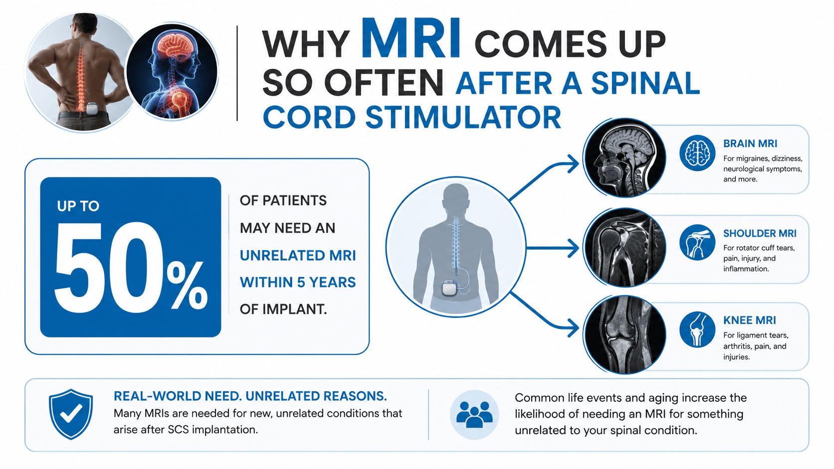
A patient may get a spinal cord stimulator for chronic back pain, do well for a while, and then need an unrelated MRI later. The reason might be a knee injury from a stumble on the stairs, a headache that needs a brain scan, or a new shoulder problem that has nothing to do with the implant. That situation is common because people keep living, aging, and developing new medical needs after implantation.
This is one reason spinal cord stimulation care has to include MRI planning from the start. A device can help with pain and still leave the patient needing imaging for a different problem months or years later.
The long-term MRI burden is not a rare edge case. A propensity-matched analysis estimated that 82% to 84% of SCS patients will need at least one MRI within 5 years, and 59% to 74% will need a non-spine MRI within 10 years (PMC analysis). The same paper projected even higher overall use over time, estimating 89% to 98% by 10 years under linear projection.
What that means in plain English
This does not mean the stimulator failed. It means the device lives in a body that may later need imaging for something else. A person can have a successful pain treatment and still need a scan for an unrelated problem later.
Practical rule: MRI planning should start before the scan is booked, not after the patient arrives at the imaging center.
That is why MRI access has become a core part of spinal cord stimulator care. The key question is not only whether the device can reduce pain. It is whether the device still allows safe, timely imaging when the rest of the body needs attention too.
Patients who understand that reality are less likely to be caught off guard later. They can ask better questions at the time of implant, keep the right paperwork, and avoid the last-minute scramble that happens when a scan is already urgent.
What MRI Conditional Really Means for Your Device
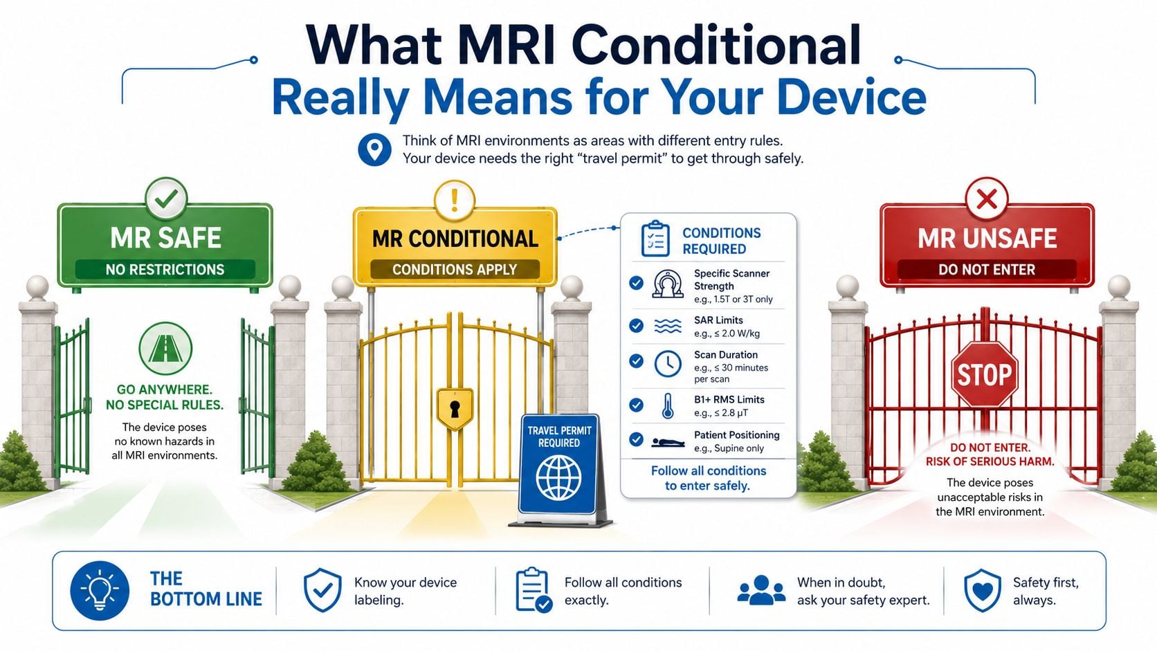
The three MRI safety labels may sound similar, but they do very different jobs. MR Safe means the device is not known to create a hazard in the MRI environment. MR Conditional means MRI can be performed only if the exact conditions in the labeling are followed. MR Unsafe means the device should not enter the MRI environment at all.
A better comparison is a car that needs specific maintenance before a long trip. A vehicle with no restrictions can go wherever it wants. A vehicle with MR Conditional labeling can make the trip only if the oil is checked, the tire pressure is right, and the route matches the owner's manual. MR Unsafe is a vehicle that should not be driven into that situation at all.
Why most modern stimulators are conditional, not safe
Modern spinal cord stimulators are often labeled MR Conditional because the device is not universally safe in every scanner, with every lead, or in every body position. MRI eligibility can depend on the exact IPG model, the lead type, the implant location, and whether the system is fully implanted and correctly configured for MRI mode (FDA labeling). Some systems permit 1.5 T whole-body MRI only, while newer platforms from some manufacturers allow selected 3 T scans under tightly defined conditions (FDA labeling). If your implant includes other hardware, such as spinal instrumentation or related components, the imaging team may need to review that hardware separately as part of the full safety check, including an assessment like the one described in spinal instrumentation guidance.
Those conditions are not decorative fine print. They include technical limits such as gradient slew rate, spatial field gradient, and RF power restrictions. If the MRI center ignores those limits, the scan can become unsafe.
Plain-language version: “Conditional” does not mean “any MRI, any time.” It means “MRI only when the exact recipe is followed.”
Why the scanner matters as much as the implant
A patient can have a device that is conditional at one field strength and not another. A scan that is allowed in one machine may be denied in a different machine, even if both rooms look identical to the patient. The difference lives in the labeling.
That is why the device card matters so much. It is not just proof of implantation, it is the first document that tells the imaging team what kind of scanner, settings, and precautions are permitted. Without that card, the words “MRI compatible” are too vague to rely on.
How an MRI Can Affect a Stimulator and Why
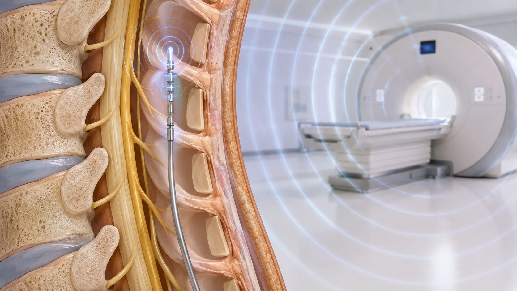
An MRI scanner uses strong magnetic fields and radiofrequency energy to build images. Those same forces can interact with an implanted stimulator, especially the lead components that carry current between the battery and the spine. The lead is not a passive wire sitting in the body. It behaves more like a conductive pathway that can concentrate RF energy in the wrong place, which is why the imaging team has to treat the whole system carefully.
The main concern is RF-induced heating at the lead electrodes. That heat can build where the scanner's energy is absorbed and focused, especially near the lead tip. This is also why MRI instructions pay such close attention to the scan setup, including the coil used, the amount of RF exposure, scan time, and the patient's exact position. If any part of that setup is off, the energy profile around the implant can change.
Three things that can go wrong
First, the lead can heat. Second, induced currents can cause painful jolting or uncomfortable stimulation. Third, electromagnetic interference can disrupt the electronics, leading to telemetry failure, a power-on reset, parameter changes, or other device malfunction.
If the stimulator includes spinal instrumentation or related hardware, the imaging team may need to review those parts too, because every component in the system can affect what the MRI team is allowed to do. Manufacturer safety guidance also warns that scanning outside the approved conditions can lead to dislodgement of components, heating of the neurostimulator, damage to electronics, and uncomfortable or severe stimulation. Some systems also require the device to be turned off or placed in MRI mode before the scan starts. MRI mode does more than shut off therapy, it prepares the system for scanning in the way the manufacturer intended.
Why the limits feel unusually strict
MRI rules for some systems can be much tighter than a routine scan. For example, published protocols for certain high-frequency systems describe whole-body average SAR ≤0.4 W/kg with 30-minute active scan limits. That kind of limit shows how tightly the scanner's energy must be controlled when a stimulator is inside the body.
The MRI team is not being difficult when it asks for exact device details. It is trying to keep the lead from concentrating energy like a heating element or carrying interference into the device electronics. The practical question is always the same. Can this specific system be scanned under this exact set of conditions, with the right programming and the right machine settings?
Your Pre-MRI Checklist Before the Scan
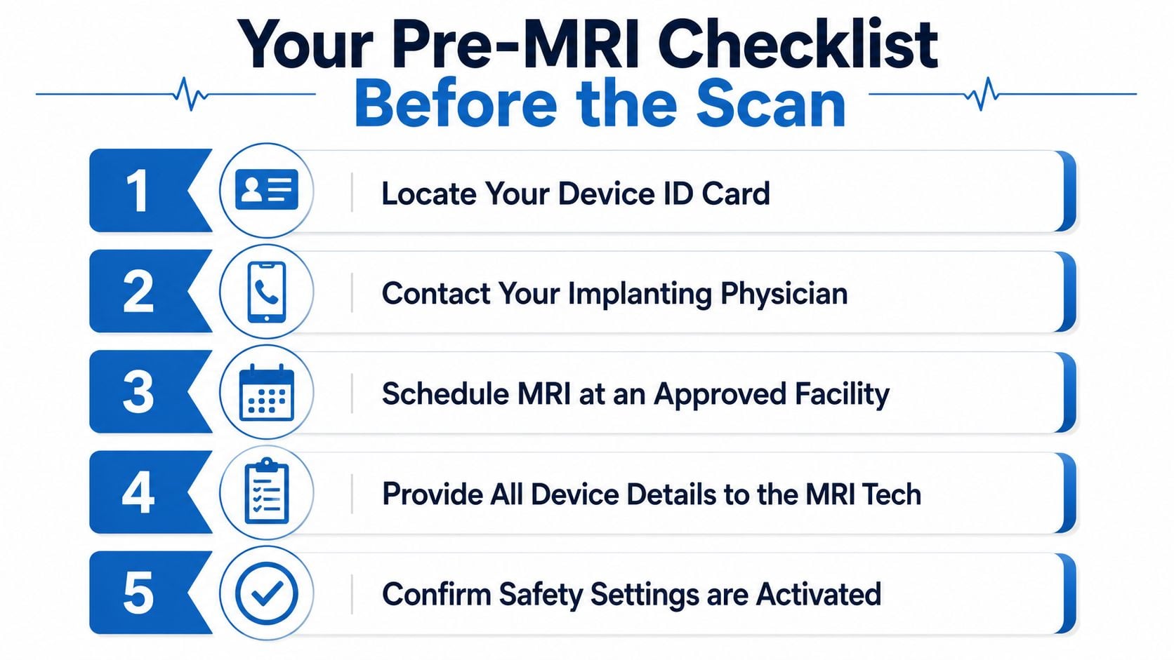
The safest MRI starts before the appointment. A patient who shows up with the device card, accurate model details, and a recent plan from the implanting clinic gives the imaging center what it needs to make a real decision. A patient who arrives with only “I have a stimulator” usually gets delayed.
The five steps that matter most
Locate the device ID card.
The card usually lists the system name, the model, and the implant details the MRI team needs.Contact the implanting physician's office before scheduling.
That office can confirm whether the full system is eligible, not just the battery pack.Verify the lead type and implant configuration.
The label can change depending on the lead and where it sits in the body.Make sure stimulation is off or MRI mode is set correctly.
“Turn it off” is not always enough. Some systems require a specific MRI mode, because that mode does more than shut down therapy.Bring written documentation to the MRI center.
The technologist and radiologist need something concrete to match against the scan they are planning.
The reason this checklist matters is simple. MRI eligibility is device-specific and protocol-specific. A patient may have the right brand but the wrong lead combination, or the right implant but the wrong scanner setup. High impedance can also block MRI-conditional mode and make a scan unsafe or impossible, so device behavior needs to be checked before the appointment when possible.
A short rule for patients to remember
If the implanting clinic cannot confirm the exact system, the MRI center cannot safely assume eligibility.
Patients often feel like they are being asked to do the clinic's paperwork for them. In reality, this paperwork is the safety system. It keeps an unnecessary denial from turning into an unsafe scan.
What to Expect on the Day of the Scan
The MRI day usually feels calmer when the prep work has already been done. The patient arrives, fills out a screening form, and tells the technologist about the stimulator right away. That gives the staff time to verify the device details and check the scan conditions before anyone enters the scanner room.
The technologist may call the implanting clinic or review the documentation on file. The radiologist then decides whether the requested scan can be done within the device's labeling and the center's protocol. This step matters because the device being “conditional” in general does not automatically mean the requested scan is approved.
Sensations that should be reported right away
A patient should speak up if the scan causes warmth, tugging, unexpected stimulation, or any sudden change in how the body feels. Those sensations can mean the device is interacting with the scanner in a way that needs attention. Normal MRI noise is expected. Sharp discomfort is not.
If the device has been placed in MRI mode, the patient should not assume therapy will stay off forever. The implanting team usually needs to return the device to normal settings after imaging. Leaving it in MRI mode longer than intended can create avoidable problems with pain control or device use.
What smooth scans have in common
When a scan goes well, the pattern is usually the same. The device details are known, the MRI team has the right label in hand, the programming is correct, and the scanner settings match the conditions. That is why the day-of experience can feel uneventful when the behind-the-scenes coordination has been done properly.
The good news is that conditional devices often do allow successful imaging when everyone follows the plan. The bad news is that none of this can be guessed at the door. It has to be checked in advance.
When MRI Conditional Still Does Not Mean MRI Approved
This is the part that often frustrates patients most. A person hears that the stimulator is “MRI-conditional,” then gets told the scan still cannot be done. That is not a contradiction. It usually means the requested scan does not match the exact rules attached to the device.
A real-world retrospective study found that only 15 of 49 cases, or 30.6%, had the entire SCS system approved for the planned MRI, and only 10.1% of implanted devices were approved for the requested MRI examination overall (2022 retrospective study). Another way to say it is that device labeling is only the starting point. The final decision depends on the exact system, the scan region, the scanner strength, and the current state of the implant.
Common reasons a scan gets denied
| Reason MRI Is Denied | What It Means | What the Patient Can Do |
|---|---|---|
| Wrong scanner strength | The requested machine does not match the device labeling | Ask whether another scanner strength is allowed |
| Lead or implant mismatch | The battery may be conditional, but the full system is not | Bring the exact model details and ask the implanting clinic to confirm the entire setup |
| MRI mode cannot be activated | The system is not ready for the scan under the required conditions | Contact the implanting office before the appointment |
| High impedance or lead issue | The device cannot enter conditional mode safely | Ask whether the device needs reprogramming, evaluation, or a different imaging plan |
| Protocol mismatch | The scan region, coil, or exposure settings do not match the label | Ask the radiology team what condition is failing |
How to push back constructively
The patient does not need to argue with the MRI technologist. A better approach is to ask for the exact reason the scan was denied and then take that reason back to the implanting clinic. That allows the clinician to check whether the problem is fixable, whether another scanner or protocol is acceptable, or whether another imaging path is safer.
Useful question: “Which part of the device label or scan protocol is not being met?”
A center may still decline the scan even when the patient feels the device is “supposed to be MRI compatible.” That is often a documentation problem, a configuration problem, or a safety problem, not a personal failure. The right response is not panic. It is to gather the missing facts and let the implanting team help decide the next step.
Alternatives When MRI Is Not an Option
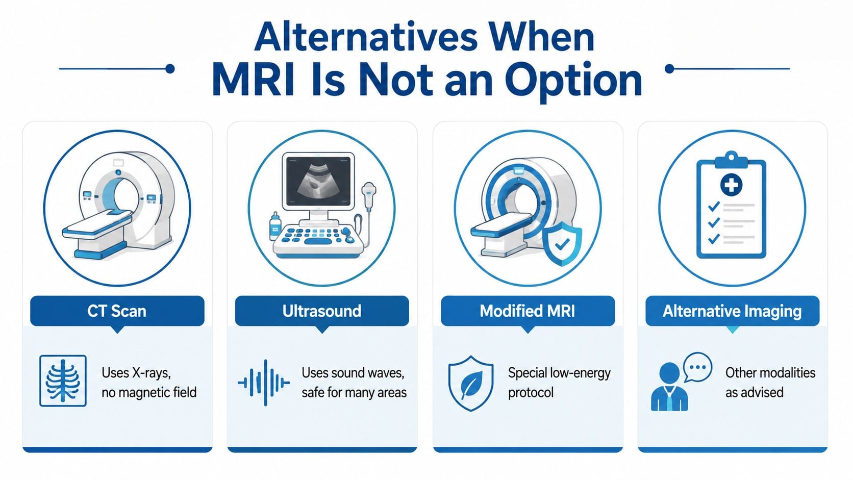
Sometimes the safest answer is no, not with this device in this configuration. That does not end the diagnostic workup. It means the ordering clinician has to choose a different path that still answers the medical question.
CT is often the most familiar alternative. It uses X-rays, so there is no magnetic field to worry about. It can be a strong option for bone detail, fractures, and some structural problems, but it uses radiation and does not show soft tissue the way MRI does.
Ultrasound uses sound waves, which makes it useful for many superficial or soft-tissue questions. It can be helpful for joints, tendons, fluid collections, and some guided procedures. It is limited for deeper structures and for problems hidden behind bone or air.
A practical way to think about the choice
- CT works well when the question is bony anatomy, bleeding, or a fast look at a problem that cannot wait.
- Ultrasound works well when the suspected issue is near the surface or when real-time guidance is useful.
- Modified MRI protocols may be possible when the device label and the scanner conditions allow a narrower, lower-energy exam.
- Other imaging pathways should be chosen by the ordering physician and the implanting clinic together, especially when the answer changes treatment.
Some centers will also consider off-label MRI under controlled conditions when the clinical need is strong and the risk is judged acceptable. Real-world data show that off-label MRI in SCS patients can be performed with relatively low complication rates, but it is not risk-free (2024 case series).
Interventional Pain Management also discusses MRI eligibility and imaging coordination for implanted devices as part of its broader care for spine and pain patients, which can help when a scan request needs a second look (Interventional Pain Management). Its radiofrequency ablation information is another example of how pain care often involves matching the right procedure to the right diagnosis rather than forcing one test or treatment to fit every case.
Your Action Plan and When to Call the Clinic
The safest path is simple. Confirm the exact device details, involve the implanting physician before the scan is scheduled, and bring the documentation to the imaging center. Those three steps solve most of the confusion around spinal cord stimulator and MRI access.
If the scan is denied, ask for the exact reason, then call the implanting clinic with that information. If the device heats, stimulates unexpectedly, or behaves differently after imaging, the clinic should be contacted right away. That kind of change deserves prompt review, not a wait-and-see approach.
For patients in New Jersey and Staten Island, a pre-MRI review with a pain management clinic can help sort out whether the system is conditional, what the scan center needs, and whether a different imaging route makes more sense. The safest MRI is the one that is planned with the implant in mind from the start.
If a spinal cord stimulator is part of the picture, Interventional Pain Management can help review the device details, coordinate with the imaging center, and talk through safer options when MRI eligibility is unclear. Visit Interventional Pain Management to request a practical pre-MRI review and get help before the scan day becomes a problem.