
The anesthesia has worn off, the foot is wrapped, and the first question arrives quickly: how long until normal walking returns? After Achilles tendon repair, recovery rarely follows one perfectly predictable path. Pain, swelling, wound healing, tendon protection, calf weakness, and confidence all influence how quickly a person moves from a splint to a boot, from a boot to shoes, and eventually back to sport.
A useful Achilles surgery recovery timeline is built around milestones rather than promises. Published rehabilitation protocols place early protection and controlled loading first, followed by motion, strength, gait retraining, and higher-level activity. The practical experience can feel less orderly. A patient may regain ankle motion while still struggling to push off, or walk in a shoe while remaining far from running readiness.
Table of Contents
- The First Days After Achilles Tendon Repair
- Early Protective Phase and Weight-Bearing Progression
- Key Recovery Milestones You Can Track Week by Week
- How Physical Therapy Builds Back Strength and Motion
- How Recovery Differs by Surgical Approach
- Returning to Walking, Driving, Work, and Sport
- Common Misconceptions and Warning Signs to Watch For
- Putting the Timeline Together and Knowing When to Seek Help
The First Days After Achilles Tendon Repair
When the anesthesia fades, the operated leg often feels distant, heavy, or numb. A bulky dressing, posterior plaster splint, or protective cast holds the ankle in a pointed position so the repaired tendon isn't placed under unnecessary tension. The patient usually leaves with crutches or another mobility aid and strict instructions about keeping weight off the foot.
The first job is protection, not exercise. The leg should be kept raised above heart level whenever possible, particularly during rest. Ice is generally placed behind the knee rather than directly over the incision or dressing, because cooling the back of the knee can help reduce discomfort without wetting or compressing the surgical site.
What pain and swelling can feel like
Numbness from anesthesia and local medication gradually fades. Around the second and third postoperative days, a throbbing ache may become more noticeable as sensation returns and swelling increases. Pain that is controlled with the prescribed medication plan and gradually becomes easier to manage is different from sudden, escalating pain that doesn't respond to medication or elevation.
Nighttime often feels difficult. The splint can press against the back of the calf, the ankle may pulse when the leg is lowered, and a trip to the bathroom can feel like a major task. A stable chair, clear pathway, and help with meals can make this period safer.
Practical rule: The repaired foot shouldn't be used as a test of courage. A slow, protected transfer is more useful than trying to walk before the surgical team allows it.
Incision checks and the emotional dip
The dressing should remain clean and dry unless the surgical team gives different instructions. Drainage that increases, a dressing that becomes saturated, spreading redness, foul odor, wound separation, or a sudden change in pain deserves a prompt call. Patients shouldn't remove sutures, staples, or dressings themselves unless they have specific instructions.
Around day four, swelling and limited mobility can create an emotional low point. The finish line feels remote, and ordinary tasks become complicated. The recovery clock is easier to manage when it's viewed as a series of small wins, such as controlling swelling, transferring safely, sleeping more comfortably, and attending the first wound review.
Early Protective Phase and Weight-Bearing Progression
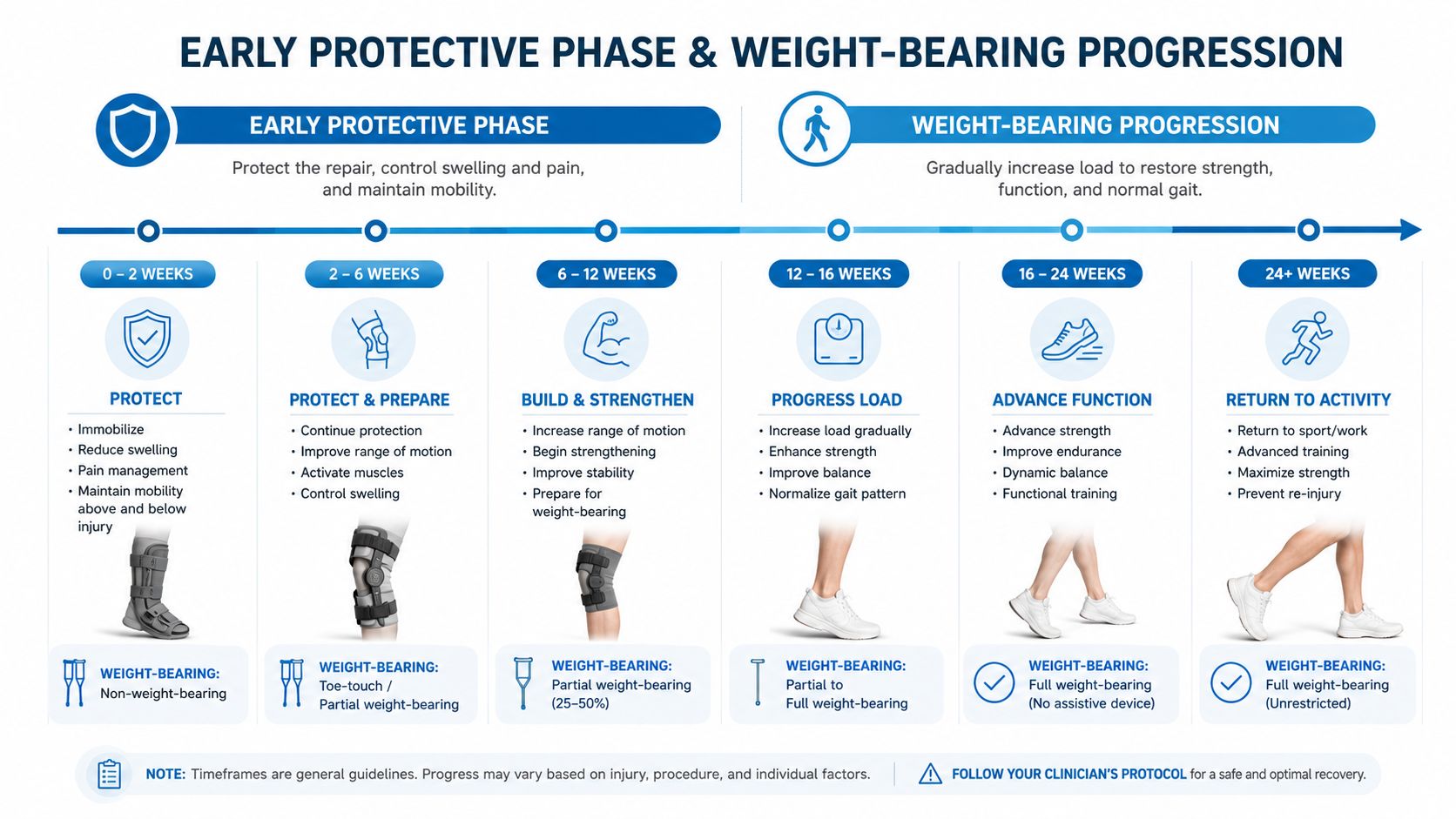
The first six to eight weeks protect the repair while the tendon begins reconnecting and adapting to carefully controlled force. A commonly used benchmark begins with zero to two weeks of non-weight-bearing in a splint or cast, followed by two to six weeks of protected weight-bearing in a boot with heel lifts. The transition toward full weight-bearing in the boot often occurs around six weeks when wound healing and gait mechanics are appropriate, as described in the postoperative Achilles rehabilitation protocol.
Weeks zero through two
The splint keeps the ankle protected and limits unwanted movement. Crutches, a walker, or a knee scooter may be needed, depending on balance, home layout, and arm strength. Floors should be cleared of loose rugs, cords, small furniture, and other trip hazards. A shower chair and a waterproof cover can reduce the risk of slipping while keeping the dressing dry.
Sleeping with the leg raised can ease nighttime throbbing. The boot or splint should remain on exactly as directed, including during sleep if the surgical team requires it. The patient shouldn't alter the ankle position, remove heel support, or begin stretching without clearance.
Weeks two through six
After the incision is checked, many patients transition into a controlled ankle motion boot with heel wedges. The wedges maintain a pointed ankle position while allowing carefully guided loading. The clinical team may remove wedges one or two at a time, but the schedule varies and shouldn't be copied from another patient's plan.
Weight-bearing usually advances gradually. A patient may begin with toe-touch contact, progress to partial weight-bearing, and then move toward full weight-bearing in the boot. Crutches remain useful until the patient can walk without a pronounced limp or unsafe compensation.
Bruising can spread downward toward the heel and foot as blood and fluid move with gravity. Swelling often increases after the leg hangs down and improves with elevation. Increasing warmth, spreading redness, persistent drainage, wound opening, or worsening pain is not something to monitor casually. Sutures or staples are removed at a follow-up visit according to incision healing, rather than on a universal calendar date.
Key Recovery Milestones You Can Track Week by Week
Milestones help patients understand progress, but they aren't deadlines. A 2024 prospective cohort study of open Achilles tendon repair found that average ankle range-of-motion recovery occurred at 6.2 to 10.1 weeks, heel-raise recovery at 12.2 to 14.2 weeks, and return to lower-extremity activity at 18.1 to 20.1 weeks. Longer immobilization delayed each milestone, showing how strongly the early protocol shapes the postoperative recovery timeline.
The numbers describe group averages, not an individual guarantee. Tendon quality, the size and location of the rupture, surgical technique, swelling, pain, and therapy participation can all shift the checkpoints.
A practical milestone map
| Week | Weight-Bearing Status | Range of Motion Target | Strength Milestone |
|---|---|---|---|
| 0 to 2 | Non-weight-bearing in a splint or cast | Protected ankle position, no forced stretching | Gentle activation only if prescribed |
| 2 to 6 | Protected weight-bearing in a boot with heel lifts | Gradual motion toward neutral under supervision | Early calf activation without forceful loading |
| Around 6 | Progression toward full weight-bearing in the boot if gait is safe | Controlled motion within the surgeon's limits | Better tolerance of supported standing |
| 12 to 14 | Walking progression according to examination and gait quality | Functional motion continues to improve | Heel-raise recovery may begin to appear, but endurance remains limited |
| 18 to 24 | More normal daily mobility for many patients | Motion should support walking and stairs | Running preparation may begin only when strength and control allow |
| 24 or later | Higher-level loading remains criteria-based | Functional range for sport tasks | Strength symmetry and calf performance guide sport clearance |
A 2023 systematic review reported a weighted mean return to full weight-bearing of 4.8 ± 6.4 weeks and return to a neutral ankle position of 4.6 ± 3.9 weeks. The same review found that early full weight-bearing within four weeks was associated with a higher re-rupture rate, 1.5% versus 0.7%, reinforcing why a faster-feeling leg isn't automatically a safer leg. The review also summarizes the 2022 GAIT consensus, which reported average non-weightbearing for 2.3 weeks, plantarflexion for the first four weeks, no stretching or eccentric loading before 12 weeks, and average return to sport at 24.4 weeks, or about six months. These findings appear in the systematic review of rehabilitation after Achilles repair.
Patients often notice motion before strength. The ankle may move better while the calf still looks smaller and struggles with stairs or push-off. Surgeon clearance to walk or start a new exercise means the tissue may tolerate that task, not that the limb has regained normal power.
How Physical Therapy Builds Back Strength and Motion
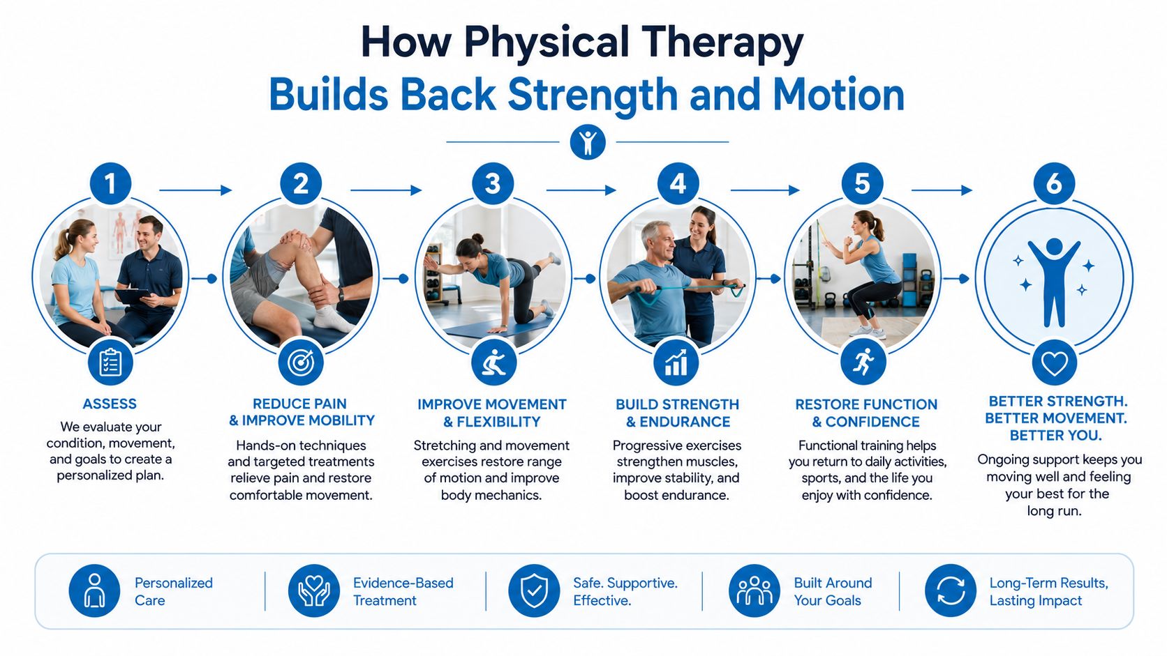
Physical therapy after Achilles repair is layered. Motion comes before demanding strength, supported strength comes before single-leg control, and controlled loading comes before jumping or rapid direction changes. The therapist adjusts the pace according to swelling, gait, tendon protection, and the quality of movement.
Early sessions may involve gentle, therapist-guided ankle motion within the approved range, basic activation, and instruction in safe transfers. The patient may feel tightness at the back of the ankle and mild soreness after a new exercise. That discomfort should settle rather than intensify from session to session.
From activation to controlled loading
As weight-bearing advances, therapy can include resistance bands, seated calf activation, balance work, and towel scrunches for foot control. These tasks may look modest, but they rebuild coordination between the foot, ankle, knee, and hip. A patient who can stand comfortably may still need weeks of practice before the repaired side accepts a smooth push-off.
Around the later strengthening phase, the therapist may introduce double-leg heel raises, then gradually shift more load to the surgical side. Heel drops on a step and eccentric loading require particular caution. Published protocols commonly delay stretching and eccentric loading until the tendon is ready, and a therapist should determine the position, range, and resistance.
The goal isn't to make the calf tired at any cost. The goal is to make the tendon and calf tolerate increasing load without losing alignment or control.
Preparing for running and sport
Plyometric progression and return-to-run work commonly fall in the 12 to 24 week window, while return-to-sport testing generally begins at 24 weeks or later. Criteria can include a limb symmetry index above 90% and strong calf performance, as described in this contemporary rehabilitation review.
Higher-level therapy may use an anti-gravity treadmill, controlled hopping, landing drills, or box-jump progressions. The psychological hurdle can be just as real as the physical one. A tendon that has been protected for months can feel vulnerable when the therapist asks for a faster step or a single-leg landing.
Skipping phases or adding load abruptly can expose a repair that isn't ready. Consistent therapy gives the care team repeated opportunities to adjust the program, address a limp, and build confidence through measurable control.
For readers managing other tendon or soft-tissue symptoms during rehabilitation, a related overview of tendinitis and bursitis can help distinguish general irritation from a postoperative recovery problem.
How Recovery Differs by Surgical Approach
The repair method can influence wound healing and early comfort, but it doesn't remove the need for prolonged tendon remodeling. Open repair uses a larger incision and may require more attention to skin healing before the boot and therapy progress. Percutaneous and minimally invasive methods use smaller access points and can reduce soft-tissue disruption, but they carry their own considerations, including possible irritation of the sural nerve.
A patient shouldn't assume that a smaller incision guarantees an earlier return to running. The tendon still needs controlled loading, calf retraining, and functional testing. The size of the tendon gap, tissue quality, chronicity of the rupture, and surgeon preference may matter more than the incision alone.
| Repair Type | Typical Boot Period | Weight-Bearing Start | Return to Sport Estimate |
|---|---|---|---|
| Open repair | Protocol-dependent, with wound healing guiding progression | Protected loading follows incision and tendon assessment | Commonly many months, based on strength testing |
| Percutaneous repair | Protocol-dependent, sometimes allowing earlier early motion | May progress sooner when the repair and wound permit | Criteria-based, not calendar-based |
| Minimally invasive or PARS-style repair | Protocol-dependent, with soft-tissue status considered | Gradual loading under surgical guidance | Often extends well beyond walking recovery |
| Gastrocnemius turndown or tendon transfer | Frequently longer protection for complex or chronic cases | May remain delayed until the reconstruction is secure | Functional return can extend toward nine to twelve months |
The table describes broad planning categories rather than fixed prescriptions. Chronic or neglected ruptures requiring tendon transfer or gastrocnemius turndown can shift the entire schedule because the reconstruction may need more protection. Patients should ask which technique was used, whether the rupture was acute or chronic, and which milestone would cause the surgeon to slow or advance the plan.
Returning to Walking, Driving, Work, and Sport
Walking recovery and sport recovery are separate achievements. A patient may walk in a boot while still lacking the calf power needed for a normal heel-to-toe stride. As the boot comes off, a heel lift may make the transition to shoes more comfortable, but the gait still needs practice.
Walking in a controlled boot often begins during the early protected phase, commonly around weeks three to six when the surgeon permits it. Unassisted indoor walking may become practical later, but a limp can persist after the patient no longer needs crutches. A normalized gait usually depends on improving calf strength, ankle motion, balance, and confidence rather than reaching a particular date.
Planning daily responsibilities
Desk work may be possible when the leg can remain raised and transportation is arranged. Standing, lifting, climbing, and other physical duties often require a longer or modified return. Employers may need clear restrictions rather than a simple “back to work” date.
Driving requires more than sitting comfortably in a vehicle. The patient must be able to enter and exit safely, control the pedals, and perform an emergency stop without hesitation. The surgical team and insurer's requirements should guide the decision, especially when the operated side controls the accelerator and brake.
A calendar can suggest when an activity might begin. A safe movement test determines whether the limb is ready.
Light jogging belongs much later than ordinary walking. Plyometric drills, running, cutting, and sport-specific work should wait until the patient demonstrates appropriate strength, landing control, and confidence. Broader outcome data report return to sport after surgical repair across a wide range, from three to 13.4 months, with recurrence reported at 0% to 5.74%, as summarized in the rehabilitation review linked earlier. That spread reflects differences in sport, patient goals, repair characteristics, and testing standards.
Common Misconceptions and Warning Signs to Watch For
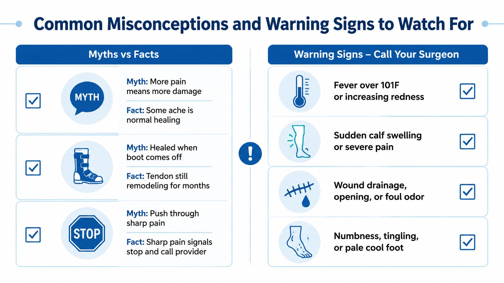
Pain doesn't automatically mean that the repair has failed, and mild aching after a new exercise can be part of rehabilitation. Sharp, escalating, or unfamiliar pain deserves more attention, especially when it appears with a pop, sudden weakness, or a change in walking ability.
The boot coming off also doesn't mean the tendon has finished healing. The tendon may tolerate ordinary walking while the calf remains weak and the tissue continues remodeling. Returning to high-impact activity because the foot feels comfortable can place more demand on the repair than the patient realizes.
Myths that can slow safe progress
- Myth, stiffness should be pushed through: Forceful stretching can place unwanted tension on a healing repair. Motion should stay within the surgeon's or therapist's limits.
- Myth, six months removes all risk: Recovery continues beyond the first half-year, particularly for running, jumping, and pivoting sports.
- Myth, stronger soreness means better training: Exercise should create a manageable response that settles. Increasing swelling, tendon tenderness, or altered gait suggests that the load needs review.
- Myth, a setback should wait for the next appointment: Sudden changes are easier to assess when reported promptly.
A same-day call is appropriate for new wound drainage, opening, spreading redness or warmth, fever above 101.5°F, sudden calf swelling, severe pain, new numbness, or a pale or cool foot. Calf swelling with chest discomfort or breathing difficulty requires emergency care rather than a routine surgical message.
Patients can document the symptom, time of onset, activity that preceded it, swelling change, skin appearance, and ability to move the ankle. That record helps the care team distinguish ordinary post-exercise soreness from a problem requiring examination. Persistent weakness or altered foot control may also warrant evaluation, particularly when it resembles a broader nerve or movement issue such as foot drop.
Putting the Timeline Together and Knowing When to Seek Help
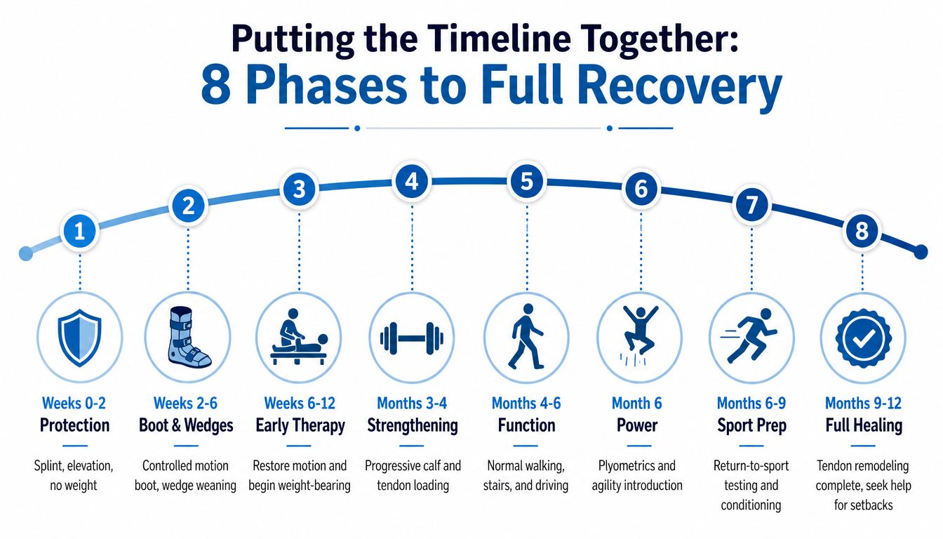
The full pathway is easier to understand as a curved arc. Protection comes first, then boot-based loading, controlled motion, strengthening, normal walking, power development, sport preparation, and longer-term tendon remodeling. Each phase depends on the one before it, so a delay in wound healing or gait control can appropriately move later milestones.
Three transitions deserve particular attention. Leaving the boot can expose weakness that wasn't obvious during protected walking. Moving from formal therapy to independent training can remove the regular feedback that kept exercise progression safe. The return-to-sport decision can feel emotionally urgent even when hop control and calf power remain incomplete.
When recovery needs a closer review
A follow-up assessment is reasonable when pain remains persistent beyond 16 weeks, calf strength is below 80% of the unaffected side, swelling continues to prevent ordinary footwear, or a single-leg heel raise still can't be completed by month five. These are escalation prompts, not diagnoses. The surgical team may identify stiffness, tendon elongation, weakness, nerve symptoms, wound problems, or another cause that needs targeted management.
Patients whose progress has plateaued can seek coordinated evaluation from orthopedic and interventional pain specialists. For ongoing tendon-related symptoms, a focused assessment of Achilles tendinitis can help separate postoperative limitations from a separate tendon condition.
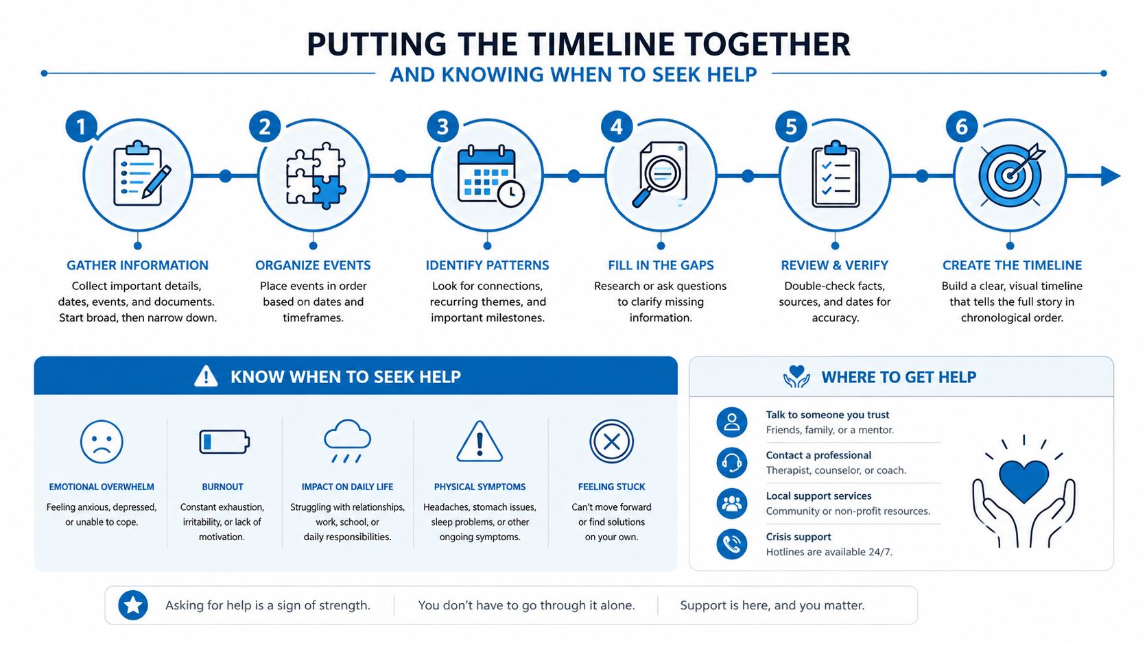
The concise version is simple: protect the repair, restore motion gradually, rebuild calf strength, test function, and treat setbacks early rather than waiting them out.
Interventional Pain Management offers coordinated evaluation for persistent pain and orthopedic concerns when Achilles recovery stalls or symptoms become difficult to interpret. Patients can visit Interventional Pain Management to request an assessment and discuss a care plan connected to current symptoms, functional goals, and the treating surgical team's recommendations.