

A patient may arrive at a spine consultation carrying two competing fears. One is the pain, numbness, or leg weakness that has survived medication, therapy, and injections. The other is the thought of a large back incision, muscle damage, and a recovery that could interrupt work and family life for months. Minimally invasive lumbar spinal fusion often sounds like the answer because the incision is smaller, but the important question is more precise: what changes with the minimally invasive approach, and what stays the same?
The approach can reduce tissue disruption and improve early recovery for appropriately selected patients. It doesn't remove the need for bone healing, hardware, decompression, or careful long-term follow-up. It also introduces trade-offs, including heavier reliance on imaging, a technically demanding learning curve, and risks that remain part of any fusion procedure.
Table of Contents
- What Minimally Invasive Lumbar Spinal Fusion Actually Means
- Core Concepts Behind the Technique
- How the Procedure Works Step by Step
- MIS Fusion Compared With Traditional Open Surgery
- Recovery Timeline and What to Expect
- Risks and Limitations Worth Knowing
- Who Makes a Good Candidate
- Common Misconceptions and Your Next Steps
What Minimally Invasive Lumbar Spinal Fusion Actually Means
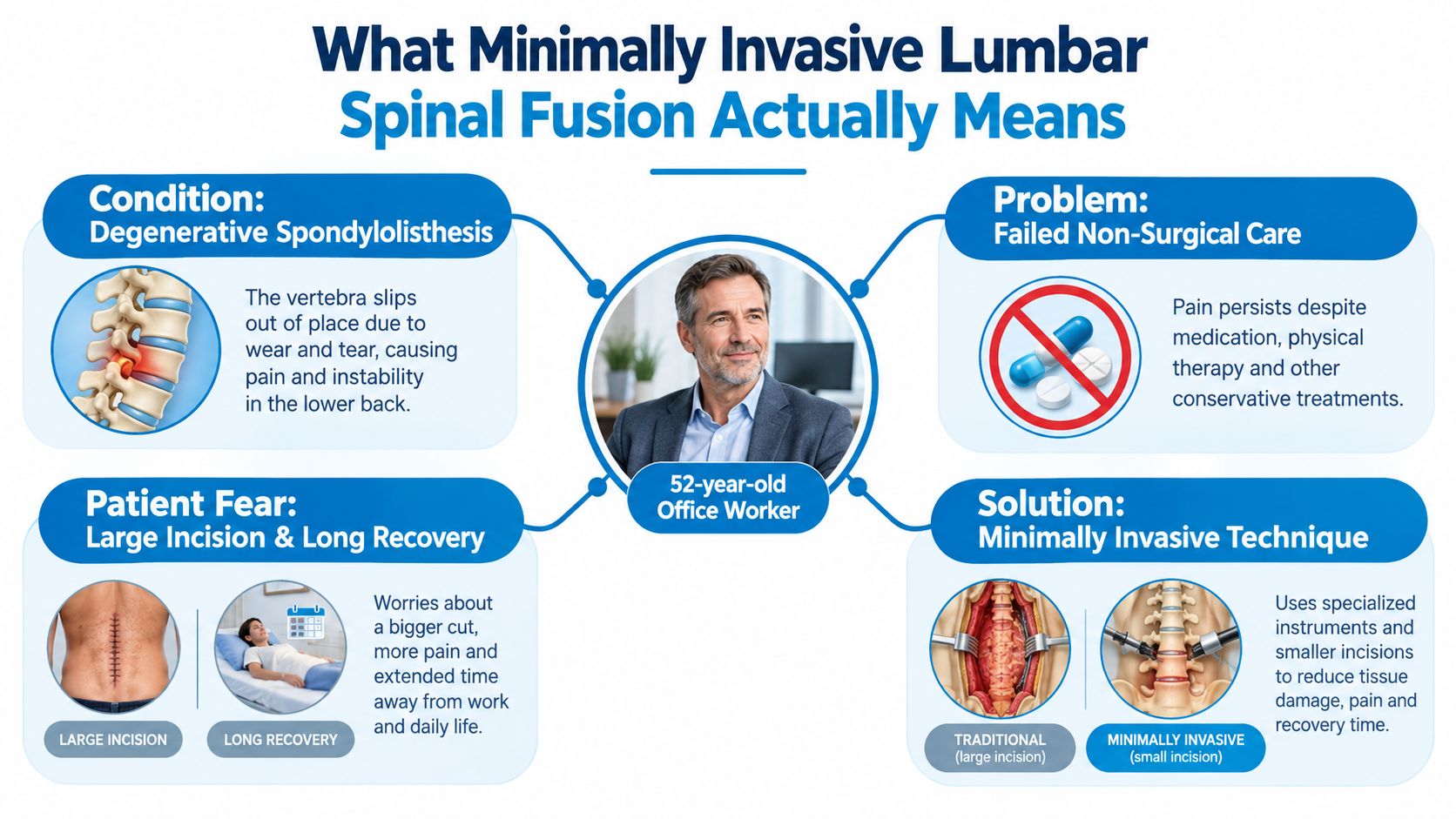
Consider a 52-year-old office worker with degenerative spondylolisthesis. The vertebra has shifted, the nearby nerve is irritated, and months of nonoperative treatment haven't restored comfortable walking or sitting. The patient is willing to discuss fusion but hesitates at the idea of a broad exposure and a prolonged hospital recovery.
Minimally invasive lumbar spinal fusion addresses that concern by changing the route to the spine. Rather than widely detaching the muscles from the back of the vertebrae, the surgeon works through smaller paraspinal corridors. Tubular retractors hold muscle fibers aside, specialized lighting improves the view, and intraoperative imaging helps guide implants through a narrow working channel.
What the smaller access route changes
The access strategy can reduce the amount of muscle disruption, blood loss, and early postoperative soreness. In a comparative five-year study, MIS and open fusion both achieved a 97.5% Grade 1 fusion rate, while MIS patients ambulated earlier, at 1.5 days versus 3 days, and experienced less initial postoperative pain, less blood loss, and shorter hospitalization. The same study found that improvements in disability and pain remained significant from six months through five years (five-year comparative MIS-TLIF follow-up).
That finding supports a useful distinction. MIS may make the early part of recovery easier, but it doesn't create a different kind of bone fusion. The surgeon still removes the painful or unstable disc, decompresses the nerve when needed, places bone graft, and stabilizes the motion segment with implants.
Practical rule: A smaller incision describes the access route. It doesn't guarantee a better long-term result.
MIS also isn't a guarantee of faster recovery for every patient. The result depends on the diagnosis, number of levels treated, bone quality, nerve compression, implant position, surgical experience, and the patient's ability to participate in healing. Open surgery may remain safer when the anatomy is severely distorted or correction requires broad exposure.
The honest summary is simple: MIS changes access mechanics and often short-term morbidity, not the underlying biology of fusion.
Core Concepts Behind the Technique
Most minimally invasive lumbar fusions use a variation called MIS-TLIF, or minimally invasive transforaminal lumbar interbody fusion. “Transforaminal” describes the route through the side of the spinal canal, near the facet joint, to reach the damaged disc without retracting the spinal cord or cauda equina directly.
The procedure relies on several components that work together.
The corridor through the muscles
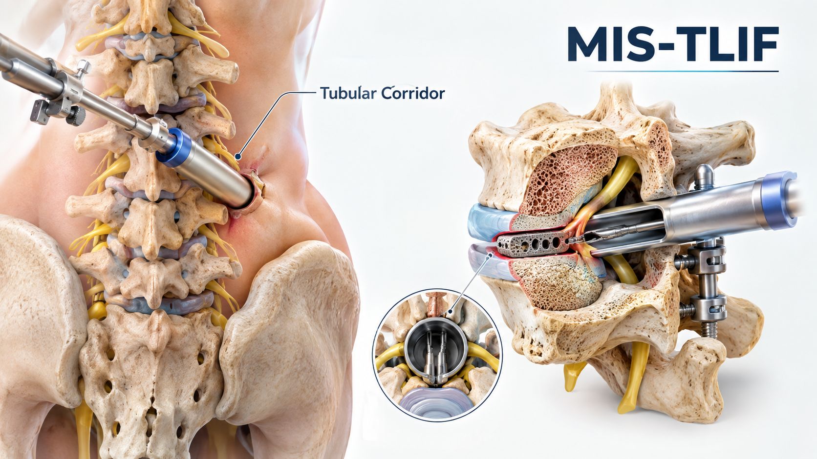
The surgeon first uses sequential dilators to separate muscle fibers gradually. A tubular retractor then maintains that passage, much like a tent pole holds fabric aside. The surgeon works through the tube instead of exposing the entire posterior spine.
This corridor is narrow by design. It can preserve more muscle than a traditional exposure, but it also limits the viewing angle and requires precise positioning. The approach is therefore not “the same operation through a smaller hole.” It demands planning, image guidance, and familiarity with the specific anatomy.
The implants that create stability
A percutaneous pedicle screw enters through a small skin puncture and travels into the vertebral pedicle under imaging guidance. Rods connect the screws, creating posterior fixation while the disc space heals.
The interbody cage is a structural spacer placed where the damaged disc was removed. It helps restore disc height and provides a space for bone graft. A useful analogy is a small support block placed between two settling shelves. The cage maintains the intended separation while bone grows around and through the grafted area.
Bone graft may come from the patient, a donor source, or a synthetic substitute. The material varies by case and surgeon preference, but its purpose remains the same, to encourage a solid bridge of bone across the treated motion segment.
A broader explanation of implant systems and fixation appears in this overview of spinal instrumentation.
How the pieces fit together
The surgeon uses the tubular corridor to remove part of the facet joint, access the disc, decompress the affected nerve, and prepare the bony endplates. The cage and graft then occupy the disc space, while screws and rods hold the vertebrae in the desired alignment.
That sequence matters because the cage alone isn't the fusion. The hardware provides immediate stability, while the graft and the patient's bone-healing response create the lasting connection. A patient considering MIS should therefore ask not only about incision size, but also how the surgeon plans decompression, alignment, grafting, and implant verification.
How the Procedure Works Step by Step
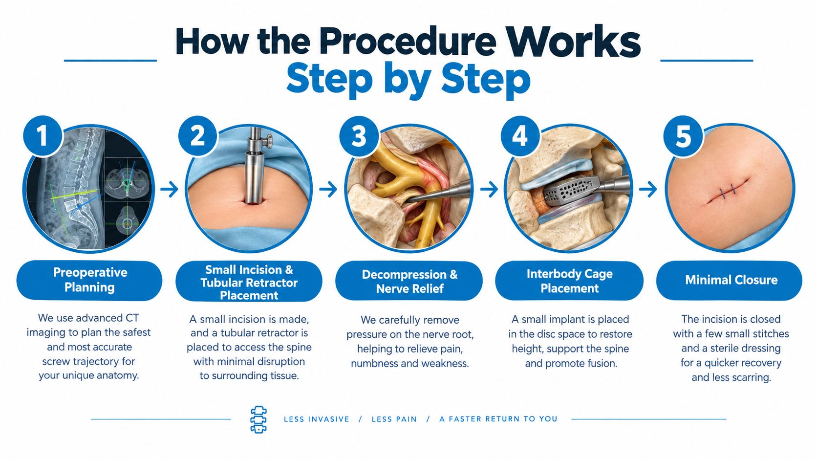
The patient experience begins before the operating room. The surgeon reviews standing X-rays and cross-sectional imaging to understand alignment, instability, disc height, and the safest screw trajectories. An anesthesia consultation addresses medical conditions, medications, airway concerns, and the plan for waking comfortably after surgery.
Once anesthesia is complete, the patient is positioned face down on a support table that protects pressure points and allows the abdomen to hang freely. The knees may be flexed to open the posterior lumbar spaces. The patient won't feel the positioning or the imaging pauses during the operation, but these details help the team work safely.
The operative sequence
Small incision and dilation: The surgeon makes a limited incision near the treated level and advances dilators through the paraspinal muscles. The tubular retractor creates the working corridor.
Access and decompression: A partial facetectomy opens the route to the disc and allows removal of bone or ligament pressing on the nerve. This is the part intended to relieve leg pain, numbness, or weakness caused by compression.
Disc removal and preparation: The damaged disc material is removed. The surgeon prepares the endplates carefully, because healthy contact between bone and graft supports the later fusion.
Cage and graft placement: The interbody cage, filled with selected bone graft, enters the disc space. Its position helps restore height and supports the intended alignment.
Screw and rod fixation: Percutaneous screws and connecting rods stabilize the vertebrae. Fluoroscopy, or live X-ray imaging, helps confirm trajectories and implant placement.
A detailed explanation of the transforaminal approach is available in this guide to TLIF surgery.
Waking up after surgery
The surgeon removes the retractor, closes the small incision, and applies a dressing. Depending on the construct and anatomy, the patient may have two small posterior incisions and several additional puncture sites for the screws. The exact pattern varies, so the number and location of marks shouldn't be treated as a promise.
In the recovery area, nurses monitor breathing, blood pressure, leg strength, sensation, pain, and urination. Walking often begins early when the medical team considers it safe. The first goal isn't athletic activity. It's controlled movement, neurological observation, and a safe transition from anesthesia to recovery.
MIS Fusion Compared With Traditional Open Surgery
The fairest comparison separates early recovery from long-term fusion performance. Minimally invasive surgery generally reduces tissue disruption, but the surgeon still performs the same essential mechanical and biological work.
A meta-analysis found that MI-TLIF involved less blood loss and shorter hospitalization than open TLIF, with similar operative time but longer fluoroscopy exposure. A systematic review reported estimated blood loss ranges of 51 to 578 mL for minimally invasive TLIF versus 225 to 961 mL for open TLIF, while hospitalization ranged from 2.3 to 10.6 days versus 2.9 to 14.6 days, respectively (meta-analysis and systematic review of MI-TLIF versus open TLIF).
| Metric | MIS-TLIF | Open TLIF |
|---|---|---|
| Tissue access | Narrow tubular corridor with less broad muscle detachment | Wider posterior exposure |
| Blood loss | Usually lower, with reported ranges varying by case | Usually higher, with wide variation by case |
| Hospital stay | Often shorter in comparative studies | Often longer in comparative studies |
| Imaging reliance | Greater fluoroscopy exposure during screw and corridor placement | May require less fluoroscopy for some steps |
| Fusion goal | Interbody fusion plus posterior fixation | Interbody fusion plus posterior fixation |
| Long-term outcome | Often comparable when indications and execution are appropriate | Often comparable when indications and execution are appropriate |
What the evidence says about durability
A comparative systematic review found equivalent two-year clinical outcomes, with similar improvement in pain and disability and possibly fewer perioperative medical complications in minimally invasive surgery. Another long-term comparison reported fusion rates of 80.5% versus 91.1%, without a statistically significant difference, and similar reoperation and adjacent-segment disease rates (comparative review of MIS and open lumbar fusion).
The trade-off is fluoroscopy. Because the surgeon works through a narrower corridor and places percutaneous screws with image guidance, the patient and operating team may experience more imaging exposure during a case. For one patient, that exposure is usually considered in the context of the procedure's benefit. For staff who perform many operations, cumulative exposure deserves strict safety controls.
The learning curve also matters. A technically demanding operation can produce different results during training than after a surgeon has developed consistent workflows. Patients should ask about experience with the exact procedure, not just whether a practice advertises “minimally invasive” care. Endoscopic methods represent another access strategy, and their indications and evidence shouldn't be assumed to match MIS-TLIF. A general overview of endoscopic surgery can help clarify that distinction.
Recovery Timeline and What to Expect
Recovery after fusion is measured in stages because the skin, muscles, nerves, and bone don't heal on the same schedule. Smaller incisions may ease the early wound and muscle recovery, but the fusion still needs time to consolidate.
The early weeks
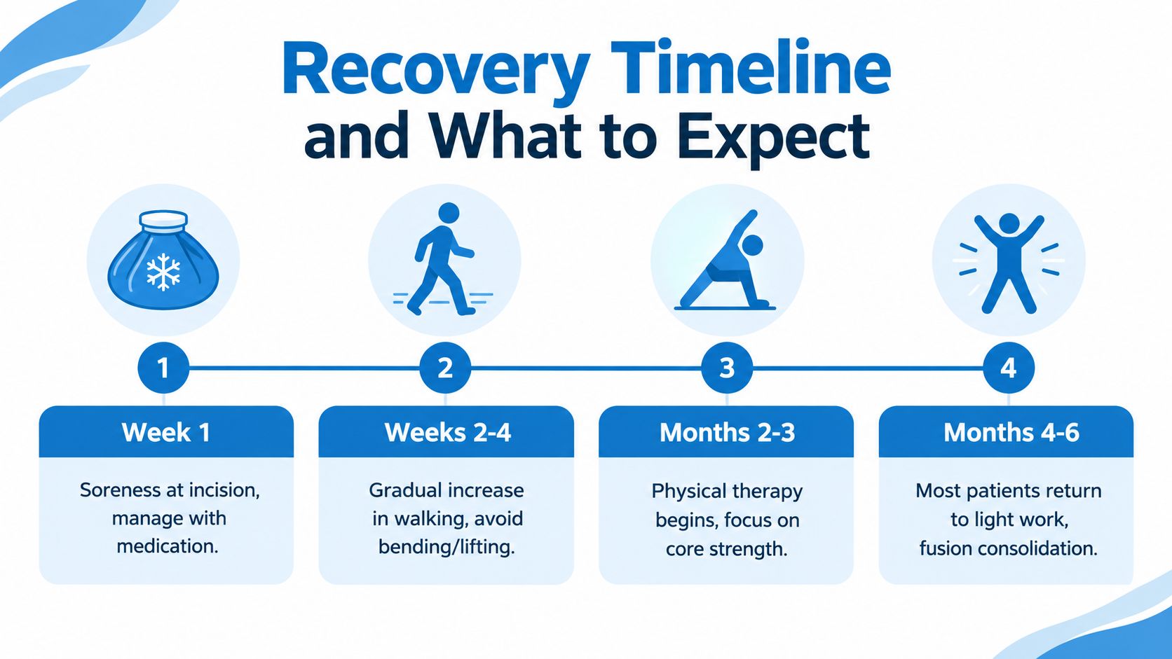
During week one, soreness around the incision and screw sites is common. Walking short distances several times a day usually matters more than prolonged bed rest. Bending, twisting, and lifting should be restricted according to the surgeon's instructions, and many patients are told to avoid lifting more than 5 to 10 pounds during the early period.
During weeks two through four, walking distance can increase gradually. Some patients taper prescription pain medication as the incision discomfort settles, but medication changes should follow the treating clinician's instructions. Driving depends on alertness, leg control, pain medication use, and the ability to brake safely, rather than on a universal calendar date.
Rehabilitation and return to activity
Physical therapy often becomes more structured after the early wound and pain have settled. The program may begin with walking mechanics, posture, transfers, and gentle trunk control before advancing toward strengthening. Desk workers may return gradually when sitting, standing, and commuting are manageable, while physically demanding jobs require a more individualized plan.
Leg pain from nerve compression can improve earlier than mechanical low back pain. The nerve may be free, but it still needs time to recover from chronic irritation. Back pain can improve more slowly as muscles regain conditioning and the fusion matures.
Recovery is not judged by the first comfortable morning. Final function and fusion status require follow-up, gradual strengthening, and patience with the biology of bone healing.
Across months three through six, activity usually progresses from basic conditioning toward more demanding strengthening. Between six and twelve months, imaging may help the surgeon assess fusion consolidation and decide whether impact exercise or unrestricted activity is appropriate. The exact clearance depends on symptoms, examination, imaging, bone health, and the treated levels.
Risks and Limitations Worth Knowing
Minimally invasive access reduces some surgical disruption, but it doesn't make fusion risk-free. The same fundamental concerns remain: nerve irritation, infection, bleeding, implant problems, non-union, adjacent-segment disease, and persistent pain.
A long-term study of MIS-TLIF reported an overall fusion rate of 97.7%, with 80% to 81% patient satisfaction at a minimum of five years. In another 34-patient series, 33 of 34 patients, or 97.1%, showed fusion at six months; the reported complications included one pulmonary embolism and one transient nerve-root pain case, with no infections or cerebrospinal-fluid leaks (published MIS-TLIF series and long-term outcomes). These results are encouraging, but a series from selected patients doesn't predict an individual outcome.
Risks related to fusion biology
A non-union occurs when the intended bony bridge doesn't become solid. Smoking, poor bone quality, medical illness, and larger constructs can affect that process. Hardware may remain intact while the bone fails to unite, or the implants may loosen when the fusion doesn't mature.
Fusion also transfers motion to the levels above and below. That doesn't mean every patient will develop symptoms, but it means a smaller incision doesn't eliminate the long-term consequences of stopping motion at one level. Recent comparative literature has cited adjacent-segment disease rates of 5% to 18% at four to fourteen years after fusion, while a 2025 study reported ten-year surgery-requiring adjacent-segment pathology of 6.1% after MI-TLIF versus 16.4% after open PLIF (recent review and comparative adjacent-segment findings).
Risks specific to the corridor
The narrow working channel can make visualization and orientation more demanding. Potential problems include cage malposition, screw breach, incomplete decompression, nerve injury, and the need to convert to a wider exposure when safety requires it.
Fluoroscopy is another consideration. MIS relies heavily on imaging to guide screw placement and instrument access, so exposure can be longer than with open techniques. A careful surgical team uses shielding, optimized imaging protocols, navigation when appropriate, and disciplined positioning.
MIS may not be the right route for severe deformity, extensive scar tissue, infection, or complex multilevel correction. The safest operation is the one that gives the surgeon adequate control of the anatomy, even when that means a larger exposure.
Who Makes a Good Candidate
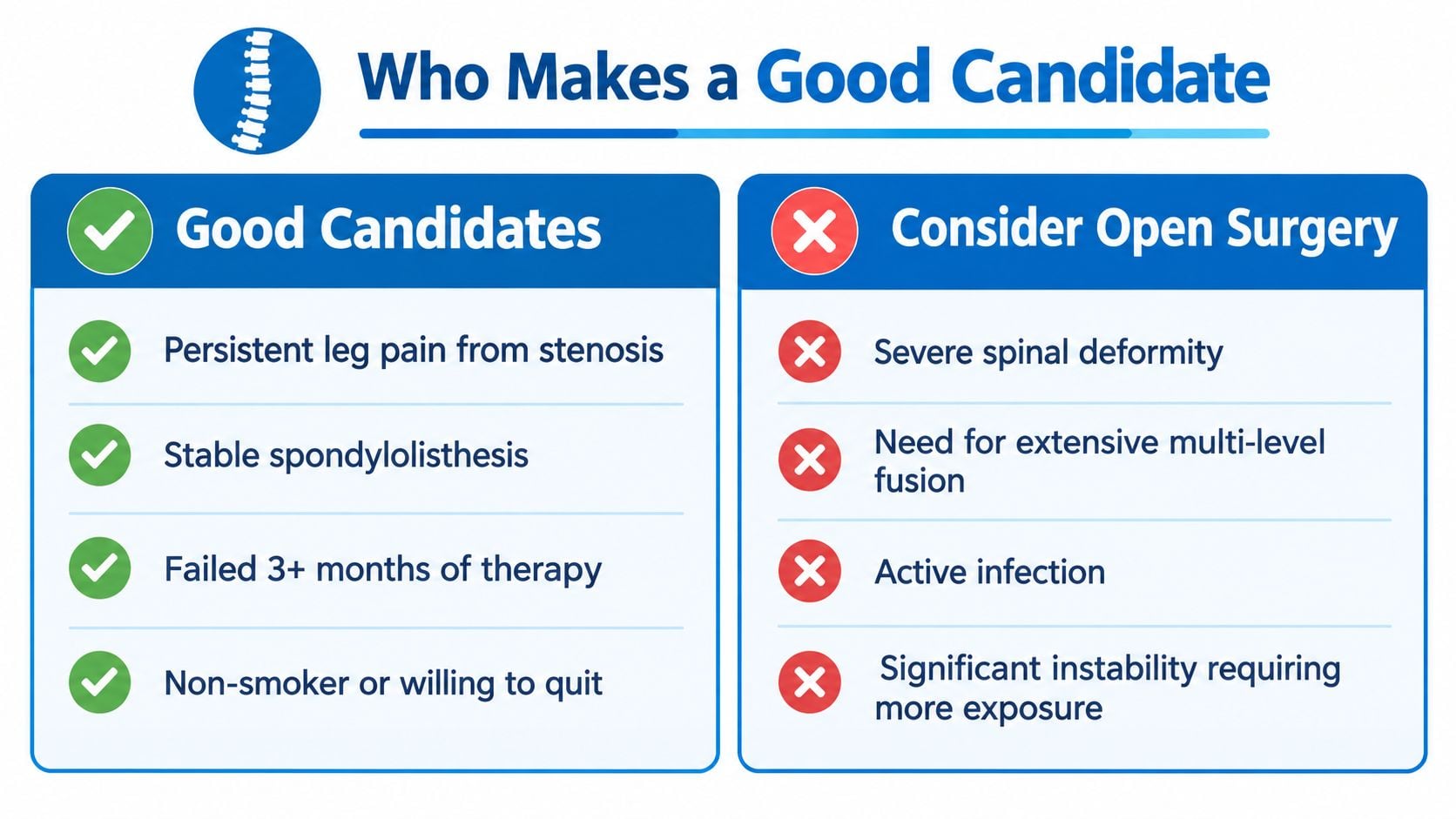
The strongest candidates usually have a clearly identified structural problem that matches their symptoms. Examples include a painful or unstable single-level condition, selected two-level disease, recurrent disc herniation with mechanical instability, lower-grade spondylolisthesis, or stenosis accompanied by deformity.
The diagnosis must also explain the patient's functional limitation. Leg pain from nerve compression may respond differently than isolated nonspecific back pain. Imaging, physical examination, symptom patterns, and the response to nonoperative treatment all matter more than the label “minimally invasive.”
Features that may support MIS
- Localized disease: A limited number of levels allows the surgeon to work through focused corridors.
- Clear instability or disc pathology: The structural reason for fusion should be visible and clinically relevant.
- Manageable anatomy: Body habitus, prior abdominal procedures, and previous posterior surgery can affect the safest route.
- Appropriate health preparation: Bone health, tobacco exposure, diabetes, and other medical conditions should be addressed before surgery.
- Realistic goals: The aim may be relief of leg pain, improved walking, or stabilization, not a promise of a completely pain-free back.
Situations that may favor open surgery
High-grade slippage, severe deformity requiring osteotomies, extensive multilevel reconstruction, active infection, distorted anatomy after prior open fusion, and poor bone quality may require broader exposure or a different reconstruction plan. In those cases, the surgeon may need more room to correct alignment, place graft, remove scar tissue, or protect the nerves.
Before a consultation, a patient can prepare a practical checklist:
- Symptoms: Which activities provoke pain, numbness, weakness, or walking limitation?
- Treatment history: Which medications, therapy programs, injections, or other measures were tried?
- Imaging: Are the latest MRI, CT, and X-ray studies available for review?
- Health factors: Are smoking, osteoporosis, diabetes, or other conditions affecting healing?
- Surgeon experience: How often does the surgeon perform the exact MIS procedure being proposed?
- Alternatives: What would happen with continued nonoperative care, decompression alone, or open reconstruction?
Common Misconceptions and Your Next Steps
MIS doesn't mean that no fusion occurs. The surgeon still removes the disc, places bone graft and an interbody cage, and stabilizes the vertebrae with screws and rods. The word “minimally invasive” describes the access route, not the absence of an implant or the absence of bone healing.
Endoscopic decompression isn't the same as MIS-TLIF. An endoscopic discectomy may remove a disc fragment through a small portal without fusing the segment. MIS-TLIF treats a different mechanical problem when decompression alone wouldn't adequately address instability or painful motion. A patient should ask which structure is causing symptoms and what the proposed operation is designed to change.
A smaller incision doesn't automatically produce a better long-term outcome. Comparative evidence indicates that early blood loss, hospital stay, and recovery may favor MIS, while longer-term pain, disability, and fusion outcomes often converge with open surgery when the indications and execution are appropriate. The operation still depends on complete decompression, sound endplate preparation, accurate implants, and successful bone healing.
Newer isn't automatically safer. Endoscopic and biportal techniques continue to develop, but the literature remains heterogeneous, with many reports based on small series and shorter follow-up. A 2026 review described rapid growth in these techniques while emphasizing technique-specific learning curves and uneven evidence (review of emerging endoscopic and biportal fusion techniques).
Questions that improve a surgical consultation
A patient can ask the surgeon:
- Why fusion: What evidence shows instability or painful motion, and why wouldn't decompression alone be sufficient?
- Why this approach: What makes MIS safer or more suitable than an open exposure for this anatomy?
- Technical experience: How frequently does the surgeon perform this exact procedure?
- Complications: What problems has the surgeon seen with cage placement, screw position, nerve symptoms, infection, or non-union?
- Recovery: What restrictions apply to work, driving, lifting, exercise, and physical therapy?
- Long-term expectations: How will the team monitor fusion and adjacent levels?
- Alternatives: What are the reasonable nonoperative, endoscopic, decompression-only, or open options?
A second opinion can clarify whether the diagnosis, proposed level, surgical approach, and recovery expectations align. The most useful decision isn't approval of a marketing label. It's agreement that the anatomy, symptoms, goals, and surgeon's technical plan fit together.
Interventional Pain Management evaluates spine symptoms through diagnostic review, nonoperative treatment, image-guided procedures, and surgical consultation when fusion or another operation may be appropriate. Adults in New Jersey and Staten Island can visit Interventional Pain Management to discuss symptoms, imaging, treatment alternatives, and a personalized next step.