

You've tried rest, stretching, medication, and perhaps even an injection, yet the pain keeps returning whenever you stand, twist, or lean backward. That pattern is frustrating, but it doesn't automatically mean the next step is another injection or a more invasive procedure. The most important question is whether the pain comes from a facet joint, and the answer usually depends on a carefully performed medial branch block, not an imaging report alone.
Facet joint pain relief is most reliable when diagnosis comes before treatment. A systematic process helps distinguish facet-mediated pain from disc pain, nerve compression, muscle pain, and other spinal conditions, while also supporting the documentation insurers often require before approving radiofrequency treatment.
Table of Contents
- Understanding Facet Joint Pain and Why It Happens
- The Diagnostic Process That Determines Your Treatment Path
- Conservative Treatment Options to Try First
- Interventional Procedures for Facet Joint Pain Relief
- Why Injections Are Not Always the Best Long-Term Solution
- What to Expect From Treatment Outcomes and Durability
- Self-Care Strategies and When to Seek Specialist Care
Understanding Facet Joint Pain and Why It Happens
A patient may describe a deep ache on one side of the lower back that worsens after standing, reaching overhead, or arching backward. Another may report neck pain that spreads toward the shoulder but doesn't travel down the arm. Both may have tried general stretching without understanding which spinal structure is being stressed.
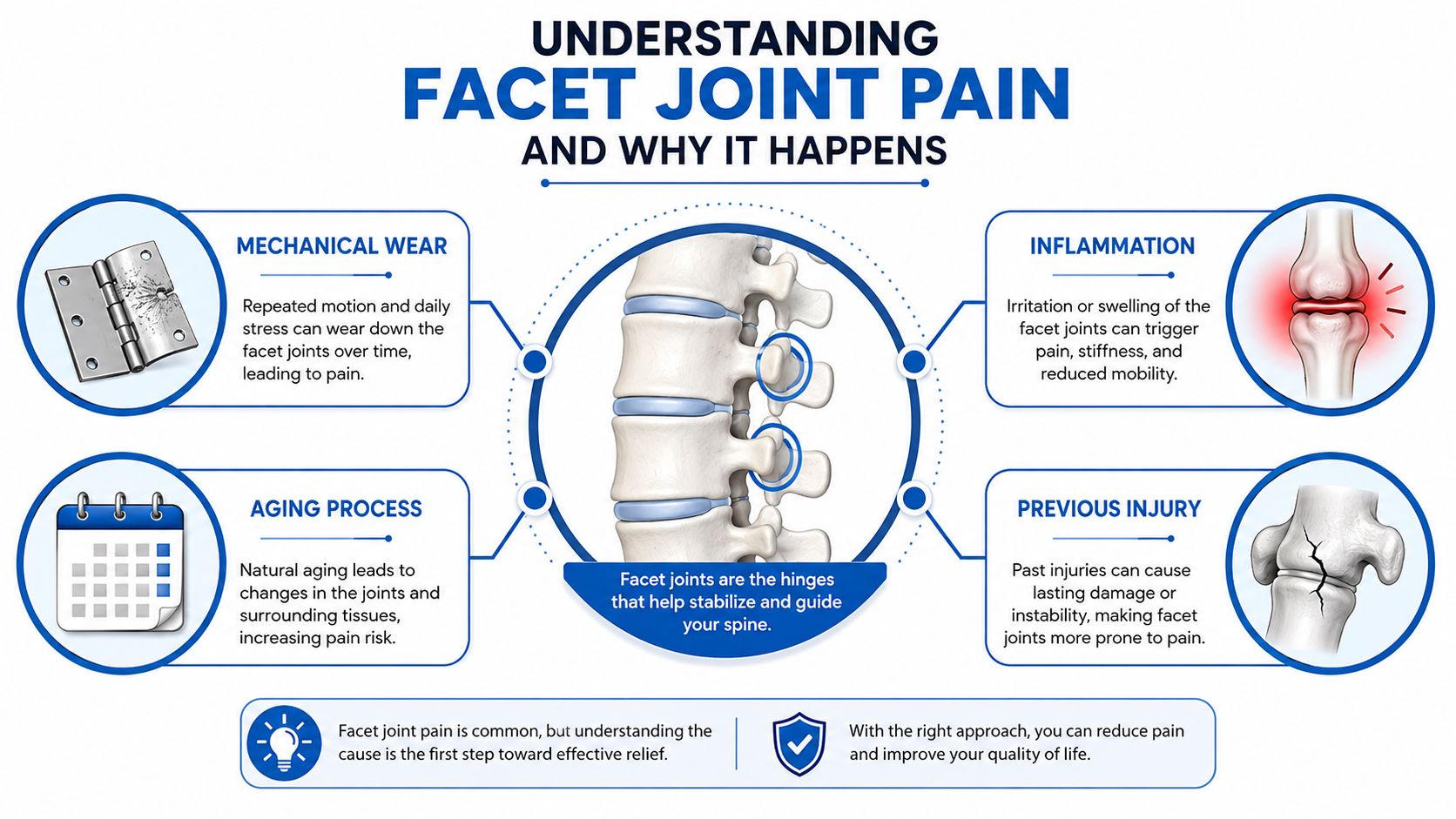
Facet joints are paired joints at the back of the spinal column. They connect neighboring vertebrae and guide movement, much like hinges on a door. Their position allows the spine to bend and rotate while limiting excessive motion. Each joint has a capsule, cartilage, fluid, and a nerve supply capable of transmitting pain.
Facet joints can become painful through mechanical wear, aging, inflammation, or previous injury. Degeneration may alter how forces move through the spine. A sudden extension or twisting injury can irritate the joint capsule. Repetitive loading, poor movement mechanics, or inflammation can also make a previously quiet joint symptomatic.
Symptoms that suggest a facet source
Facet pain is usually axial, meaning it stays mainly in the neck, mid-back, or lower back. It may refer into the buttock, hip, upper thigh, shoulder, or area around the shoulder blade, but it typically doesn't follow the narrow, electric path associated with a compressed nerve.
Common clues include:
- Movement-related pain: Symptoms often increase with backward bending, rotation, prolonged standing, or positions that compress the back of the spine.
- Localized tenderness: Examination may reproduce pain near the affected joints rather than along a nerve pathway.
- Stiffness: Symptoms can be more noticeable after rest or prolonged sitting and may ease with gentle movement.
- Regional referral: Pain can spread into nearby areas without causing a clear pattern of numbness, tingling, or weakness.
These clues are useful, but they don't prove the diagnosis. Facet pain often overlaps with disc degeneration, muscular strain, spinal stenosis, and nerve irritation. Imaging can also show joint wear in people who have no pain, while a painful joint may not look dramatic on a scan. That's why a structured examination and confirmatory block matter before committing to a procedure.
The Diagnostic Process That Determines Your Treatment Path
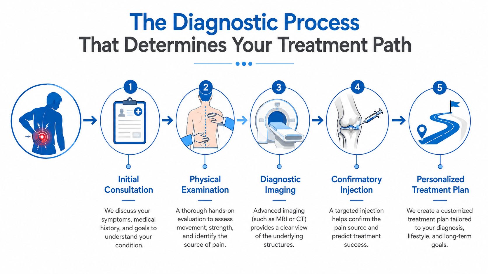
Before discussing an injection or procedure, establish whether a facet joint is producing the patient's familiar pain. Skipping this step can direct treatment at the wrong structure, lead to radiofrequency ablation that does not address the main pain source, or result in an insurance denial when required diagnostic documentation is missing.
The evaluation begins with the history. Pain location, duration, aggravating movements, relieving positions, previous injuries, neurological symptoms, and effects on daily function help define the pattern. Examination assesses spinal motion, tenderness, posture, strength, sensation, reflexes, and findings that may indicate nerve-root involvement.
What imaging can and cannot prove
X-rays show alignment and degenerative changes. MRI evaluates discs, nerves, soft tissues, and other possible pain sources. CT provides detailed views of bone and joint anatomy. These studies help rule out competing diagnoses and guide safe needle placement, but they do not prove that a particular facet joint is generating pain.
A diagnostic medial branch block usually provides the more direct test. Medial branch nerves transmit pain signals from facet joints. With image guidance, the clinician places a small amount of local anesthetic near the nerves supplying the suspected joints. The patient then records whether the usual pain improves during the medication's expected period of action.
Practical rule: Imaging can show an abnormal-looking joint. A controlled block tests whether that joint is responsible for the patient's familiar pain.
Why controlled and dual blocks matter
A single block may appear positive for reasons unrelated to the facet joint. Anesthetic can spread to nearby structures, muscle relaxation can temporarily change symptoms, fear of movement may decrease, or the patient may alter activity during the observation period.
One evidence synthesis reported lumbar facet pain prevalence estimates of 27% to 40% when an 80% pain-relief threshold was used, while false-positive rates ranged from 27% to 47%. Those findings explain why a response must be interpreted carefully rather than treated as automatic proof.
Many protocols therefore use dual comparative medial branch blocks. Two separate blocks use anesthetics with different durations or controlled conditions. Reproducible, substantial relief of the patient's usual pain supports the diagnosis more strongly than vague improvement.
Patients can review how these injections fit into an image-guided evaluation through medial branch nerve block care. The required threshold, number of blocks, and documentation standards vary by payer and by the requested treatment. A clear pain diary should record familiar pain intensity, activity tolerance, and the duration of relief. Examination findings and medication records also help connect the diagnostic response to medical necessity.
A careful block is not an administrative obstacle. It helps determine whether a later procedure has a reasonable target and gives the patient a clearer basis for weighing benefit, risk, and durability.
Conservative Treatment Options to Try First
Most patients should begin with a focused conservative plan unless symptoms suggest an urgent condition. The aim isn't to rest until the pain fades. It's to reduce joint loading, restore controlled movement, and improve the muscles and habits that support the spine.
Build support without provoking compression
Physical therapy commonly emphasizes core stabilization, hip mobility, flexibility, posture, and gradual strengthening. Exercises such as pelvic tilts, supported bridges, controlled abdominal activation, and gentle flexion-based mobility may be appropriate for some patients, but exercise selection should match the examination. A movement that helps one patient can aggravate another, particularly when disc or nerve symptoms coexist.
Short, frequent practice is often more useful than occasional strenuous sessions. The patient should monitor symptoms during the exercise and afterward, progressing only when the movement doesn't produce escalating pain, new numbness, or weakness.
Reduce avoidable stress during daily activities
Ergonomics matter because facet irritation often responds to repeated positions rather than one isolated event. Useful adjustments include:
- Sitting changes: Use back support, keep frequently used items within reach, and change position regularly instead of remaining fixed for long periods.
- Lifting mechanics: Keep loads close to the body, avoid combining heavy lifting with rotation, and ask for help with awkward objects.
- Sleep positioning: Side sleeping with support between the knees or back sleeping with support under the knees may reduce uncomfortable extension.
- Activity modification: Temporarily reduce deep back bending, repetitive twisting, high-impact loading, and prolonged standing if those actions reproduce symptoms.
Heat may relax guarded muscles and improve comfort before movement. Ice can be useful after an acute flare for some patients. For people seeking a non-prescription approach to soothe muscle tension naturally, topical comfort measures can complement, but not replace, diagnostic evaluation.
Use medication with a safety plan
Over-the-counter anti-inflammatory medication may help selected patients, but stomach, kidney, cardiovascular, bleeding, pregnancy, and medication-interaction risks must be considered. Topical products may be suitable when systemic exposure is a concern, while prescription medication requires an individualized review.
Conservative care should be active and measurable. If pain continues to limit sleep, work, walking, or basic activity despite consistent therapy, the clinician can reassess the diagnosis and discuss image-guided options. Escalation shouldn't be based only on the calendar. It should reflect persistent functional limitation, a credible pain pattern, and failure of a properly delivered non-invasive plan.
Interventional Procedures for Facet Joint Pain Relief
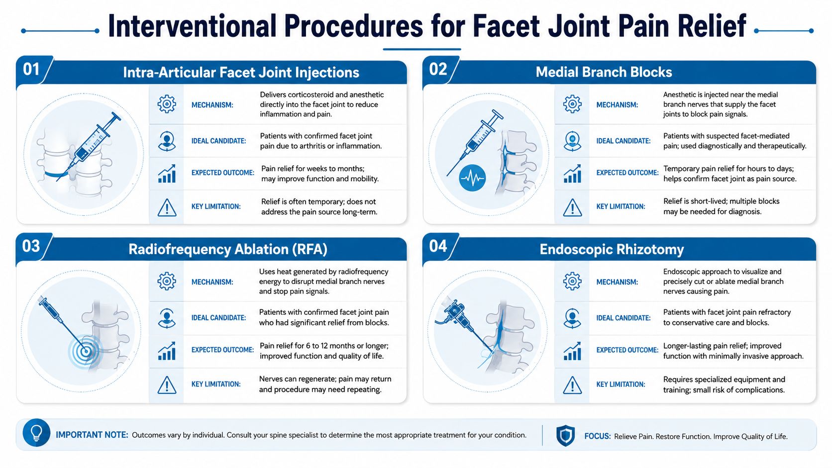
A patient may arrive expecting an injection, yet the more important first question is whether the facet joint is generating the pain. Injections, medial branch blocks, and radiofrequency ablation use image guidance, but they serve different purposes and should not be treated as interchangeable.
Intra-articular facet injections
An intra-articular injection places anesthetic, often with corticosteroid, inside the suspected facet joint. It can calm an inflamed joint and provide short-term relief, particularly when inflammation appears prominent or an acute flare needs treatment. The procedure may also be considered when direct access to the joint is clinically appropriate.
Relief may not last. Evidence reviews have found moderate support for short-term benefit and limited support for long-term benefit from these injections (evidence review of facet injections). A temporary response does not prove that repeated injections will provide durable control, and it does not replace a careful diagnostic plan.
Medial branch blocks
A medial branch block places local anesthetic near the small nerves carrying pain signals from the facet joints. Its primary purpose is diagnostic. Relief of the patient's familiar pain during the expected anesthetic window supports the possibility that those nerves, and the linked facet joints, are involved.
The quality of this step affects everything that follows. The clinician should document the baseline pain, the specific movements that reproduce it, the response after the block, and the degree and duration of relief. Many insurers require defined responses to diagnostic blocks before approving radiofrequency ablation. Skipping or poorly interpreting this step can lead to treatment of the wrong structure, denied coverage, and weaker long-term results.
A positive block alone does not guarantee that ablation is appropriate. The symptoms must fit, competing causes such as untreated radicular pain must be considered, and the documentation must meet the payer's requirements.
Radiofrequency ablation
RFA applies controlled thermal energy to selected medial branch nerves, reducing pain transmission from a confirmed facet source. It does not restore cartilage or reverse degeneration. Its potential benefit depends on accurate diagnosis and appropriate patient selection.
A systematic review and meta-analysis reported Level II evidence and a moderate recommendation for lumbar medial branch RFA in chronic axial spinal pain of facet origin (RFA systematic review and meta-analysis). Temporary soreness or altered sensation can occur afterward. Follow-up should assess pain, function, activity, and medication use rather than relying on pain scores alone.
Patients can review the procedural approach in this overview of radiofrequency ablation treatment.
Emerging approaches
Endoscopic neurotomy and pulsed radiofrequency continue to receive attention, but access, insurance coverage, and guideline support vary. A recent observational study of a three-tined cannula technique reported favorable pain and improvement outcomes, without severe complications in that study (study of a three-tined RFA technique). Those findings are informative, not a guarantee for an individual patient. The diagnostic block remains the foundation for choosing any procedure.
Why Injections Are Not Always the Best Long-Term Solution
A patient may feel better after a facet injection, then have the pain return as the medication wears off. That pattern does not establish that repeated injections are the right answer. The word “injection” can describe two different purposes: calming a short inflammatory flare inside the joint or testing whether facet-related pain is present.
The evidence review cited above found moderate support for short-term relief and limited support for long-term benefit from intra-articular facet injections (review of injection evidence). If relief is brief, repeating the same treatment without confirming the pain source can postpone a better plan and increase exposure to medication-related risks. Therapeutic corticosteroid injections for facet pain should therefore be considered in light of the diagnosis, expected duration of benefit, and available alternatives.
Coverage rules also separate diagnostic medial branch blocks from therapeutic injections. One current policy framework considers therapeutic intra-articular facet injections generally not medically necessary for facetogenic axial low back pain, while supporting thermal RFA after positive dual diagnostic medial branch blocks (interventional pain management coverage guidance). Proper blocks matter because they test whether the medial branch nerves are transmitting pain from the suspected facet joints. Their results can affect both treatment selection and insurance approval.
A less invasive procedure is not automatically the better long-term procedure. Diagnostic accuracy often matters more than how simple the treatment sounds.
RFA is still not appropriate for every patient. Selection generally requires a predominantly axial pain pattern and meaningful, reproducible relief from diagnostic medial branch blocks. Another untreated condition must not explain the symptoms better. Prominent radicular pain, progressive neurological deficits, infection, instability, or another urgent spinal problem may require a different treatment path.
Before agreeing to an injection or procedure, ask four questions: Which structure is suspected? How was it confirmed? What benefit should the treatment provide? What will we do if relief is temporary or absent? Clear answers reduce the risk of repeating a procedure because it produced brief improvement.
What to Expect From Treatment Outcomes and Durability
Facet joint pain relief is rarely a one-time cure. The underlying joint may remain degenerated, and the treated nerve can eventually recover function. A realistic plan therefore includes both the expected benefit and the next decision if symptoms return.
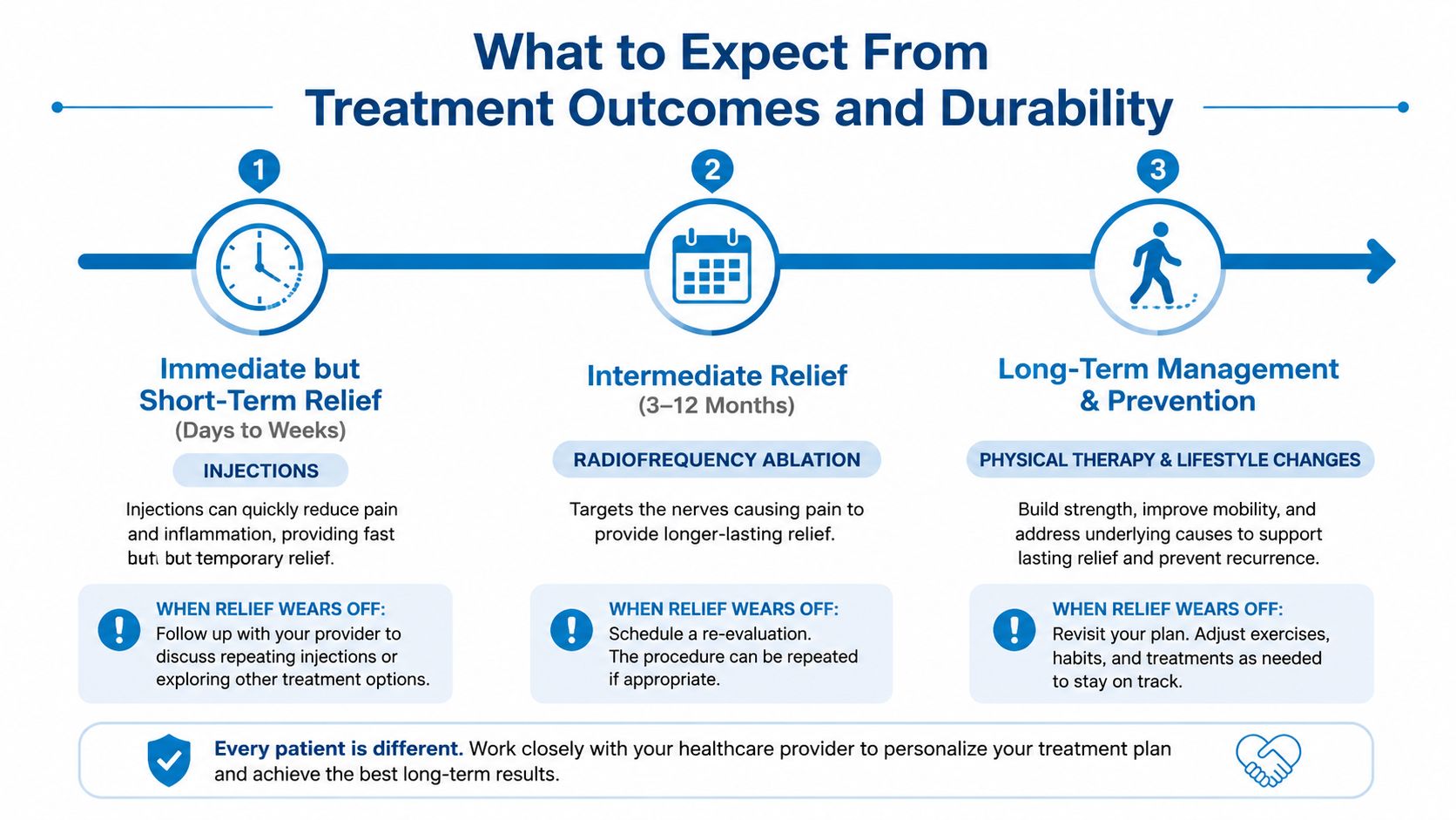
Injection relief is usually measured in a shorter window, often days or weeks in clinical discussions, although individual responses vary. Diagnostic blocks are intentionally temporary because their purpose is to test pain generation. They shouldn't be judged by the same durability standard as RFA.
RFA can provide longer relief for an appropriately selected subset of patients. In a cohort selected with dual comparative blocks showing at least 80% relief, at least 50% pain reduction was reported by 63.2% of patients at 6 to 12 months, 65.6% at 12 to 24 months, and 44.1% beyond 24 months (cohort study of RFA durability). These findings support careful selection, but they don't promise a fixed duration for every patient.
When relief wears off
Recurrence doesn't necessarily mean the original procedure failed. It may reflect nerve recovery, progression of adjacent pain generators, a new injury, or a diagnosis that was incomplete from the start. The clinician should reassess the pain pattern before repeating treatment.
CMS coverage rules note that no more than 2 RFA sessions per spinal region are reimbursed in a rolling 12-month period, and repeat treatment generally requires meaningful relief from the earlier procedure (CMS RFA coverage policy). Coverage rules vary by payer, so patients should verify requirements before scheduling.
A practical follow-up plan tracks walking tolerance, sleep, work tasks, medication use, and pain intensity. If standard RFA stops providing useful relief, the clinician may revisit diagnostic blocks, consider a different technique where supported, or refer for surgical evaluation when imaging and symptoms indicate instability, severe compression, deformity, or another structural problem. Surgery shouldn't be selected solely because facet pain has lasted a long time.
Self-Care Strategies and When to Seek Specialist Care
Patients can begin with gentle walking, comfortable spinal mobility, and a supervised strengthening plan. Avoid movements that repeatedly worsen symptoms, especially deep backward bending, forceful rotation, and heavy lifting in an extended posture. Heat before activity may ease stiffness, while ice after a flare may improve comfort.
Workstations should allow frequent position changes. A supportive chair, a screen near eye level, and brief movement breaks can reduce sustained loading. Sleep positioning can also help, but increasing pain, new weakness, or symptoms extending along an arm or leg require clinical review rather than continued self-treatment.
Emergency evaluation is appropriate for new bowel or bladder dysfunction, saddle numbness, rapidly progressing weakness, fever with severe spinal pain, major trauma, or unexplained systemic illness. A scheduled specialist consultation is warranted when pain persists despite structured conservative care, repeatedly disrupts sleep or work, or has a pattern suggesting a facet, disc, or nerve source.
Patients should choose a clinician trained in interventional pain medicine, confirm that image guidance is used, and bring imaging reports, medication lists, prior treatment records, and a concise pain diary. Before an advanced procedure, the office and insurer should clarify whether conservative therapy, diagnostic blocks, relief thresholds, and repeat-block requirements are documented. Interventional Pain Management offers diagnostic evaluation, image-guided nerve blocks, and radiofrequency procedures as part of a broader assessment that can also involve orthopedic and spine services.
Patients with persistent back or neck pain can request a structured evaluation that identifies whether the facets are driving symptoms before proceeding to treatment. Visit Interventional Pain Management to discuss diagnostic medial branch blocks, conservative care, and image-guided options with a team that can connect diagnosis, treatment planning, and insurance navigation.