
A tender bump on the inside of the foot can turn an ordinary walk, training session, or day in tight shoes into a persistent source of pain. A teenager may notice it during a growth spurt, while an adult runner may feel it after increasing mileage or changing footwear. The bump may look like a minor anatomical quirk, but soreness in this location can involve both the bone and the tendon that supports the arch.
Accessory navicular syndrome occurs when an extra bone or cartilage fragment near the navicular becomes painful. The accessory navicular itself is often harmless. Symptoms usually develop when pressure from footwear, repetitive loading, growth-related changes, or irritation of the nearby posterior tibial tendon turns a quiet anatomical variant into a mechanical problem.
Table of Contents
- Why Your Medial Arch Hurts and What This Guide Will Answer
- The Anatomy Behind Accessory Navicular Syndrome
- Symptoms That Point to the Accessory Navicular
- Conditions That Mimic Accessory Navicular Syndrome
- How Clinicians Confirm the Diagnosis
- Conservative Care That Actually Works
- Surgical Options and How Surgeons Choose
- Recovery, Outcomes, and When to Seek Specialty Care
Why Your Medial Arch Hurts and What This Guide Will Answer
Pain along the medial arch, the inside edge of the foot facing the other foot, has several possible sources. The navicular bone, posterior tibial tendon, nearby joints, nerves, and plantar fascia can all produce symptoms in a similar area. That overlap explains why a person may search for “arch pain” yet still have no explanation for a tender bump.
Accessory navicular syndrome is one possibility. An accessory navicular is an extra ossicle, meaning a small bone or partly formed bone, beside the navicular. Because it may sit close beneath the skin, a shoe counter or strap can press on it. Walking, running, jumping, or prolonged standing can also stress the connection between the accessory bone and the main navicular.
The finding on an X-ray and the cause of pain are not automatically the same. Having an accessory navicular doesn't automatically mean having the syndrome. A 2024 systematic review pooling 39 studies, 11,015 patients, and 36,837 feet estimated accessory navicular prevalence at 17.5% in patient-based analyses and 12.6% in foot-based analyses (systematic review of accessory navicular prevalence). Many people have no symptoms and discover the variant only after imaging for another problem.
The main questions are practical:
- Does the symptom pattern fit? Pain location, activity triggers, swelling, tenderness over the bump, and signs of posterior tibial tendon irritation matter more than the X-ray finding by itself.
- What should happen first? Footwear changes, reduced aggravating activity, inserts, focused rehabilitation, and sometimes temporary immobilization can decrease stress on the area.
- When is surgery reasonable? The choice depends on how long pain persists, the condition of the posterior tibial tendon, the way the arch and foot move under load, and the person's functional goals. Some people need only pressure relief and support, while others have tendon or flatfoot mechanics that require a different plan.
Seek prompt clinical assessment for sudden severe pain, a new deformity, inability to bear weight, or numbness. These symptoms may indicate an injury or nerve problem rather than uncomplicated accessory navicular syndrome.
The Anatomy Behind Accessory Navicular Syndrome
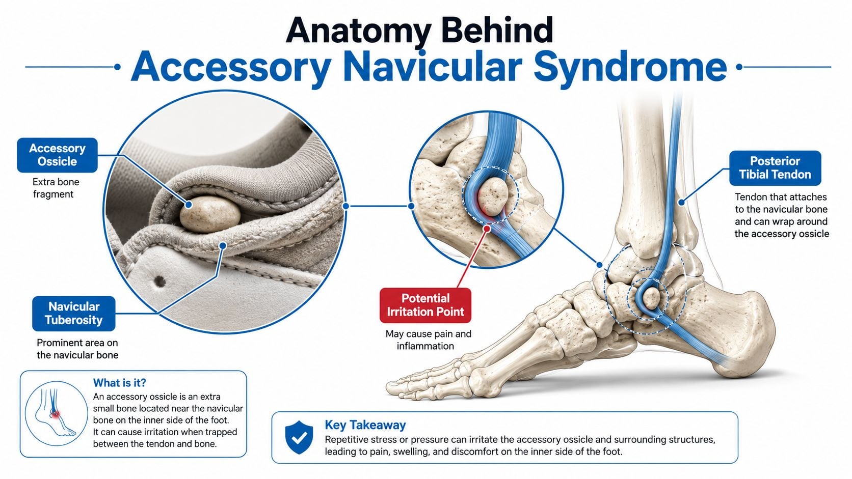
A person may have had a small extra bone inside the foot since birth, then develop pain after a change in activity or shoe pressure. It can behave like a pebble caught in a folded seam. The bone itself is not automatically a problem. Repeated pressure, force through the arch, or irritation of nearby tissue can turn an otherwise quiet anatomical variation into a painful one.
The navicular sits in the middle of the foot, near the top of the arch. The posterior tibial tendon attaches close to it and helps hold up the arch, control the foot as it accepts weight, and assist with push-off. Pain can therefore involve more than the bony prominence. The accessory bone, its connection to the navicular, and the tendon may all contribute.
The three anatomical types
Clinicians commonly describe the accessory navicular using the Geist classification:
- Type I is a small ossicle within the distal fibers of the posterior tibial tendon, close to its attachment. It may function like a small bone associated with the tendon.
- Type II is a larger fragment joined to the navicular by fibrocartilage or fibrous tissue called a synchondrosis. Under load, movement at this connection can make it the painful interface.
- Type III is a prominent, horn-shaped extension of the navicular. The accessory fragment has generally fused with the main bone, leaving a broad prominence on the inner side of the foot.
Type II often receives particular attention because its synchondrosis can behave like a small stressed joint. Clinicians also assess the posterior tibial tendon where it attaches, since tendon irritation and painful bone contact can occur together.
The anatomical variant is not automatically a deformity. It may occur in both feet, and a 2024 review reported bilateral involvement in 50.0% of patients (accessory navicular prevalence and bilateral involvement). The same review reported prevalence of 38.4% in East Asian populations and 8.0% in North American populations. These differences show why an X-ray finding needs context. Symptoms, examination findings, tendon function, and flatfoot mechanics help determine whether the accessory bone is incidental or part of a broader spectrum of arch stress. Some feet need only pressure relief, while tendon involvement or progressive flatfoot mechanics may require a more involved plan.
Symptoms That Point to the Accessory Navicular
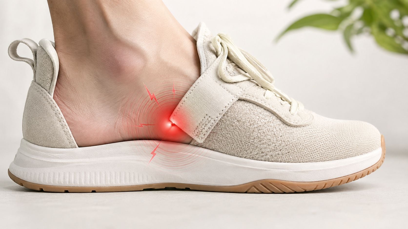
A person may notice pain after a long walk, a run, court sports, or repeated jumping. The discomfort usually centers on a tender prominence along the inside of the midfoot, just above the arch. Shoe pressure can rub the area directly, leaving it sore during activity or afterward.
These activities increase tension through the posterior tibial tendon and, when present, the connection between the accessory navicular and the main navicular. Tight straps and firm shoe counters create a separate problem: direct compression over the bump. Swelling, redness, or warmth may follow activity, though visible changes are not required for the area to be painful.
Symptoms can first appear during adolescence, as growth changes the fit between the bones, tendon, arch, and footwear. A Japanese pediatric radiology study found accessory navicular in 15.1% of children, with pain reported in 20.8% of affected patients (pediatric accessory navicular findings). Adults may notice symptoms after overuse, a training change, a foot or ankle injury, or a gradual loss of arch support.
What tendon irritation feels like
The posterior tibial tendon helps support the arch and assists with push-off. Its irritation may feel like this:
- Arch aching: Pain travels from the bony prominence along the inner side of the foot.
- Post-activity swelling: The area becomes more tender after extended standing or walking.
- Reduced push-off: Running, climbing stairs, or rising onto the toes feels painful or weaker.
- Fatigue with standing: The foot seems to work harder to hold its shape.
The painful structure may be the type II synchondrosis, the tendon insertion, or both. MRI can show marrow edema in the accessory navicular and navicular tuberosity, as well as thickening or enhancement where the tendon attaches (MRI findings in painful accessory navicular syndrome). This helps explain why accessory navicular syndrome exists on a spectrum. Pressure relief may be enough when shoe contact is the main issue, while tendon involvement or flatfoot mechanics can sustain symptoms and require broader treatment.
Practical rule: A visible bump supports the diagnosis, but it does not prove that the bump causes the pain. The examination must match the tender spot with the person's loading pattern and tendon function.
Seek timely medical assessment for sudden severe pain, a visibly changed foot shape, inability to take steps, marked swelling after an injury, or numbness. These features may point to a fracture, tendon failure, nerve compression, or another problem needing a different response.
Conditions That Mimic Accessory Navicular Syndrome
A medial arch bump can make accessory navicular syndrome seem obvious, but several conditions can produce similar pain. Diagnosis depends on the exact tenderness, the foot's movement, the onset, and the imaging pattern.
Posterior tibial tendon dysfunction may cause pain along the tendon behind the inner ankle and into the arch. Weakness with a single-leg heel rise, progressive loss of arch height, or increasing hindfoot turning can make tendon dysfunction more likely than isolated synchondrosis irritation. Some people have both problems, so the accessory navicular may be one part of a broader tendon-and-arch disorder. Readers comparing tendon-related symptoms can review this clinical overview of tendinitis and bursitis.
Tarsal coalition is an abnormal connection between certain foot bones. It often produces a stiff, less adaptable flatfoot, restricted hindfoot motion, and recurrent ankle symptoms. Accessory navicular pain is more commonly centered over the medial prominence, while coalition-related discomfort may involve a broader pattern of stiffness and motion limitation.
Navicular stress fracture deserves particular caution in runners and jumping athletes. Pain may feel deep in the midfoot and become worse with impact, sometimes without a prominent tender bump. A clinician may consider a stress injury when pain escalates with loading or persists despite rest.
Plantar fasciitis usually causes pain under the heel or along the bottom of the arch, especially with the first steps after rest. That distribution differs from the focal medial bony tenderness typical of accessory navicular syndrome.
Medial plantar nerve irritation can produce burning, tingling, or electric discomfort rather than a purely sore prominence. Numbness or radiating symptoms shift attention toward nerve involvement.
These distinctions matter because an insert that relieves pressure on a bump may not address a coalition, tendon tear, fracture, or nerve problem. A careful evaluation prevents the X-ray finding from becoming an automatic diagnosis.
How Clinicians Confirm the Diagnosis
A painful bump along the inner arch does not identify its own cause. Diagnosis starts with a focused history. The clinician asks when the pain began, whether activity or footwear brings it on, whether an injury came first, and whether the arch has changed. Age, sports, work demands, and previous treatment help separate a short-lived irritation from a continuing mechanical problem.
The examination answers the first questions
The foot is assessed while the patient stands and walks. The clinician checks arch height, heel alignment, midfoot flexibility, swelling, shoe pressure points, and tenderness around the medial navicular. Pressing directly over the accessory navicular or its connection to the main navicular may reproduce the familiar pain.
A single-leg heel-rise test checks posterior tibial tendon function. Difficulty rising onto the toes may reflect pain, weakness, or both. That finding matters because the tendon acts like a supporting cable for the arch. If it is overloaded or impaired, treatment must address tendon capacity and flatfoot mechanics, not only pressure over the bony prominence.
The pattern can range from a tender accessory bone with otherwise flexible mechanics to a more involved problem with tendon dysfunction and a collapsing arch. That spectrum helps explain why one patient may improve with pressure relief while another needs a broader treatment plan.
Weight-bearing radiographs show structure under load
Plain X-rays provide the first structural view. Weight-bearing images show how the bones line up while the foot supports the body, allowing the clinician to assess the accessory navicular, main navicular, arch alignment, and other bony causes of pain. An oblique view can make the accessory fragment easier to see.
The practical questions include:
- Is an accessory navicular present?
- What type and shape does it have?
- Does the foot show a flexible or more rigid flatfoot pattern?
- Could another bone injury explain the pain?
An X-ray shows anatomy, but it may not show which structure is actively irritated. Persistent pain with incomplete or unrevealing radiographs may call for further assessment, as with suspected foot and ankle fractures.
MRI clarifies the pain generator
MRI is considered when symptoms persist, synchondrosis inflammation is suspected, or the posterior tibial tendon needs closer evaluation. Bone marrow edema near the accessory navicular supports active irritation. Tendon thickening or abnormal signal can indicate tendon disease and help explain why flatfoot mechanics are contributing.
MRI may reveal clinically relevant findings that plain radiographs do not show, particularly in children with painful flatfoot. Ultrasound can also help assess tendon integrity and movement around the accessory fragment. The scan is therefore selected to answer a specific question, rather than treated as an automatic next step.
Conservative Care That Actually Works
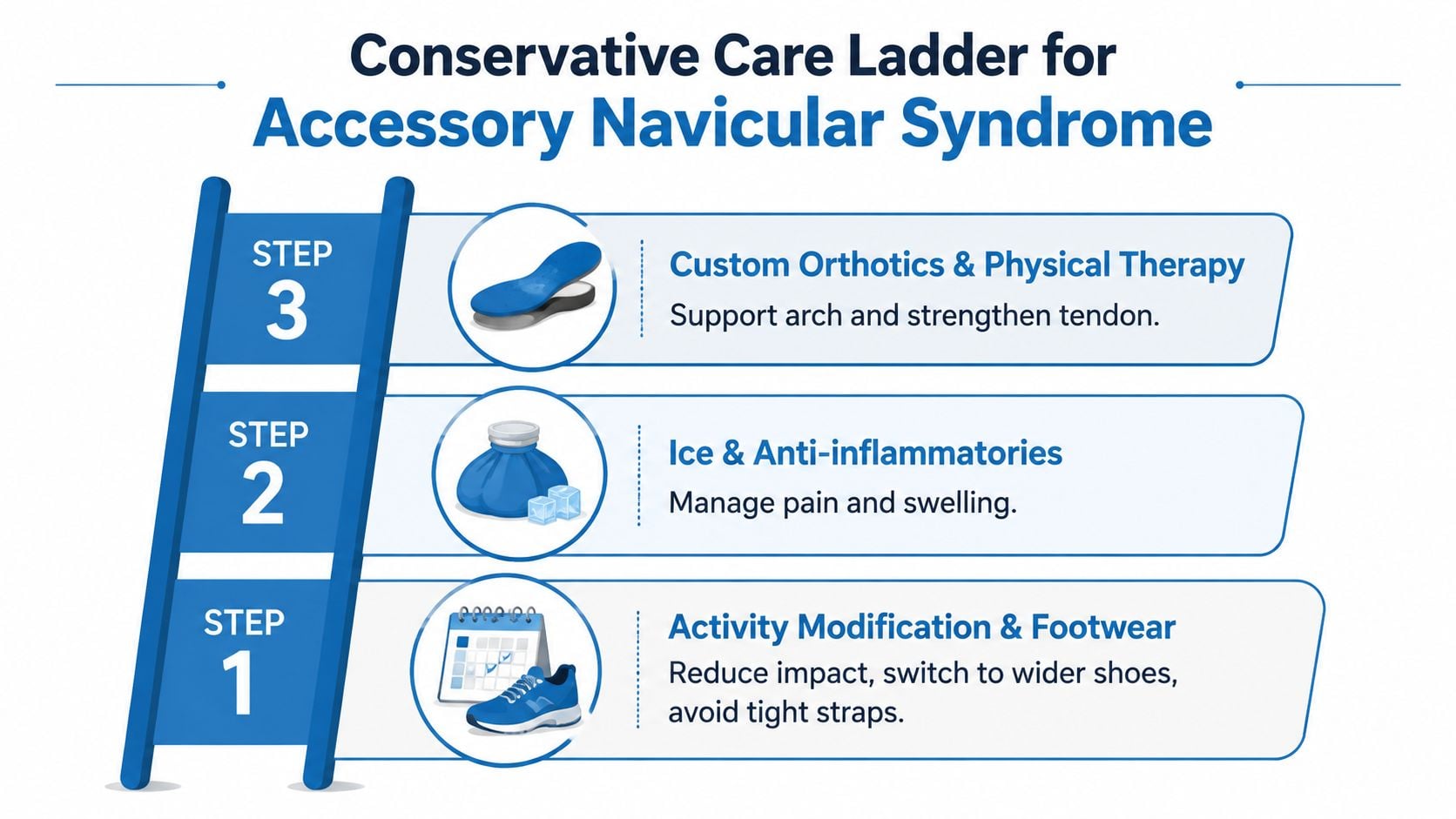
Non-surgical care should reduce the forces irritating the synchondrosis and help the posterior tibial tendon tolerate normal activity. The best plan usually progresses from simple pressure relief to more structured support, rather than starting with the most invasive option.
Start by removing the aggravating load
For a runner, that may mean temporarily replacing running with lower-impact conditioning. For a teenager, it may involve reducing jumping drills while maintaining activity that doesn't reproduce pain. Wider shoes, softer medial counters, and looser straps can prevent direct rubbing over the prominence.
Ice can help after activity when swelling or soreness is present. Pain medication may be appropriate for some patients, but medical history, kidney function, stomach health, allergies, and other medicines should guide that decision.
Add support when mechanics keep provoking symptoms
An off-the-shelf insert may provide enough arch support for mild symptoms. A custom orthotic can be shaped to support the arch while creating a relief area beneath the accessory navicular. The goal isn't to force the foot into a rigid position. It's to reduce excessive strain and make ordinary walking less provocative.
Physical therapy addresses the structures that must share the load. A program may include calf flexibility, progressive posterior tibial tendon loading, intrinsic foot strengthening, balance work, and gradual return to impact. A short period in a walking boot can be considered when ordinary steps remain painful despite activity and footwear changes.
A corticosteroid injection placed into the painful synchondrosis may provide short-term relief in selected cases, but it must be performed with an accurate diagnosis and appropriate guidance. Readers considering this option can review information about corticosteroid injections.
Conservative care options at a glance
| Option | Target | Typical Onset |
|---|---|---|
| Activity modification and footwear changes | Direct pressure and repetitive loading | Relief may begin as the irritant is removed |
| Ice and clinician-approved medication | Pain and post-activity swelling | Short-term symptom control |
| Orthotics | Arch support and pressure redistribution | Improvement develops as walking adapts to the insert |
| Physical therapy | Tendon capacity, calf flexibility, and foot control | Progress is gradual with consistent loading |
| Immobilization | Persistent pain during ordinary steps | Used for a protected healing interval selected by the clinician |
| Synchondrosis injection | Localized pain at the painful interface | Usually intended for short-term relief |
A treatment should be reconsidered when pain remains functionally limiting despite a consistent, properly directed trial. Continuing to increase activity through worsening pain can obscure whether the plan is helping.
Surgical Options and How Surgeons Choose
Surgery isn't a single solution for every painful accessory navicular. The operation should match the pain generator, the posterior tibial tendon's condition, the flexibility of the arch, and the patient's goals.
Bone-focused procedures
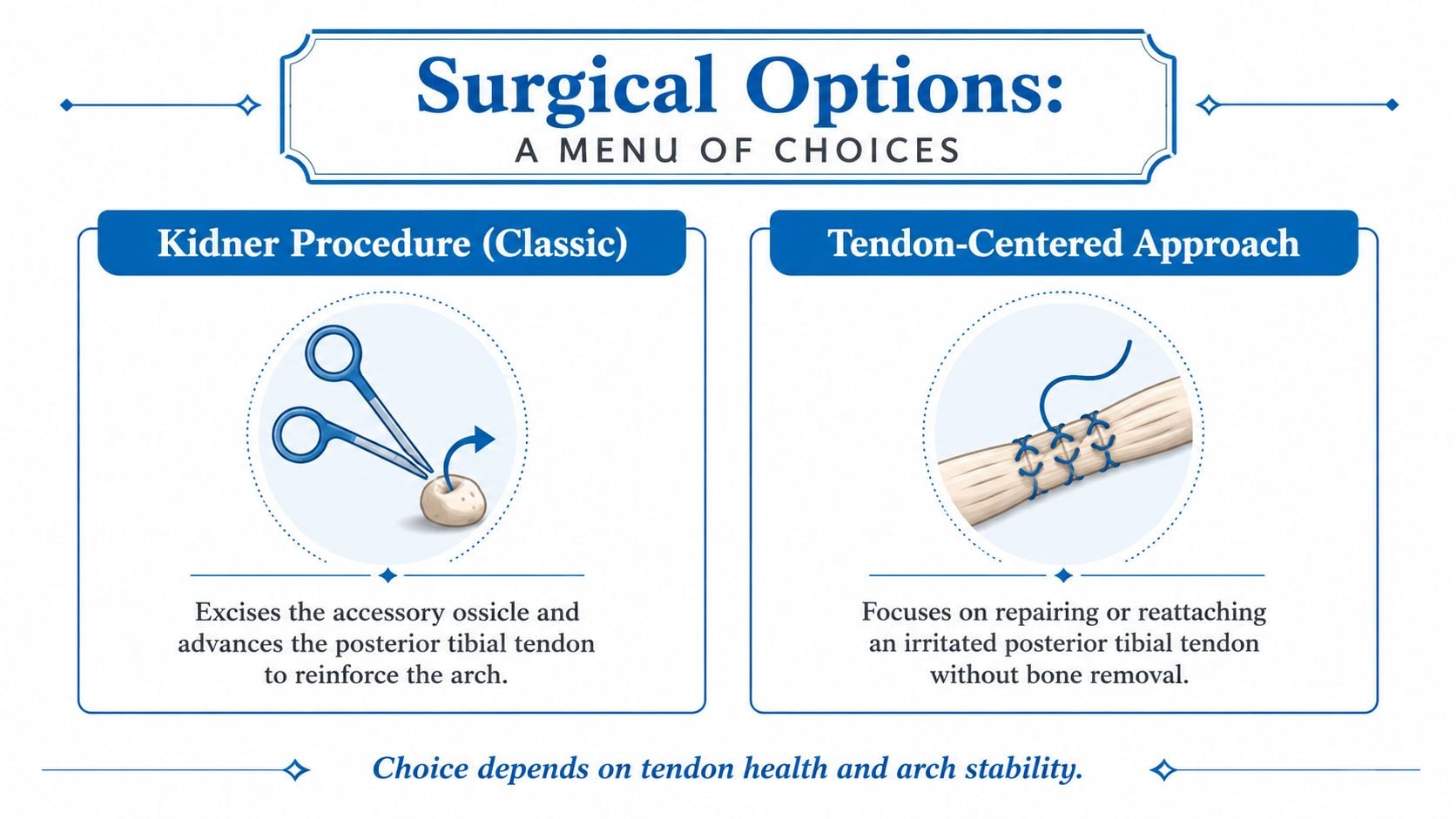
The classic Kidner procedure removes the accessory ossicle and repositions or advances the posterior tibial tendon onto the navicular. It can make sense when the extra bone is the primary pressure point and the tendon needs a more stable attachment.
Simple excision removes the painful fragment without the same tendon advancement concept. That may be considered in selected anatomy, but the surgeon must determine whether removing the fragment could weaken tendon function or leave an unstable insertion.
Fixation or fusion techniques preserve the accessory fragment and stabilize it against the navicular. These approaches may be considered when the synchondrosis is the dominant pain generator and the anatomy allows the fragment to contribute to the medial column.
Arch mechanics can change the operation
A flexible flatfoot may continue to overload the tendon after the accessory bone is removed. In that setting, a surgeon may discuss an additional procedure, such as a medializing heel osteotomy or tendon reconstruction, when examination and imaging show that the arch requires more support.
This is the central decision point: pain relief alone may not correct the mechanics that created the pain. A patient with a stable arch and healthy tendon may need a more limited operation, while a patient with tendon weakness or progressive arch collapse may need a broader reconstruction.
Recent review data found a mean surgical complication rate of 9.02% and a failure rate of 4.23%, with no major outcome differences between Kidner-type procedures and other surgical approaches (systematic review of surgical treatment). These figures support realistic counseling. Surgery often improves pain and function, but it carries meaningful risks and doesn't guarantee that every symptom will disappear.
The same literature emphasizes that no single operation has been established as the definitive choice for every patient. The surgeon's recommendation should therefore be based on tendon health, arch stability, imaging, activity requirements, and what the patient wants to return to doing.
Recovery, Outcomes, and When to Seek Specialty Care
Recovery depends on the operation, tendon involvement, arch mechanics, and the person's healing progress. Early care commonly includes protection from full loading, followed by a gradual transition toward walking and rehabilitation. Running, jumping, and sport should return only after strength, motion, balance, and pain have improved sufficiently for the treating clinician to approve progressive impact.
A practical recovery discussion should include more than a calendar. The patient should know what the foot can safely do at each stage, which symptoms are expected, and which findings require a review. Persistent swelling, increasing pain, wound concerns, or new weakness should not be dismissed as routine recovery.
Individuals with symptoms improve when treatment addresses the actual source of irritation. However, a meaningful minority need further evaluation because the tendon, synchondrosis, and arch mechanics weren't all addressed by the first treatment.
When specialty care makes sense
A foot and ankle evaluation is reasonable when:
- Pain persists: Symptoms continue despite a consistent trial of activity modification, footwear changes, support, and rehabilitation.
- The arch changes: The foot appears flatter, the heel turns outward, or walking mechanics progressively worsen.
- The tendon feels weak: A single-leg heel rise becomes difficult, painful, or impossible.
- The diagnosis remains uncertain: MRI or another imaging study may be needed to distinguish tendon disease, coalition, fracture, or nerve irritation.
- Daily function is restricted: Ordinary walking, work, exercise, or sleep remains affected.
Common questions
Can the bump be removed without surgery? The bone itself won't disappear through padding, inserts, or therapy. Non-surgical care can reduce pressure and inflammation, which may make the bump comfortable without removing it.
Does the condition return later? The congenital variant remains part of the anatomy, but treatment can reduce the painful irritation. Recurrent symptoms may reflect persistent flatfoot mechanics, tendon weakness, a new activity load, or an alternate diagnosis rather than the simple return of a removed bone.
Interventional Pain Management offers coordinated evaluation for persistent musculoskeletal pain, including orthopedic assessment, diagnostic review, image-guided procedures, and referrals when surgery or rehabilitation is appropriate. Patients with ongoing medial foot pain, tendon weakness, or uncertain imaging findings can visit Interventional Pain Management to request an evaluation and discuss a treatment plan customized to their symptoms and activity goals.