
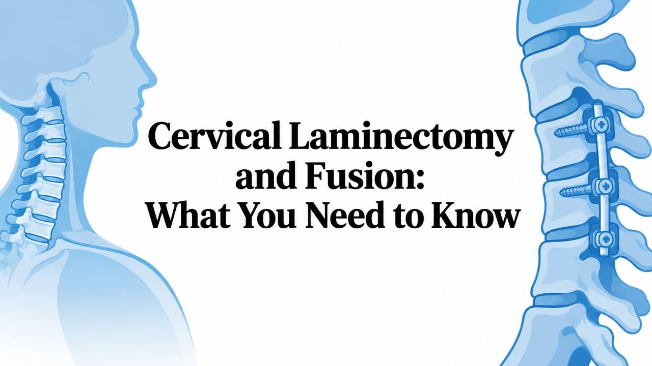
A coffee mug slips from a patient's hand for the third time in a week. At first, the clumsiness seems like carpal tunnel syndrome or simple fatigue. Then the patient notices a stiff, unsteady walk, trouble with buttons, and a tendency to misjudge steps. Those symptoms can reflect cervical myelopathy, a condition in which the spinal cord is compressed in the neck.
Cervical laminectomy and fusion is one operation used to relieve that pressure and stabilize the spine. It can improve function, but it isn't automatically the right choice for every person with cervical stenosis. The decision depends on the number of compressed levels, the spine's alignment, the source of compression, the patient's health, and the value placed on preserving neck motion.
Table of Contents
- What Cervical Laminectomy and Fusion Actually Does
- Who Is a Good Candidate for This Procedure
- How the Surgery Is Performed Step by Step
- How It Compares to Laminoplasty and Other Options
- Benefits and Risks You Should Weigh Honestly
- Recovery Timeline and Rehabilitation Milestones
- Practical Questions to Ask Before You Commit
What Cervical Laminectomy and Fusion Actually Does
The cervical spine contains the spinal cord, which carries signals between the brain and the rest of the body. Arthritis, bone spurs, thickened ligaments, disc degeneration, or ossification of the posterior longitudinal ligament can narrow the canal around that cord. When compression affects the cord, symptoms may include hand clumsiness, balance problems, weakness, stiffness, and changes in reflexes.
The word laminectomy refers to removing the lamina, the bony plate forming the back part of each vertebra. In simple terms, the surgeon removes the “roof” over the narrowed spinal canal. This creates more room behind the cord, allowing it to move away from the pressure when the spine's alignment is suitable.
Fusion adds a second goal. Screws and rods hold the treated vertebrae in a stable position while bone graft encourages the levels to heal together. A wide decompression without stabilization can allow the neck to gradually bend forward, a deformity called post-laminectomy kyphosis. A neurosurgical evidence review reported that cervical laminectomy with fusion improved functional outcomes in cervical spondylotic myelopathy and ossification of the posterior longitudinal ligament, without the late deformity associated with laminectomy alone in that review. The same review reported postoperative neurological improvement in roughly 70% to 95% of patients undergoing cervical decompression procedures, with fusion success rates of 89% to 99% for single-level fusions and 72% to 90% for dual-level fusions. The historical evidence review is available through PubMed.

How it differs from other decompressions
A laminoplasty keeps the lamina attached and opens it like a door, enlarging the canal while preserving more motion. An anterior cervical discectomy and fusion approaches the spine from the front, removing a disc or bone spur that is directly pressing backward against the cord or a nerve. A corpectomy removes part of a vertebral body when compression extends beyond a disc space.
The posterior fusion operation is particularly useful when several levels need decompression and the surgeon wants to protect alignment. It also means reduced motion across the fused segments, more hardware, and a recovery that may be more demanding than motion-preserving surgery.
The central question isn't simply whether the cord needs more room. It's whether decompression should be paired with stabilization for that particular neck.
Who Is a Good Candidate for This Procedure
The strongest candidate usually has multilevel cervical stenosis with clinical myelopathy, not just neck pain or an isolated pinched nerve. Typical clues include worsening hand dexterity, gait imbalance, increased reflexes, leg stiffness, or weakness that matches compression seen on MRI. The operation can decompress several levels from behind, making it useful when the narrowing is spread across the cervical spine.
Alignment often determines the recommendation more than symptom intensity alone. A neck with normal forward curvature, called lordosis, or a relatively straight alignment may allow the spinal cord to drift backward after posterior decompression. A neck with severe kyphosis bends the cord forward over the deformity, so a posterior-only operation may fail to address the mechanical problem. Flexion-extension X-rays can reveal abnormal movement or instability, while MRI can show the levels of compression and signal changes within the cord.
Patients seeking an evaluation for suspected spinal stenosis can review general condition information through spinal stenosis care guidance, but treatment still requires a physical examination and imaging review.
Candidate selection at a glance
| Factor | Favors Fusion | Argues Against |
|---|---|---|
| Number of levels | Multilevel compression requiring broad posterior decompression | One dominant level with a focal anterior lesion |
| Alignment | Preserved lordosis or a straight, stable cervical profile | Severe kyphosis or major forward bending |
| Symptoms | Gait imbalance, clumsy hands, weakness, or cord-related reflex changes explained by MRI | Symptoms that don't match the imaging |
| Stability | Existing instability or concern that decompression may weaken the posterior elements | Stable anatomy with low deformity risk |
| Imaging | Multilevel dorsal or circumferential narrowing | Compression best reached from the front |
| Medical fitness | Able to tolerate anesthesia, prone positioning, and fusion recovery | Active infection or medical conditions that make a longer operation unsafe |
An MRI cord signal change can indicate that the spinal cord has been under stress, although it doesn't predict recovery perfectly. The surgeon must also account for bone quality, medications, smoking status, balance, shoulder function, and the patient's ability to participate in rehabilitation.
A person with mild, stable symptoms may reasonably seek additional evaluation before committing to surgery. Progressive weakness, worsening balance, or declining hand function deserves faster reassessment because waiting can allow cord injury to become harder to reverse.
How the Surgery Is Performed Step by Step
The operation begins after anesthesia is administered and the patient is positioned face down. The head is secured in a Mayfield clamp, which helps prevent movement, and neuromonitoring leads are placed before the incision. The team checks the position carefully because the neck must remain protected while the surgeon works from behind.
A midline incision exposes the targeted cervical levels. The paraspinal muscles are gently moved aside to reveal the laminae. The surgeon confirms the operative levels with imaging before beginning bone removal.
Decompression and preparation
Using a high-speed drill and Kerrison rongeurs, the surgeon removes the lamina over the compressed levels. The thickened ligamentum flavum may also be removed if it contributes to narrowing. The aim is to take pressure off the dural sac and spinal cord without disturbing the cord itself.
The surgeon then prepares the lateral masses or, in selected anatomy, the pedicles for fixation. The joint surfaces and outer bone are decorticated, meaning the surface is lightly prepared to encourage graft healing. Local bone collected during decompression or prepared allograft may be placed along the lateral gutters.
Screws, rods, graft, and closure
Lateral mass or pedicle screws are inserted into the selected vertebrae. Rods connect the screws, creating a stabilizing framework that holds the cervical spine in the desired position. Details about the broader role of implants and fixation appear in spinal instrumentation information.
Bone graft rests beside the rods and screws, where it can gradually become a solid bridge between the treated levels. The surgeon checks alignment and hardware placement, then closes the muscle, fascia, and skin in layers. A drain may be left temporarily to reduce fluid collection beneath the incision.
The operation treats two linked problems: decompression gives the spinal cord room, while instrumentation and graft provide stability during healing. The hardware doesn't instantly create a biological fusion. Bone healing develops over time, which is why lifting limits and follow-up imaging matter after discharge.
How It Compares to Laminoplasty and Other Options
There isn't a universal winner among posterior cervical procedures. Laminoplasty preserves more motion because the lamina is hinged open rather than removed and fused. It can suit a patient with multilevel compression, acceptable alignment, and no major instability who values motion preservation. It has less ability to correct a kyphotic neck.
Laminectomy alone removes the posterior bony roof without placing fusion hardware. It may be reasonable in carefully selected patients with favorable alignment and low instability risk. The tradeoff is concern about later deformity or instability, especially when a broad decompression removes important posterior support.
A 2019 cohort illustrates how commonly surgeons use fusion in this setting. Among 3,796 patients, 2,397, or 63%, underwent cervical laminectomy and fusion, while 1,399, or 37%, underwent laminoplasty. The cohort data are reported in PubMed. A separate matched analysis found that patients who had laminectomy without immediate fusion later converted to fusion at rates of 3.2% at 2 years, 5.0% at 5 years, and 5.7% at 10 years, also reported in that source.
Comparing the practical tradeoffs
| Procedure | Alignment Correction | Motion Preserved | Typical Recovery | Reoperation Risk |
|---|---|---|---|---|
| Laminectomy and fusion | Stabilizes alignment and can limit further deformity | Less motion across fused levels | More involved because of fusion and posterior muscle healing | Hardware, nonunion, adjacent-level disease, or residual symptoms can require revision |
| Laminoplasty | Preserves the existing alignment but doesn't reliably correct kyphosis | More cervical motion is retained | Often less fusion-specific recovery burden | Later instability or deformity remains possible in unsuitable anatomy |
| Laminectomy alone | Doesn't provide structural correction | Motion is preserved initially | Usually avoids fusion-related restrictions | Later conversion to fusion is possible |
| Anterior discectomy and fusion or corpectomy | Can correct focal or segmental alignment problems | Motion is reduced at fused levels | Depends on the number of levels and anterior reconstruction | Further surgery may be needed for persistent, recurrent, or adjacent-level disease |
An anterior approach directly reaches ventral disc and bone compression. It can be a strong option for a focal lesion or significant kyphosis, but treating many levels from the front can require a more extensive reconstruction. Patients can explore laminoplasty treatment information when motion preservation is part of the discussion.
A 2024 meta-analysis found better mJOA improvement, less loss of cervical lordosis, and a lower complication rate with laminectomy and fusion than with laminectomy alone in the analyzed CSM studies. The meta-analysis results are available through PubMed. A separate systematic review reported similar neurological recovery, pain improvement, and complication rates between laminectomy alone and laminectomy with fusion, reinforcing that patient selection changes the answer.
Benefits and Risks You Should Weigh Honestly
The main benefit of cervical laminectomy and fusion is neurological protection and recovery, not guaranteed elimination of neck pain. Myelopathy surgery aims to relieve pressure on the spinal cord, halt deterioration, and give walking, balance, dexterity, and strength a chance to improve. Pain caused by separate facet, muscle, or shoulder conditions may not disappear because the canal is decompressed.
In a consecutive cohort of 58 patients with cervical spondylotic myelopathy, the mean mJOA score improved from 13.2 before surgery to 16.1 afterward, an average gain of 2.9 points. Eighty-five point five percent improved clinically, the neurological recovery rate was 56.6%, and the overall complication rate was 10.3%. These cohort findings are reported in PubMed.
The specific complication patients ask about
C5 palsy is a temporary or persistent weakness affecting the shoulder and deltoid region after cervical decompression. In that cohort, transient C5 palsy occurred in 6.9% of cases, and all reported cases resolved. More recent radiographic work found C5 palsy in 16.8% of 125 patients after cervical laminectomy with posterior instrumented fusion, showing that reported rates vary by study population and definition. The later radiographic findings are available through Jefferson's repository.
Other risks include infection, wound problems, bleeding, spinal fluid leakage, nerve or cord injury, blood clots, anesthesia complications, nonunion, screw or rod failure, and adjacent-segment degeneration. Hardware can cause local irritation, and persistent or recurrent symptoms can lead to revision surgery.
A realistic goal: Surgery may stop the decline and improve function, but it can't erase established spinal cord injury. Recovery often reaches a plateau, and some deficits may remain.
The long-term alignment question matters because postoperative neurological improvement has been associated with alignment measures after surgery in recent radiographic work. A 2024 review also reported that laminectomy and fusion preserved cervical lordosis better than laminectomy alone in its analysis. These findings support alignment as a clinical goal, not merely a measurement on an X-ray.
Recovery Timeline and Rehabilitation Milestones
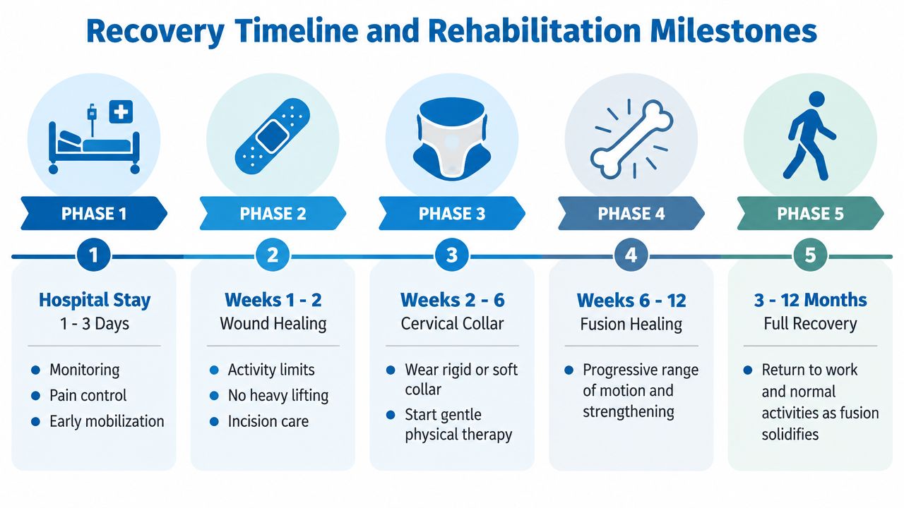
Recovery varies with age, medical health, the number of treated levels, preoperative weakness, balance impairment, and the extent of cord injury. Hospital care commonly focuses on neurological checks, pain control, wound observation, and safe movement. A person may walk soon after surgery, but early walking doesn't mean the fusion is complete.
Early healing
During the first couple of weeks, the priority is protecting the incision and avoiding activities that load the neck or pull on the posterior muscles. The surgical team may prescribe a rigid or soft collar, depending on the construct, bone quality, and surgeon preference. Short, frequent walks are usually more useful than prolonged bed rest, while heavy lifting, forceful bending, and twisting should wait for clearance.
Between the early healing phase and the next several weeks, therapy may begin gently. The exact plan differs, but common goals include:
- Shoulder blade control: Scapular stabilization can reduce compensatory shoulder and upper-back strain.
- Neck support: Deep neck flexor retraining builds controlled support without aggressive motion.
- Walking quality: Gait and balance exercises address the neurological effects of myelopathy.
- Daily independence: Therapy can rehearse transfers, dressing, stair use, and safe body mechanics.
Aggressive range-of-motion work too early can place unnecessary stress on healing tissues and instrumentation. Rehabilitation should follow the surgeon's restrictions rather than a generic exercise program.
Later progress and warning signs
Over the following months, strengthening and functional activity usually expand as follow-up examinations and imaging support progression. Neurological recovery can continue gradually, especially when balance and hand coordination were impaired before surgery. Driving and work decisions depend on neck control, medication use, reaction time, job demands, and the treating team's clearance.
Urgent contact with the surgical team is appropriate for new or worsening weakness, changes in swallowing or breathing, fever, increasing wound drainage, loss of coordination, or a new severe headache. These symptoms may signal a complication that needs prompt assessment rather than observation at home.
Practical Questions to Ask Before You Commit
A consultation should answer more than “Is surgery needed?” It should explain why this operation fits the patient's anatomy better than laminoplasty, laminectomy alone, or an anterior approach. The patient should leave knowing what the surgeon expects to improve, what may remain, and what could require another procedure.
Useful questions include:
- Anatomy: Which levels are compressed, and is the pressure mainly in front of or behind the spinal cord?
- Alignment: Does the neck have lordosis, straight alignment, instability, or kyphosis on standing and movement X-rays?
- Expected benefit: Is the primary goal to stop progression, improve walking, restore hand function, reduce pain, or accomplish several of these?
- Recovery: Will a collar be needed, what restrictions apply, and when can driving, work, and exercise resume?
- Complications: How does the surgeon counsel patients about C5 palsy, infection, nonunion, hardware failure, and residual myelopathy?
- Alternatives: Why is fusion preferable to laminoplasty or laminectomy alone in this specific case?
- Surgeon experience: How often does the surgeon perform posterior cervical fusions, and how are complications and revisions tracked?
A second opinion is especially reasonable when symptoms are mild, imaging and symptoms don't match, the neck is kyphotic, or another surgeon recommends a different operation. The delay should be discussed promptly if weakness or balance is worsening.
When waiting becomes less comfortable
New gait instability, rapidly worsening hand clumsiness, progressive weakness, or bowel and bladder changes warrant urgent medical reassessment. These signs don't automatically dictate one operation, but they make watchful waiting less reassuring because spinal cord dysfunction can progress.
A reasonable summary is straightforward. Cervical laminectomy and fusion fits many patients with multilevel myelopathy, suitable alignment, and a meaningful risk of postoperative instability. Laminoplasty may better serve someone who has appropriate alignment and wants to preserve motion. An anterior operation may be more logical for focal front-sided compression or kyphosis. Continued nonoperative care may remain appropriate when symptoms are mild, stable, and carefully monitored.
Interventional Pain Management evaluates neck and spine conditions through history, examination, and imaging, and its services include nonoperative interventions, orthopedic assessment, and spine surgery options when clinically indicated. Patients can visit Interventional Pain Management to request an evaluation and discuss whether cervical laminectomy and fusion or another treatment path fits their symptoms and anatomy.