
A 42-year-old office worker opens a patient portal after several weeks of back and leg pain. The MRI impression is brief, but one phrase stops the scrolling: “central disc extrusion at L4-L5.” The word extrusion can sound like a surgical emergency, especially when the report also mentions nerve compression or canal narrowing.
That reaction is understandable, but the MRI phrase is a description of disc shape and location, not a verdict about the future. A central disc extrusion can cause severe symptoms, mild symptoms, or no symptoms at all. The sensible next step is to connect the scan with the person's pain pattern, strength, sensation, reflexes, and daily function.
Many patients can begin with conservative care while clinicians monitor the neurological picture. The important questions are not only, “How large is the extrusion?” but also, “Which structures are affected, are symptoms progressing, and is function improving?” Translating the report into decisions, rather than anxiety, creates a clearer path forward.
Table of Contents
- When the MRI Report Says Central Disc Extrusion
- What a Central Disc Extrusion Actually Means
- Symptoms a Central Location Tends to Produce
- How Doctors Diagnose It on Imaging
- Conservative Care and What to Try First
- Injections and Surgical Options If Symptoms Persist
- Prognosis and the Resorption Question
- Questions Patients Ask After the Diagnosis
When the MRI Report Says Central Disc Extrusion
After reading “central disc extrusion,” a patient may picture a loose fragment pressing directly on the spinal cord and assume surgery is urgent. In the lumbar spine, the anatomy is more specific. A central fragment projects toward the middle of the spinal canal, where it may contact the thecal sac or the nerve roots passing downward. Depending on its size, direction, and relationship to those structures, it can cause symptoms in both legs, pain that alternates sides, or mainly back pain.
The term extrusion describes the shape of the disc material. It means material has moved beyond the usual disc boundary in a pattern that meets radiology criteria. The fragment may remain attached to the original disc by a narrow neck, or it may travel upward or downward within the canal. Radiologists describe morphology, such as bulge, protrusion, extrusion, or sequestration, separately from location, such as central, paracentral, foraminal, or extraforaminal. This distinction is outlined in this radiology overview of disc extrusion.
Why the scan and the symptoms can disagree
An MRI shows structure, not the full experience of pain. It cannot directly measure how irritated a nerve is, how recently a finding developed, or how much it limits a person's activities. Research also suggests that the relationship between MRI findings and symptoms is only moderate. Pure central protrusions or extrusions may cause few symptoms or none, while a centrolateral lesion that compromises a foramen may be more likely to produce clear nerve pain, as discussed in this review of MRI findings and symptoms.
The scan still matters, but it must be matched with the examination. Progressive weakness, new bowel or bladder changes, numbness in the saddle area, or rapidly worsening symptoms in both legs require prompt medical assessment. Stable pain with preserved strength often leaves room for activity modification, rehabilitation, and observation.
Practical rule: An alarming MRI phrase deserves a careful clinical review, not an automatic operation.
The plan usually follows the person's progress over several weeks. Pain may ease as inflammation and nerve irritation settle, and the fragment may gradually resorb. When function is improving, repeat imaging may not change management. If weakness progresses, or disabling symptoms continue despite appropriate care, an intervention becomes more reasonable. The word “extrusion” sets the anatomical question. The examination and the symptom trend help answer the treatment question.
What a Central Disc Extrusion Actually Means
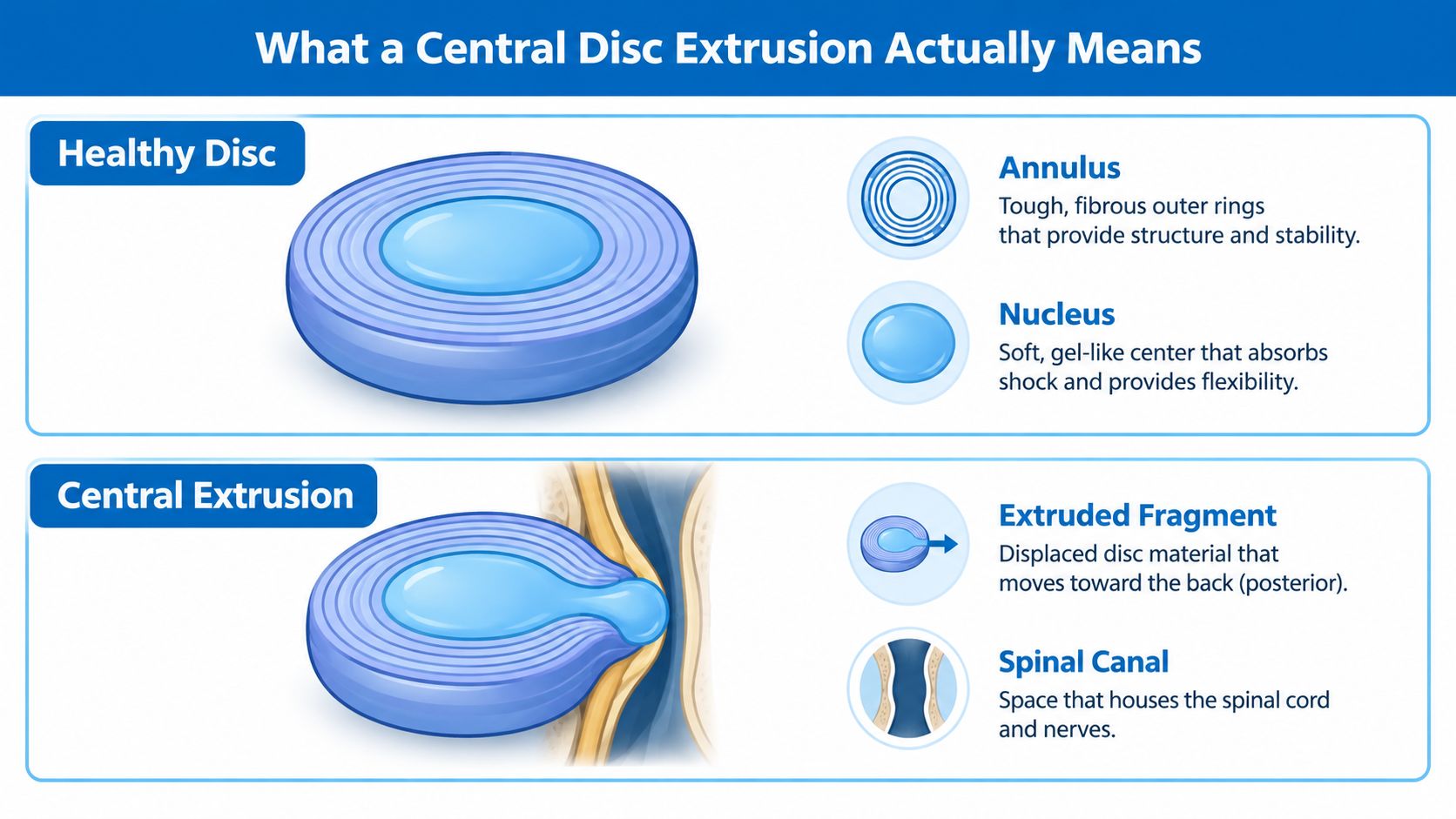
A lumbar disc sits between two vertebral bodies and helps distribute load during movement. Its outer layer, the annulus fibrosus, consists of strong, concentric collagen rings. Inside those rings is the nucleus pulposus, a softer, gelatinous center that helps the disc absorb and transfer forces.
A thin structure called the posterior longitudinal ligament runs along the back of the vertebral bodies and provides additional restraint behind the discs. The spinal canal lies just behind the disc, containing the thecal sac and nerve-root structures. This arrangement explains why a backward-moving fragment can matter even when the original problem began inside the disc.
The before-and-after picture
In a healthy disc, the nucleus remains contained by the annulus. With a bulge, the disc extends beyond its usual boundary over a broad area, while the outer fibers generally remain continuous. A protrusion is more focal, but the displaced material remains contained and its base is wider than the portion extending outward.
With a central disc extrusion, nucleus material passes through disrupted annular fibers and moves posteriorly toward the canal midline. By standard nomenclature, the displaced material is wider than its neck at the origin, or it has migrated cranially or caudally. Imaging descriptions commonly identify the fragment as larger than a protrusion and may show interruption of the posterior longitudinal ligament.
A sequestration is different again. It means a fragment has separated from the parent disc and is no longer connected by a neck. That distinction helps radiologists describe what they see, but it doesn't independently determine whether surgery is necessary. The location, neural compression, examination findings, and clinical course still carry greater practical weight.
Patients seeking a broader explanation of the terminology can review this guide to a herniated or bulging disc. The useful takeaway is simple: extrusion describes how disc material has displaced, while “central” describes where it has gone.
Symptoms a Central Location Tends to Produce
A central disc extrusion projects toward the center of the spinal canal rather than toward one side. That position can indent the thecal sac, the protective covering around the nerve-root bundle, and may affect both traversing roots. Symptoms therefore don't always follow the familiar one-sided pattern associated with classic sciatica.
Some patients have central low back pain with no leg symptoms. Others experience pain, tingling, or numbness in both legs, symptoms that alternate sides, or a diffuse aching and burning pattern that doesn't match one neat dermatome. A central lesion can also cause one-sided symptoms if the fragment is slightly off-center or if surrounding inflammation affects one root more than the other.
A paracentral extrusion sits beside the midline and more often contacts a particular traversing root. That pattern commonly produces recognizable unilateral radicular pain. A foraminal or far-lateral lesion reaches the opening where the exiting root travels, so the pain and weakness map can occur higher in the limb and may differ from the pattern produced by a central lesion. Patients can learn more about the broader relationship between nerve irritation and leg pain in this guide to sciatica and a pinched nerve.
Location changes the expected pattern
| Location | Structure compressed | Typical symptoms |
|---|---|---|
| Central | Thecal sac and potentially both traversing roots | Low back pain, bilateral or alternating leg symptoms, diffuse nerve pain, or no symptoms |
| Paracentral | Usually one traversing nerve root | More typical one-sided sciatica, numbness, or weakness |
| Foraminal | Exiting nerve root within the foramen | Focal radiating pain, sensory change, or weakness in the exiting-root distribution |
| Far-lateral | Exiting root outside or near the foramen | Higher, sharply localized radicular symptoms and possible motor deficits |
Two people can have nearly identical MRI wording and very different experiences. One may have pain mainly from chemical inflammation around a nerve, while another may have mechanical compression. A third may have the same visible extrusion but symptoms from a facet joint, muscle problem, hip condition, or another source.
When the pattern is urgent
Certain symptoms require immediate medical evaluation rather than routine follow-up:
- Saddle anesthesia: New numbness around the inner thighs, buttocks, or genital region.
- Bowel or bladder change: Difficulty starting urination, loss of control, or a new inability to sense normal bladder or bowel function.
- Bilateral weakness: Weakness in both legs, especially when it is worsening.
- Rapid progression: A noticeable decline in walking, foot control, or leg strength over a short period.
These features raise concern for severe central neural compromise, including possible cauda equina involvement. Pain alone can be intense without creating that emergency pattern, which is why the neurological examination matters so much.
How Doctors Diagnose It on Imaging
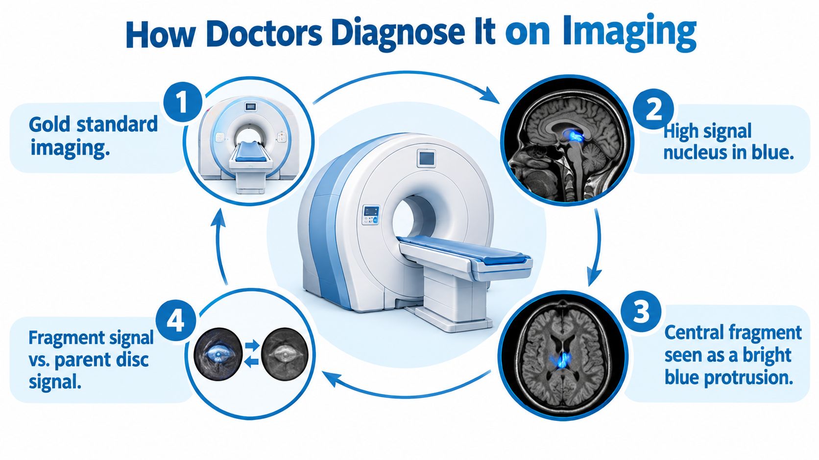
MRI is the preferred imaging test for a central disc extrusion because it shows the disc, thecal sac, nerve roots, and surrounding soft tissues in detail. Sagittal images show the spine from front to back and help identify whether a fragment has migrated upward or downward. Axial images show the canal from above and are especially important for confirming whether the fragment is central, paracentral, foraminal, or lateral.
T2-weighted sequences help distinguish fluid-rich structures and show the relationship between the fragment and the thecal sac. Radiologists also compare the fragment's signal with the parent disc and assess whether the thecal sac is indented or whether nerve roots are displaced. The report may describe the fragment's size, direction, level, migration, and degree of canal compromise.
When other scans enter the discussion
CT can show bone detail and may help when MRI isn't possible. A CT myelogram combines CT imaging with contrast around the thecal sac and can provide useful information in selected patients who cannot undergo MRI, although it is more invasive. The choice depends on the patient's medical history, implanted devices, prior operations, and the specific diagnostic question.
The scan also needs a clinical comparison. A fragment that touches a nerve root is more convincing when the patient has weakness, sensory loss, reflex change, or radiating pain in the matching distribution. A dramatic-looking report without matching symptoms may not explain the person's complaint.
Red flags that change the timetable
Urgent imaging or urgent assessment becomes more important with:
- Bilateral leg symptoms, particularly when they are new or worsening.
- Progressive weakness in the foot, ankle, knee, or hip.
- Saddle numbness or new bowel and bladder dysfunction.
- Recent trauma, especially when symptoms involve multiple root levels.
- Fever, systemic illness, or unexplained weight loss, which may point to causes beyond routine disc herniation.
Without those concerns, repeating MRI too soon often doesn't change management. A follow-up scan may add value when symptoms worsen, a new neurological deficit appears, or the clinical course fails to match the initial plan. Timing should be individualized rather than dictated by the MRI label alone.
Conservative Care and What to Try First
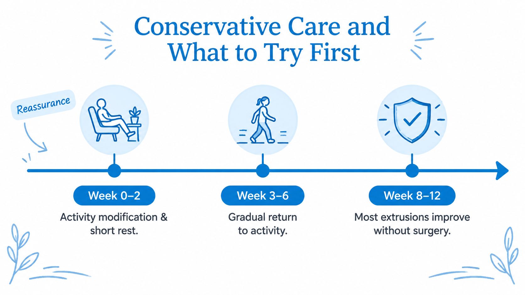
Most symptomatic lumbar disc herniations improve without surgery. A 2024 review describes 60% to 90% of patients responding to nonoperative treatment, and StatPearls notes that 85% to 90% of symptomatic cases resolve within 6 to 12 weeks without surgery, as summarized in this clinical review of lumbar disc herniation. Those figures describe broad lumbar disc-herniation populations, so an individual plan still depends on strength, sensation, pain severity, and function.
The first phase isn't complete bed rest. Short periods of rest can help during a severe flare, but prolonged inactivity often reduces conditioning and makes the return to movement harder. A practical early plan usually limits painful lifting, repeated bending, and prolonged sitting while preserving brief, comfortable walks and ordinary movement.
A staged approach
During the early flare, the priority is symptom control. Frequent position changes, supported sleep positions, and a temporary reduction in aggravating activity can be more useful than forcing stretches that reproduce leg pain. Medication decisions should be made with a clinician, particularly for people with kidney disease, stomach ulcers, blood thinners, liver disease, or other medical risks.
As symptoms settle, physical therapy can restore movement and confidence. Treatment may include lumbar stabilization, exercises matched to the patient's directional preference, gentle nerve gliding, and a gradual return to walking or swimming. Aggressive hamstring or lumbar stretching can aggravate an irritated nerve in some patients, so the therapist should use symptom response to guide progression.
If radicular pain remains dominant, a clinician may discuss anti-inflammatory medication or other pain-relieving options. Acetaminophen and nonsteroidal anti-inflammatory drugs may be considered when medically appropriate. Short courses of neuropathic-pain medication, such as gabapentin or duloxetine, may be discussed when nerve pain remains prominent, but these medicines require individualized prescribing and monitoring.
Everyday factors matter
Sleep quality, ergonomic seating, smoking cessation, and gradual weight management can support recovery and reduce mechanical strain. None of these measures can physically pull a fragment back into the disc overnight. They can, however, make rehabilitation more tolerable and help a patient return to normal activity without repeatedly provoking symptoms.
An image-guided epidural injection may be considered when leg pain prevents participation in therapy. It generally serves as a bridge to movement and rehabilitation, not as a guaranteed cure or a substitute for neurological monitoring.
Injections and Surgical Options If Symptoms Persist
Intervention becomes more reasonable when pain remains disabling after a genuine conservative trial, when a patient can't progress in rehabilitation, or when a neurological deficit persists or worsens. The MRI morphology helps identify a target, but the decision also depends on functional limitation and examination findings.
An epidural steroid injection can deliver medication near irritated nerve structures. A transforaminal approach follows the path toward a particular exiting or traversing root, while an interlaminar approach places medication through the posterior epidural space and may be useful when symptoms are broader or central. Relief can be temporary, partial, or absent. When it helps, the main value is often creating enough comfort for walking, sleep, and physical therapy.
Endoscopic discectomy may suit selected patients with an accessible fragment and persistent radicular symptoms. Microdiscectomy remains a common decompression procedure for ongoing nerve-root pain caused by a herniated fragment. Fusion is a different operation, generally considered when instability, recurrent herniation, or substantial degenerative structural change adds a reason to stabilize the motion segment rather than remove the fragment alone.
Matching the procedure to the problem
| Option | Best patient profile | Expected recovery | Key consideration |
|---|---|---|---|
| Epidural steroid injection | Persistent radicular pain limiting rehabilitation | Pain relief may support activity over a limited or variable period | It doesn't remove the extrusion |
| Endoscopic discectomy | Selected accessible fragments with persistent nerve symptoms | Often a minimally invasive recovery pathway | Anatomy and surgeon experience determine suitability |
| Microdiscectomy | Persistent, function-limiting radiculopathy with matching MRI findings | Recovery focuses on wound healing, walking, and gradual activity | The goal is nerve decompression, not removal of every degenerative change |
| Decompression with fusion | Herniation combined with instability, recurrence, or major degeneration | More extensive recovery than decompression alone | Fusion adds stabilization but also adds surgical scope |
In one study of large central extruded disc herniations, 67% of patients had successful nonoperative outcomes, supporting selective rather than automatic surgery, according to this PubMed clinical reference. The same reference describes an endoscopic interlaminar series involving 27 patients, with a mean operative time of 41 minutes, postoperative hospitalization of 5 days, and recurrence of 7.4%. Those figures apply to specific treatment groups and shouldn't be treated as a personal forecast.
The surgical question is usually not “Does the scan look large?” It is “Does the patient have a persistent, explainable problem that decompression is likely to improve?”
Patients preparing for an authorization request may also need stronger prior authorization documentation so the clinical findings, failed treatments, imaging, and functional limitations are presented clearly. A spine consultation should address expected benefits, risks, alternatives, and the possibility that pain from another source may remain after decompression. Information about microdiscectomy can help patients prepare for that discussion.
Prognosis and the Resorption Question
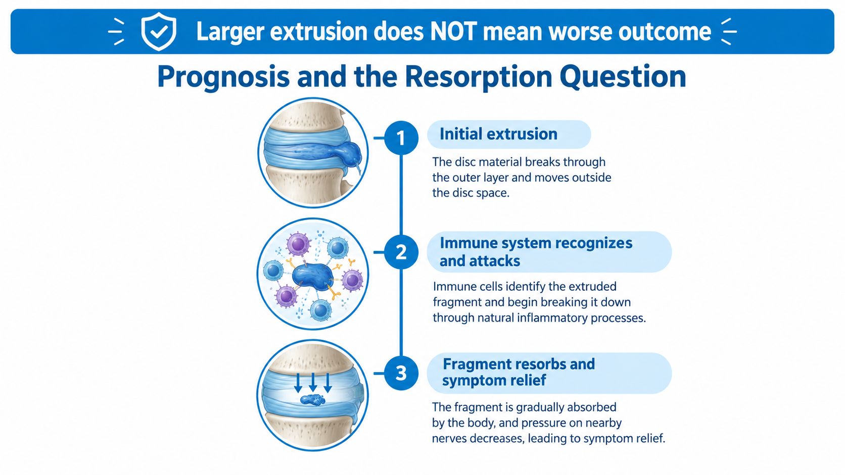
An alarming MRI does not always predict a poor outcome. A large extrusion may create a better biological opportunity for resorption than a smaller, contained bulge because displaced disc material sits outside its usual environment. The immune system can recognize it, and macrophages help break down and clear the fragment over time.
A 2024 systematic review reported an overall lumbar disc-herniation resorption incidence of 76.6%, with resorption often occurring during the first 6 months of conservative treatment. Ruptured herniations also showed higher resorption rates than contained lesions, according to this systematic review summary. A later review reported regression probabilities of approximately 70.4% for extrusions and 93.0% for sequestrations. These findings explain why a visibly displaced fragment is not automatically a worse prognostic sign.
Why symptoms often improve first
Pain can ease before an MRI shows clear shrinkage. Inflammation may settle, the nerve may become less reactive, and movement may improve while disc material remains visible. The reverse can also occur. A fragment may become smaller, yet pain may continue because the nerve endured prolonged irritation or another pain source is present.
The timeline is usually judged by function, not by the scan alone. Someone who is steadily walking farther, sleeping better, and regaining strength may not need repeat imaging to confirm that the fragment has disappeared. New or worsening weakness, persistent disabling radiculopathy, or a changed symptom pattern can justify renewed imaging and a different treatment plan.
A useful prognosis asks what the patient can do, rather than whether the MRI looks normal. A visible extrusion may become an incidental finding after symptoms resolve. A partially persistent fragment does not, by itself, show that conservative care has failed.
Questions Patients Ask After the Diagnosis
Does the extrusion definitely explain the pain?
No. The finding may explain the symptoms when the level, side, and compressed structure match the clinical pattern. It becomes less convincing when the pain distribution doesn't fit, the neurological examination is normal in a conflicting way, or another condition better explains the complaint.
A central extrusion can be asymptomatic, and even a visible contact with the thecal sac doesn't prove that every symptom comes from that fragment. Clinicians compare the MRI with the history, strength testing, sensation, reflexes, gait, and provocative maneuvers before deciding how much weight to give the report.
When should repeat MRI be considered?
Routine repeat scans aren't always useful when symptoms are steadily improving. New or progressive weakness, worsening bilateral symptoms, bowel or bladder dysfunction, saddle numbness, significant trauma, fever, or an unexplained decline in health should prompt urgent reassessment.
A repeat MRI may also be reasonable when disabling symptoms remain unchanged after an appropriate period of care and the result would affect a decision about injection or surgery. The purpose is to answer a management question, not to collect images for reassurance alone.
Should the patient stop all activity?
Usually, no. Complete inactivity can increase stiffness and deconditioning. Most patients benefit from gentle movement that doesn't intensify leg pain, with temporary limits on heavy lifting, repeated bending, twisting under load, and long uninterrupted periods of sitting.
A useful response rule is to judge the next day, not only the moment. If a walk or exercise produces a manageable sensation that settles promptly, the activity may be appropriate. If it causes escalating leg pain, new numbness, or loss of strength, the plan needs review.
Can the extrusion recur?
Recurrence is possible, especially when the disc remains structurally vulnerable, but an MRI alone can't predict an individual recurrence. Gradual conditioning, safe lifting mechanics, smoking cessation, and attention to repeated symptom triggers may support a durable return to activity. A recurrence of pain also doesn't automatically mean a new extrusion, since muscles, joints, and nerves can produce overlapping symptoms.
What should patients ask a surgeon?
A second opinion can be useful when surgery has been recommended, particularly if the symptoms are stable and the rationale isn't clear. Patients can ask:
- Which symptom is the operation expected to improve?
- Does the neurological examination match the MRI level?
- What happens if conservative care continues?
- What procedure is proposed, and why that procedure rather than an injection or decompression alone?
- Which risks, recovery limits, and recurrence concerns apply to this specific case?
- What finding would make surgery urgent rather than elective?
Those questions shift the conversation from the frightening word in the report to the actual decision. Progressive weakness, bowel or bladder dysfunction, and saddle anesthesia require immediate evaluation. Stable symptoms with preserved function often allow a thoughtful, monitored treatment course.
Interventional Pain Management evaluates central disc extrusion through the full clinical picture, combining examination, imaging review, medication planning, rehabilitation guidance, injections, and surgical referral when appropriate. Patients with persistent back or leg symptoms can visit Interventional Pain Management to request an evaluation and discuss a plan based on symptoms, neurological findings, and functional goals.