
A runner stops halfway through a downhill stretch and rubs the inside of the knee. Later that day, a fifty-year-old feels a sharp twinge stepping off a curb. Both people may describe the same problem, but the tissue causing it could be very different.
The location of the pain matters. So does the timing, the activity that triggered it, and whether the knee swells, catches, locks, or feels unstable. Understanding those clues can help explain the causes of medial knee pain before a clinical examination confirms the diagnosis.
Table of Contents
- Why the Inside of the Knee Hurts More Than People Expect
- The Medial Knee in Plain Language
- The Most Common Structural Causes of Medial Knee Pain
- Less Common Causes Worth Knowing About
- How Doctors Narrow Down the Diagnosis
- Treatment Options From Conservative Care to Surgery
- Red Flags, Prevention, and When to Seek Care
- Putting It All Together for Your Next Steps
Why the Inside of the Knee Hurts More Than People Expect
A runner feels pain at the inner knee after planting a foot and twisting. An older adult notices a gradual ache while walking. Both symptoms sit on the same side, yet the irritated tissue may be different. The key clues are where the pain is felt and how the knee was loaded.
The inner side is the medial compartment, where the thighbone and shinbone meet. It transfers force as the leg bends, straightens, rotates, and absorbs impact. Walking can place greater compression through this compartment, so cartilage wear, altered alignment, and repeated loading may produce symptoms there. Cleveland Clinic explains the loading and structural reasons behind inner-knee pain.
Location narrows the possibilities. Pain directly along the joint line, the crease between the bones, can involve the medial meniscus or the joint surfaces. A blow that pushes the knee inward may strain the medial collateral ligament, or MCL. A planted-foot twist can irritate or tear the meniscus. Pain a few finger widths below the joint line points more toward the pes anserine tendons and bursa, especially after repeated stairs, running, or squatting.
The timeline adds another layer. Sharp pain after a single twist suggests a different process from a gradually increasing ache and stiffness. These patterns can overlap, so the pain map and timeline work together rather than acting as a diagnosis on their own.
Why imaging can confuse the picture
An MRI can reveal a meniscal change without proving that it causes the pain. A major review reported that meniscal tears affect an estimated 12% of adults. In younger adults, they may follow acute twisting trauma. In older adults, they may develop gradually alongside osteoarthritis. The JAMA review explains the difference between traumatic and degenerative meniscal pathology.
The same review cited a community study in which 35% of people older than 50 had MRI evidence of a meniscal tear, while 61% of those tears caused no symptoms. An age-related scan finding may be present while cartilage loss or broader medial-compartment degeneration produces the discomfort. Imaging needs to match the examination, pain location, and loading history.
Practical rule: Ask “Where exactly does it hurt, and what was the knee doing when the pain began?” before asking what the scan shows.
The Medial Knee in Plain Language
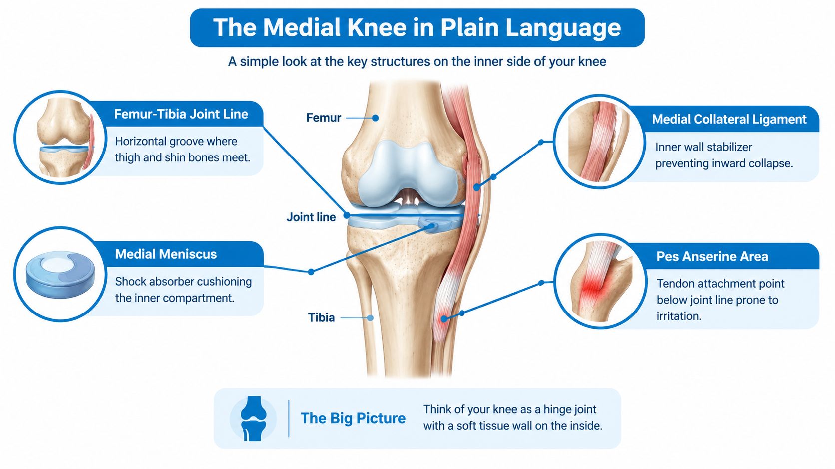
A simple mental model makes the anatomy easier. The knee works mainly like a hinge, with the femur above and tibia below. On the inner side, several structures form a protective and load-sharing wall.
Start with the joint line. Run a finger along the crease between the end of the thighbone and the top of the shinbone. This horizontal groove is where the medial meniscus sits between the bones. Pain directly on this line raises concern for the meniscus or the surfaces of the joint itself.
The MCL lies on the inner side of the knee and acts like a strong support strap. It limits excessive inward movement of the lower leg relative to the thigh. A force that pushes the knee inward, such as a direct impact or sudden change of direction, can strain this ligament.
Inside the joint, the medial meniscus forms a curved, cushion-like pad. It spreads load, improves the fit between the rounded femur and flatter tibia, and helps the knee manage rotation. A forceful twist can injure it, while gradual wear can weaken it over time.
The area below the joint line
Move a few finger widths below the joint line, toward the inner upper shin. That's where the pes anserine area is found. Three tendons join there and help control hip and knee movement. A small bursa can become irritated when the tendons repeatedly rub or carry more load than they can tolerate.
The joint also has smooth cartilage covering the contact surfaces of the femur and tibia. When that cartilage and the surrounding bone change, the medial compartment may become painful with weight-bearing and stiff after inactivity.
Alignment influences all of these tissues. A leg that directs more force through the inner compartment can increase compression there, while valgus stress or twisting can challenge the MCL and meniscus. This is why the same activity, such as running downhill, may provoke different structures in different people.
The Most Common Structural Causes of Medial Knee Pain
Four structural problems explain much of the discussion around inner-knee pain: medial compartment osteoarthritis, medial meniscus injury or degeneration, MCL injury, and pes anserine bursitis or tendinopathy. Begin with the pain's exact location. Pain at the joint line points toward structures inside the joint, while pain several finger widths below it points more toward the tendons and bursa on the upper shin. The pattern and loading trigger then narrow the possibilities, although no single symptom confirms a diagnosis.
| Cause | Pain Location | Typical Pattern | Common Trigger | Typical Profile |
|---|---|---|---|---|
| Medial compartment osteoarthritis | Along the inner joint line or across the medial knee | Gradual ache, stiffness, reduced mobility, possible grinding | Walking, stairs, prolonged standing | Adults with progressive joint degeneration or prior injury |
| Medial meniscus injury or degeneration | Directly at the medial joint line | Pain with rotation, possible catching, locking, or swelling | Twisting on a planted foot or repeated loading | Athletes after trauma, or adults with degenerative changes |
| MCL sprain or tear | Along the inner ligament, often above or around the joint line | Local tenderness, pain with sideways stress, possible instability | Direct impact or force pushing the knee inward | Active people involved in contact or cutting sports |
| Pes anserine bursitis or tendinopathy | Below the medial joint line on the upper shin | Localized ache and tenderness, worse with repeated bending | Running, stairs, squatting, repetitive loading | Runners and adults with increased medial soft-tissue load |
MCL pain follows a force
An MCL injury commonly follows a recognizable event. The outside of the knee may take a blow, or the foot may remain planted while the lower leg shifts outward. The resulting pain usually tracks along the inner ligament, rather than feeling deep inside the joint. During an examination, a clinician may apply controlled valgus stress, gently pushing the knee inward, to check for ligament pain or looseness.
A mild sprain can be painful without causing major instability. A larger tear may create a feeling that the knee gives way, especially during cutting or changing direction. Persistent instability deserves an examination instead of rest alone. For a related overview of torn meniscus symptoms and evaluation, remember that meniscal and MCL problems still require different physical tests.
Meniscal pain is usually joint-line pain
The medial meniscus becomes more likely when tenderness sits precisely in the crease between the thighbone and shinbone. Twisting, pivoting, or squatting may bring on the pain, and catching, locking, swelling, or pain with rotation adds support to that possibility. Degenerative meniscal changes can also develop gradually, without one clear injury, especially alongside osteoarthritis.
A scan can show a tear, but the examination must establish whether that finding matches the person's symptoms. Degenerative tears may cause no pain, so treatment should not be based on imaging alone.
Pes anserine symptoms sit lower
Pes anserine irritation is usually felt below the joint line, on the inner upper shin rather than directly in the knee crease. The spot may be tender when pressed. Repeated bending and straightening, climbing stairs, running, or standing from a chair can increase the ache.
This location helps separate pes anserine symptoms from meniscal pain, even though both occur on the inner side. People seeking broader, non-surgical background can browse osteoarthritis remedies, while keeping in mind that general information cannot identify which structure is painful in one individual.
Arthritis is a loading problem, not just an age label
Medial compartment osteoarthritis often builds gradually. Walking, stairs, and prolonged standing can produce an ache, while stiffness after rest, reduced walking tolerance, or grinding may develop as the joint changes. Previous injury can also alter how force travels through the knee.
The medial compartment works like the inner side of a hinge. If alignment or damaged joint surfaces send more body weight through that side, compression rises during loading. That helps explain why symptoms may appear with walking or downhill movement even without a new injury. To browse osteoarthritis remedies, use general guidance only as background, not as a substitute for locating the painful structure.
Less Common Causes Worth Knowing About
Not every inner-knee complaint is a routine sprain, meniscal issue, or arthritis flare. Some conditions create a familiar pain location but require a different response. The timeline often provides the first warning.
| Cause | Hallmark Features | Typical Patient | Key Red Flag |
|---|---|---|---|
| Medial tibial plateau stress fracture | Deep, progressively worsening pain with loading, sometimes pain at night | Runners, endurance athletes, or people with sudden training increases | Pain that persists at rest or makes weight-bearing difficult |
| Synovial plica syndrome | Anteromedial ache, snapping, or a rubbing sensation during repeated bending | Active people with repetitive flexion and extension | Persistent mechanical symptoms despite reducing activity |
| Referred hip pathology | Vague medial knee pain with little local tenderness or swelling | Teenagers with hip disorders or adults with hip degeneration or labral problems | Hip stiffness, altered walking, or inability to bear weight |
| Crystal or inflammatory arthritis | Swelling, warmth, marked tenderness, and restricted movement | Adults with inflammatory or crystal-related disease | Fever, redness, rapid swelling, or systemic illness |
A stress fracture can begin. The pain may first appear during running and then become easier to trigger as the bone becomes less tolerant of load. Night pain or discomfort that continues when the knee is unloaded deserves faster evaluation, especially in an endurance athlete or an adult with reduced bone strength.
A synovial plica is a fold of joint lining that can become irritated. It may produce anteromedial snapping or aching after repeated knee flexion. Because the pain is near the inner front of the knee, people can mistake it for a meniscal problem. The absence of major swelling doesn't make persistent snapping irrelevant.
The hip can be the hidden source
The hip and knee share nerve pathways, so hip disease can be felt near the knee. In teenagers, a slipped capital femoral epiphysis can present with poorly localized knee discomfort. In adults, hip osteoarthritis or a labral problem may produce knee symptoms even when the knee examination is relatively quiet.
A clinician will therefore assess hip movement when knee findings don't explain the complaint. A stiff or painful hip, a limp, or pain reproduced by hip rotation shifts the diagnostic focus upward.
Inflammation changes the urgency
Gout, pseudogout, rheumatoid arthritis, and septic arthritis can all cause knee pain, but the surrounding signs matter. A rapidly swollen, hot, red knee with severe pain and reduced motion isn't a condition to test with repeated workouts. Fever or a generally unwell feeling increases the need for prompt assessment.
For an overview of tendon and bursa problems, readers can review tendinitis and bursitis information, while recognizing that warmth, systemic symptoms, and rapid swelling require a clinician's judgment.
How Doctors Narrow Down the Diagnosis
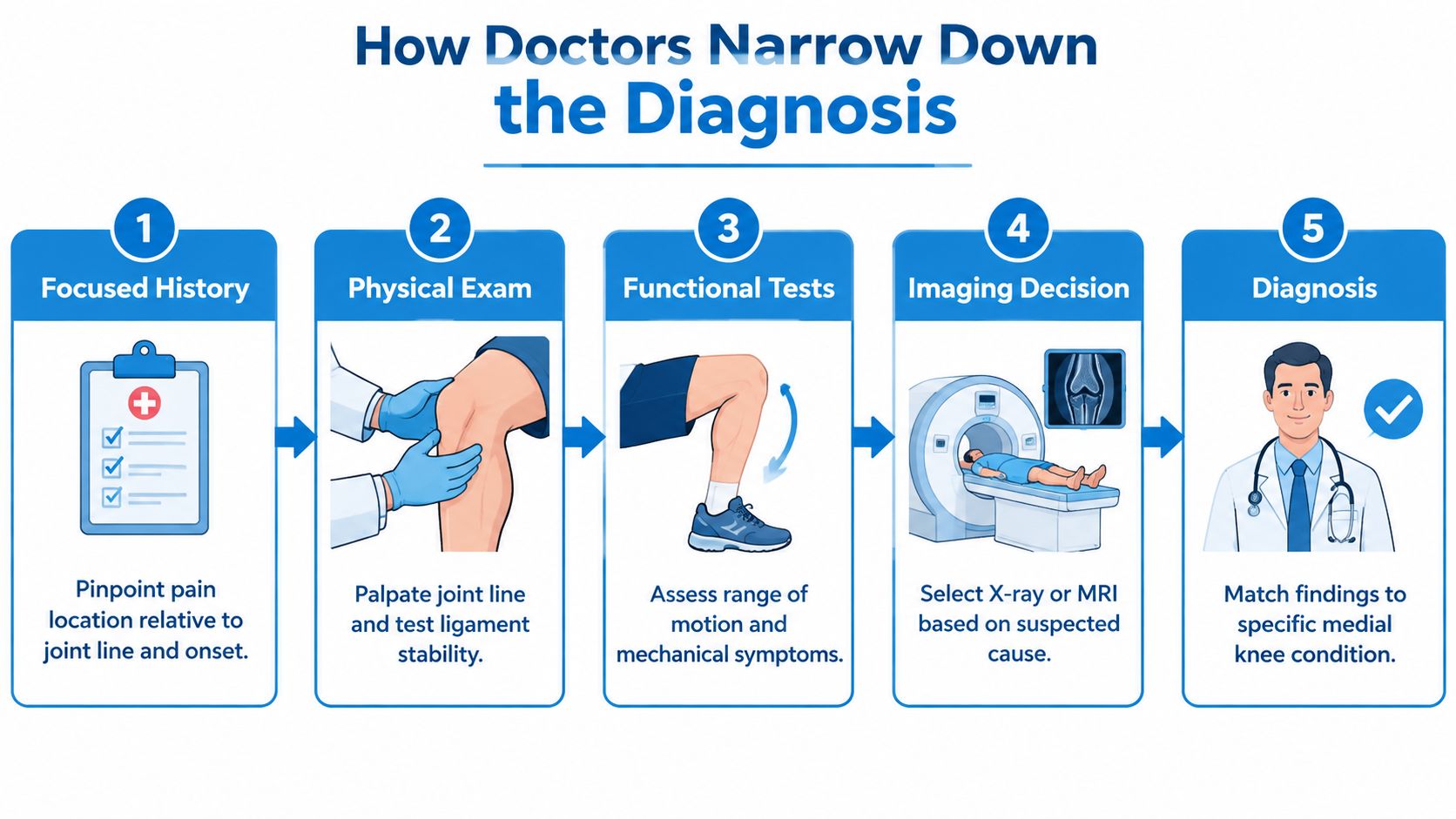
A sports-medicine clinician starts with the story, not the scan. The key questions include whether the pain sits on the joint line or below it, whether the onset followed a twist or impact, and whether swelling appeared immediately or later. Locking, catching, instability, night pain, recent training changes, and pain at rest all change the level of concern.
The examination then maps the symptoms to anatomy. The clinician palpates the joint line, traces the MCL, and checks the pes anserine region below the joint. Range of motion, walking mechanics, alignment, strength, and swelling provide additional clues.
What the manoeuvres test
Each physical test answers a specific question rather than proving a diagnosis by itself.
- Valgus stress test: Applies controlled inward stress to examine MCL pain or looseness.
- McMurray test: Combines knee movement and rotation to look for symptoms associated with meniscal injury.
- Apley test: Uses compression and rotation to help distinguish joint or meniscal pain from ligament discomfort.
- Thessaly test: Assesses symptoms during weight-bearing rotation, although a painful or unstable knee may make it inappropriate.
- Ober test: Examines lateral thigh and hip flexibility, which can influence lower-limb mechanics and symptom interpretation.
Imaging follows the clinical question. Weight-bearing X-rays can show joint-space loss, alignment, and bony changes associated with osteoarthritis. MRI gives more detail about the meniscus, MCL, cartilage, and other deep structures. Ultrasound can help examine superficial tendons and bursae, while bone-focused imaging may be considered when a stress fracture is suspected.
Laboratory testing can help separate inflammatory or infectious disease from a mechanical problem. In selected cases, a diagnostic injection may help identify whether pain comes from a particular joint or nearby structure, but it should support, not replace, the history and examination. For suspected ligament damage, MCL tear evaluation can help readers understand why stability testing matters.
Treatment Options From Conservative Care to Surgery
Treatment should match the structure involved and the way the knee is being loaded. A mild new problem may settle with relative rest, ice, short-term anti-inflammatory medication when medically appropriate, and temporary changes to running, stairs, or deep squats. A clinician or pharmacist should review medication safety, particularly for people with other medical conditions or existing prescriptions.
Footwear and training habits can also matter. A runner may need to reduce downhill mileage, vary surfaces, or adjust cadence and stride under professional guidance. A practical resource on selecting running shoes for medial knee pain can support that conversation, but footwear won't correct every structural cause.
Rehabilitation restores load tolerance
Physical therapy is often central because pain relief alone doesn't change how the knee handles force. A program may address quadriceps and hamstring strength, hip abductors, core control, balance, and gradual return to the movements that caused symptoms. MCL rehabilitation usually progresses loading carefully, while pes anserine care may emphasize hip and lower-limb mechanics.
Personalized gait retraining offers another way to address medial-compartment loading. Recent evidence reported that individualized foot-angle changes can reduce pain, lower knee loading, and might slow progression in medial compartment osteoarthritis. A Stanford summary from 2025 described pain relief comparable to medication in mild-to-moderate medial compartment osteoarthritis. The Stanford summary explains the gait-retraining approach and its potential role.
Braces, sleeves, and orthoses may support an injured MCL or reduce stress on a painful arthritic compartment. Their value depends on alignment, stability, fit, and the underlying diagnosis.
Injections and surgery have specific roles
Corticosteroid injections may provide short-term relief during an arthritis or bursitis flare. Hyaluronic acid, platelet-rich plasma, and aspiration may be considered in selected situations, but suitability and expected benefit vary. An acutely swollen joint may need aspiration and laboratory analysis rather than an injection designed only to reduce symptoms.
Surgery becomes more reasonable when a repairable meniscal injury causes persistent mechanical blockage, a severe MCL injury leaves ongoing instability, or focal arthritis remains disabling despite appropriate rehabilitation. Options can include meniscal repair or debridement, ligament reconstruction, osteotomy, or compartment-specific replacement. The decision depends on examination findings, imaging, activity goals, and the condition of the rest of the knee.
Interventional Pain Management evaluates musculoskeletal and joint conditions through medical assessment, imaging review, non-invasive care, image-guided procedures, and orthopedic referral when indicated. The appropriate treatment remains diagnosis-dependent.
Red Flags, Prevention, and When to Seek Care
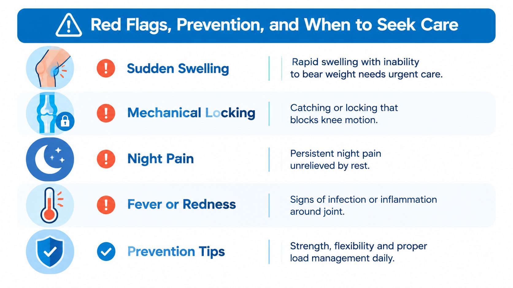
Most mild mechanical knee pain doesn't require emergency treatment, but certain patterns should change the plan. Sudden swelling combined with inability to bear weight may indicate a substantial injury. A knee that locks and blocks movement, rather than feeling briefly stiff, needs assessment.
Night pain that doesn't ease with rest deserves attention, particularly when it steadily worsens. Fever, warmth, redness, or rapid swelling can signal infection or inflammatory disease. High-energy trauma, visible deformity, or new neurological symptoms also justify urgent care.
Prevention is load management
The goal isn't to avoid movement. It's to help the knee tolerate movement without sudden spikes in demand.
- Progress gradually: Increase running, hills, speed work, and jumping in manageable stages rather than changing several demands at once.
- Build support: Strengthen the quadriceps, hamstrings, hip abductors, and core so the leg controls inward motion and rotation more effectively.
- Warm up deliberately: Begin with easy movement, then add activity-specific drills before faster running or cutting.
- Vary the workload: Cross-training can maintain conditioning while giving a repeatedly irritated medial compartment a different demand.
- Check equipment and surfaces: Well-fitted footwear and sensible surface variation may reduce avoidable stress, although neither replaces diagnosis.
- Keep mobility work consistent: Gentle post-run mobility can help maintain comfortable hip, thigh, and knee movement.
Pain that continues beyond two weeks, recurrent swelling, repeated giving way, or worsening limitation should prompt a clinical appointment. People should seek faster evaluation when pain appears at night, follows significant trauma, or prevents normal weight-bearing. Early assessment can prevent an active person from repeatedly loading the wrong structure.
Putting It All Together for Your Next Steps
A useful self-check begins with four questions:
- Where is the pain? Directly on the medial joint line suggests the meniscus or medial joint surfaces. Pain below that line points more toward the pes anserine area.
- How did it begin? A twist or sideways force raises concern for meniscal or MCL injury. A gradual activity-related ache fits better with overload or degeneration.
- What else is happening? Locking, catching, swelling, instability, warmth, fever, night pain, and inability to bear weight all change the urgency.
- What is the safest next step? Mild new pain may justify relative rest, ice, and reduced loading. Persistent mechanical symptoms call for examination and structured rehabilitation. Red flags require prompt medical care.
The scan matters, but it should match the pain map and physical findings. Accurate diagnosis helps prevent months of trying treatments aimed at the wrong tissue, while early conservative care often gives mechanical knee problems a better chance to settle.
Interventional Pain Management can evaluate medial knee pain through focused examination, imaging review, rehabilitation planning, and interventional or orthopedic care when clinically appropriate. Patients in New Jersey and Staten Island can visit Interventional Pain Management to request an evaluation and discuss the next step for persistent, unstable, or activity-limiting knee pain.