

You're sitting in a spine surgeon's office with an MRI showing a lumbar disc fragment pressing on a nerve. The consent form lists discectomy and microdiscectomy, and both sound like reasonable answers. The key question isn't which operation has the smaller incision. It's which operation gives the surgeon the safest, clearest route to fully decompress the affected nerve, while fitting the patient's anatomy, surgical history, and plans for the next month.
Both procedures remove herniated disc material. Both aim to relieve radiating leg pain, numbness, or weakness caused by nerve compression. The difference lies in how the surgeon reaches the spine, how much muscle and bone must be disturbed, and whether the smaller corridor provides adequate access.
Table of Contents
- When You Are Facing Herniated Disc Surgery
- How Each Procedure Actually Works
- Who Is a Candidate for Each Approach
- Outcomes and Complication Rates Compared
- Recovery Timelines and Return to Daily Life
- Cost, Setting, and Surgeon Experience Factors
- Making the Right Choice for Your Situation
When You Are Facing Herniated Disc Surgery
You have leg pain that limits sleep, work, or walking. Your MRI shows a lumbar disc fragment near a nerve, and the consent form lists discectomy and microdiscectomy. The right choice depends less on incision size than on the fragment's shape and location, the surgeon's training, the risk of another herniation, and what you need to do during the first month after surgery.
A discectomy, often called an open discectomy, uses a traditional posterior approach. The surgeon makes a midline incision, moves the paraspinal muscles aside, creates a small opening through the lamina when needed, and removes the disc material compressing the nerve root. The incision and bone work vary with the spinal level, disc position, body habitus, and amount of decompression required.
A microdiscectomy reaches the same target through a smaller working corridor. The surgeon uses an operating microscope or another magnification system, often with tubular retractors that separate muscle fibers rather than broadly detaching them. Magnification can improve the view of the nerve root and disc fragment, but it does not guarantee safer or more complete treatment in every case.
The shared objective matters more than the label. Neither procedure repairs the entire disc or prevents another herniation. Both remove the material currently irritating the nerve. Patients can review the condition and nonsurgical options in this guide to herniated and bulging discs, then discuss whether persistent symptoms justify decompression.
Why surgery enters the conversation
A meaningful trial of conservative care usually comes first. Treatment may include activity modification, medication, physical therapy, and image-guided injections. Surgery becomes more compelling when leg-dominant pain, numbness, or weakness persists and the MRI finding matches the physical examination.
The symptom and imaging match must be clear. An MRI can show disc abnormalities that are not causing pain. The surgeon should identify the affected nerve, explain why the fragment is responsible, and state what decompression can realistically improve.
Practical rule: Choose the operation that gives the surgeon complete nerve decompression with the least unnecessary tissue disruption. Do not choose based on the smallest skin opening alone.
Microdiscectomy became a widely used minimally invasive refinement of lumbar discectomy in the late twentieth century. Early technical development was credited to Yaşargil and Caspar in Europe, then popularized in the United States by Williams and Wilson. A 2002 review described more than 3,000 microdiscectomies, with good-to-excellent results in nearly 90% of patients. Most returned to work within one month. Reported rates for dural tears, discitis, or root injury were under 2%, with a 5% reoperation rate. The Journal of Neurosurgery review
That history supports microdiscectomy for a straightforward first-time lumbar herniation. Open discectomy remains sensible when the fragment's morphology, scar tissue, spinal stenosis, prior surgery, or the surgeon's training makes broader access safer and more reliable. The best choice should also match your recovery demands, especially what you expect to do 30 days after surgery.
How Each Procedure Actually Works
The two procedures share the same endpoint but create different access routes. Open discectomy prioritizes a broader field. Microdiscectomy prioritizes a narrower, magnified corridor.
Exposure and tissue handling
With open discectomy, the surgeon makes a midline incision and performs subperiosteal dissection, separating paraspinal muscle from the underlying bone. A partial laminectomy or hemilaminotomy may create the bony window. The surgeon then identifies the affected nerve root and extracts the offending fragment with instruments such as pituitary rongeurs.
Microdiscectomy uses a smaller skin incision and sequential dilation through the paraspinal muscles. Rather than broadly stripping muscle from bone, tubular retractors spread fibers along their natural planes. The microscope, or magnified loupes with a headlight, enlarges the operative field so the surgeon can work around the nerve root with a limited exposure.
That distinction can reduce muscle disruption, but it doesn't eliminate the need for judgment. A fragment that has migrated far upward or downward, sits behind bone, or is surrounded by scar tissue may not be easier to reach through a tube. Magnification helps only when the corridor points toward the target.
What the surgeon sees
Open surgery offers direct line-of-sight access across a wider field. This can help when the case involves a large sequestered fragment, bony overgrowth, stenosis, or anatomy altered by previous surgery.
Microdiscectomy provides a close, illuminated view of the nerve root and disc space. The surgeon can often remove the fragment while preserving more surrounding muscle and limiting bone removal. Yet a smaller corridor can become a disadvantage if the fragment lies in a blind spot or requires a wider decompression.
An experienced surgeon should change the approach when visibility or nerve safety demands it. The procedure name matters less than whether the surgeon can see the entire path of the compressed nerve.
| Variable | Open Discectomy | Microdiscectomy |
|---|---|---|
| Primary access | Wider midline exposure with muscle retraction | Smaller corridor with tubular dilation |
| Visualization | Direct view, sometimes with loupes | Operating microscope or magnified optics |
| Muscle handling | More broad retraction or detachment | Fiber-splitting approach designed to limit disruption |
| Bone removal | Partial laminectomy or hemilaminotomy as needed | Often limited bone removal, depending on the target |
| Best technical advantage | Instrument freedom and broad access | Precision through a smaller exposure |
| Main limitation | Greater soft-tissue disruption | Restricted working angle in complex anatomy |
For readers reviewing the procedure itself, the microdiscectomy explanation from Interventional Pain Management outlines how surgeons approach lumbar nerve compression. In practice, anesthesia and operating time depend on the patient and the surgeon's technique. Both procedures can be completed efficiently in experienced hands, and neither should be selected solely because a brochure promises a shorter operation.
Who Is a Candidate for Each Approach
The common candidate has a lumbar disc herniation that corresponds with a specific nerve-root problem. The usual pattern is radiating leg pain, numbness, or weakness that persists despite nonsurgical care, combined with imaging that shows compression at the same level and side.
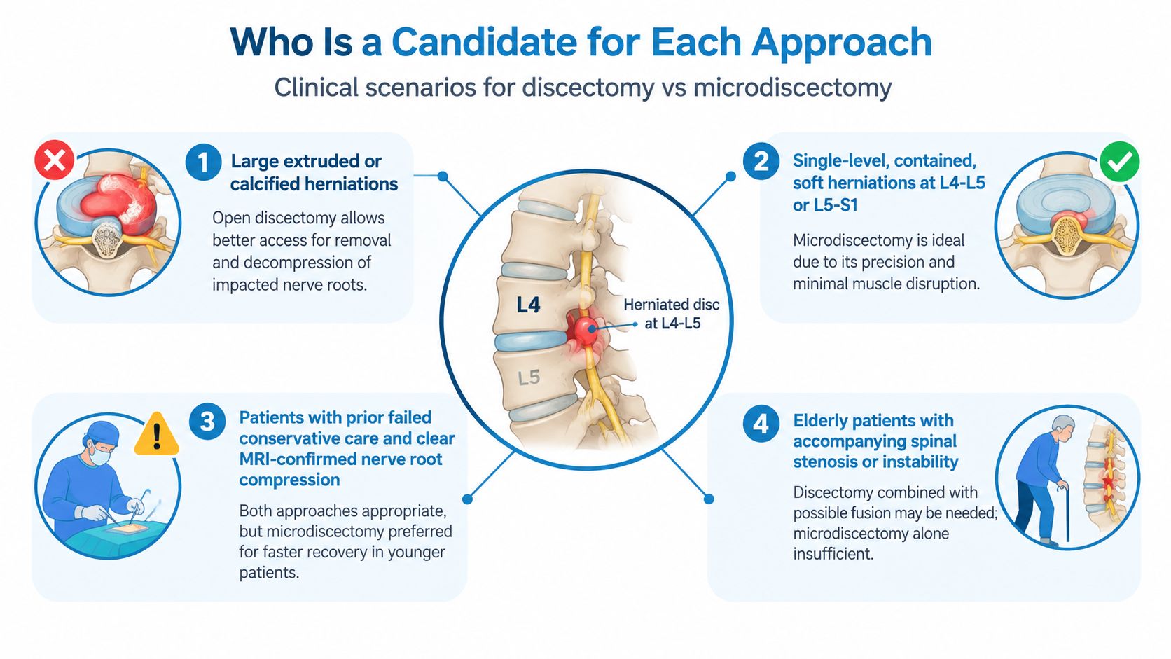
A contained or modest paracentral herniation in a patient undergoing first-time surgery is often well suited to microdiscectomy. The smaller corridor can preserve more paraspinal tissue while giving the surgeon enough access to remove the fragment. A patient whose job, caregiving responsibilities, or athletic routine makes early mobility especially important may reasonably favor that option when the anatomy is uncomplicated.
Situations that favor broader access
Open discectomy deserves serious consideration when the fragment has migrated substantially, is very large, or is calcified. A wider exposure can give the surgeon more freedom to work around bone and protect the nerve instead of forcing instruments through an awkward angle.
Recurrent herniation creates a different problem. Scar tissue can obscure normal planes and increase the difficulty of separating the nerve from surrounding structures. The question then becomes less about incision size and more about revision experience, visualization, and the surgeon's ability to enlarge the exposure safely.
Other factors can shift the balance:
- Concurrent stenosis: A patient who needs additional bony decompression may benefit from an approach that allows more complete work at the same operation.
- Multiple levels: Multilevel symptoms or compression may require an open or staged plan rather than a small single-level corridor.
- Body habitus: A deeper operative field can make tubular access more technically demanding, although body size alone shouldn't dictate the choice.
- Previous surgery: Prior work at the same level can distort anatomy for either technique and calls for a surgeon comfortable with revision cases.
Red flags change the priority
New bowel or bladder dysfunction, saddle numbness, or rapidly worsening weakness requires urgent medical assessment. Suspected cauda equina syndrome is an emergency, and the need for prompt decompression outweighs enthusiasm for a particular incision.
Surgeon training is a candidacy factor too. A high-volume microdiscectomy surgeon may offer a safer, more predictable result than a low-volume surgeon performing an open procedure, and the reverse can be true for a complex revision or migrated fragment. Patients should ask which technique the surgeon performs most often for the exact morphology shown on the MRI.
Outcomes and Complication Rates Compared
The evidence doesn't support the idea that one procedure wins every category. Microdiscectomy generally offers the advantage of a smaller exposure and may improve early recovery measures, while open discectomy remains effective when broader access is necessary.
A comparative analysis reported success rates of 86.8% for microdiscectomy versus 77.8% for open discectomy. The same analysis reported discitis in 0.4% of microdiscectomy patients versus 2.6% after open discectomy. A systematic review of microdiscectomy techniques found any-complication rates of about 12.5% for open approaches and 10.8% for percutaneous microdiscectomy. Comparative evidence in the PMC review
Those findings favor microdiscectomy in selected patients, but they shouldn't be read as a guarantee. Results depend on patient selection, disc location, surgeon skill, definitions of complications, and whether the operation achieved complete decompression.
Where microdiscectomy has a defensible edge
The practical advantages usually appear early:
- Less muscle disruption: A fiber-splitting corridor may reduce the local soft-tissue burden.
- Lower exposure-related morbidity: Smaller access can reduce wound and tissue problems in appropriate cases.
- Functional recovery: Patients may mobilize and resume routine activities sooner when postoperative muscle pain is limited.
- Comparable core decompression: The goal remains removal of the fragment, not preservation of an attractive scar.
A 2023 meta-analysis of 4 randomized controlled trials involving 523 patients compared tubular microdiscectomy with conventional microdiscectomy. Tubular surgery produced a statistically better Oswestry Disability Index result, but the analysis found no significant differences in operating time, blood loss, hospital stay, pain scores, reoperation, recurrence, dural tear, or overall complications. The 2023 meta-analysis
That result is useful because it separates functional improvement from marketing claims about every perioperative measure. A narrower approach may help function without producing a dramatic difference in every surgical variable.
| Metric | Open Discectomy | Microdiscectomy |
|---|---|---|
| Leg-pain relief | Effective when the nerve is fully decompressed | Effective when the nerve is fully decompressed |
| Early tissue recovery | Can be slower because of broader muscle handling | Often more favorable in suitable anatomy |
| Discitis in one comparative analysis | 2.6% | 0.4% |
| Any complication in a systematic review | About 12.5% | About 10.8% for percutaneous technique |
| Reoperation and recurrence | Must be discussed for the individual case | Not automatically eliminated by smaller access |
| Complex migrated or scarred fragment | May provide better working freedom | May require conversion or a broader corridor |
For a patient, the statistical difference translates into a simple question: does the smaller approach provide complete decompression without forcing the surgeon to work around an unsafe angle? If yes, microdiscectomy is usually the sensible starting point. If no, an open approach may offer the better risk-benefit balance.
Recovery Timelines and Return to Daily Life
A patient may wake after surgery with less leg pain but still struggle to sit, sleep, or move confidently. The recovery plan depends on nerve healing, preoperative weakness, job demands, pain control, and how much muscle was disturbed. Microdiscectomy often offers earlier comfort and mobility, while the difference may narrow as both approaches heal.
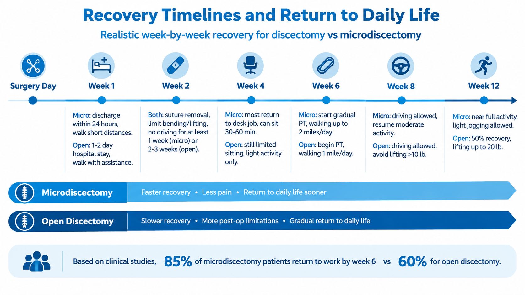
Once anesthesia has cleared, patients are generally expected to walk with assistance. A straightforward case may allow discharge the same day or after observation. More extensive surgery can require a longer stay. Follow the surgeon's restrictions, especially if the operation included additional decompression. A calendar cannot account for the actual work performed.
The first weeks
During the first week, short walks are usually encouraged. Bending, lifting, and twisting remain restricted. With less muscle disturbance, microdiscectomy patients often find it easier to change positions, sleep, shower independently, and reduce medication. Open-procedure patients may have more incisional and muscular soreness even when nerve pain has improved.
By about the second week, inspect the wound and increase walking gradually. Do not test strength with household lifting. Driving depends on medication use, reaction time, comfort while sitting, and the surgeon's approval. If work duties may change, review guidance on communicating with your employer after injury and document medical restrictions clearly.
Work and activity
Return to desk work when you can sit, stand, and walk without worsening symptoms. The 2002 review reported that most microdiscectomy patients returned to work within one month. That figure reflects the authors' reported experience, not a promise for every patient or occupation.
Heavy labor, repetitive bending, and unrestricted twisting need a slower progression. Physical therapy may start after the wound settles and the surgeon confirms that the nerve and spine can tolerate strengthening. Running and demanding sports should resume in stages, based on neurological recovery and movement control rather than impatience with the incision.
Your 30-day reality matters more than incision size. Ask which movements your job requires, how long you can sit, and whether the planned corridor supports complete decompression. Less leg pain does not always mean full recovery. Guarding, fatigue, and fear of reinjury can persist, so clear restrictions, follow-up, and gradual exposure to normal movement help rebuild confidence.
Cost, Setting, and Surgeon Experience Factors
The advertised procedure price rarely represents the complete financial decision. Total cost can include the surgeon, anesthesia, operating room, imaging, facility fee, pathology when applicable, medications, follow-up, and rehabilitation. An outpatient microdiscectomy may avoid an overnight charge, while an open procedure in a hospital may involve broader facility resources, but clinical necessity should drive the setting.
Insurance contracts can influence which site is proposed. Patients should request a written estimate and ask whether the surgeon, anesthesiologist, facility, and postoperative services are in network. A lower facility quote isn't a bargain if the approach cannot safely address the herniation or if an unexpected admission creates additional cost.
Surgeon experience outranks branding
A surgeon who routinely performs microscope-assisted decompressions on the relevant anatomy may be a stronger choice than a surgeon who performs the technique occasionally. Conversely, a surgeon with substantial revision and open-decompression experience may be better equipped for a migrated, calcified, recurrent, or stenotic case.
The consultation should address:
- Case-specific volume: How often does the surgeon perform the proposed operation for similar disc locations and morphologies?
- Conversion planning: If the smaller corridor doesn't expose the fragment adequately, will the surgeon enlarge the approach?
- Revision capability: How does prior surgery change the plan, particularly around scar tissue and the nerve root?
- Outcome tracking: Does the practice monitor reoperation, infection, dural tear, and patient-reported recovery?
Patients evaluating broader spine surgery services should also clarify whether the practice can coordinate nonsurgical care, injections, decompression, and more complex procedures when the diagnosis changes.
A practical decision matrix
| Decision factor | More favorable to open discectomy | More favorable to microdiscectomy |
|---|---|---|
| Anatomy | Large, migrated, calcified, or multilevel disease | Straightforward single-level fragment |
| Prior surgery | Revision with distorted planes | First-time operation |
| Facility | Hospital resources needed for added decompression | Efficient outpatient pathway when clinically suitable |
| Recovery priority | Complete access outweighs early muscle comfort | Early mobility and less tissue disruption matter |
| Surgeon expertise | Surgeon's strongest technique is open revision work | Surgeon routinely performs the indicated minimally invasive approach |
For patients dealing with an injury claim or work-related restrictions, a plain-language microdiscectomy guide for workers' compensation cases can help organize questions about treatment documentation and functional limitations. It shouldn't replace the surgeon's individualized recommendation.
Long-term spine health deserves a candid answer. Preserving paraspinal muscle and limiting unnecessary bone removal are logical goals, especially for younger patients, but the available comparison doesn't justify promising that microdiscectomy will prevent future degeneration or fusion. Complete decompression and appropriate treatment of the underlying problem come first.
Making the Right Choice for Your Situation
For a younger patient with a single-level, contained paracentral herniation, no prior surgery, and a clear nerve-root match on MRI, microdiscectomy is the recommended starting point when an experienced surgeon can safely access the fragment. The smaller corridor, reduced muscle disruption, and likely early mobility advantage fit a patient who needs to return to work, caregiving, or activity without sacrificing decompression.
Open discectomy deserves priority when the fragment is large, central, migrated, calcified, or accompanied by stenosis requiring broader bone work. Higher body mass, recurrent herniation, significant bony overgrowth, or altered anatomy from prior surgery can also make the wider approach more practical. A patient shouldn't accept “minimally invasive” as a substitute for a clear explanation of how the surgeon will reach the entire fragment.
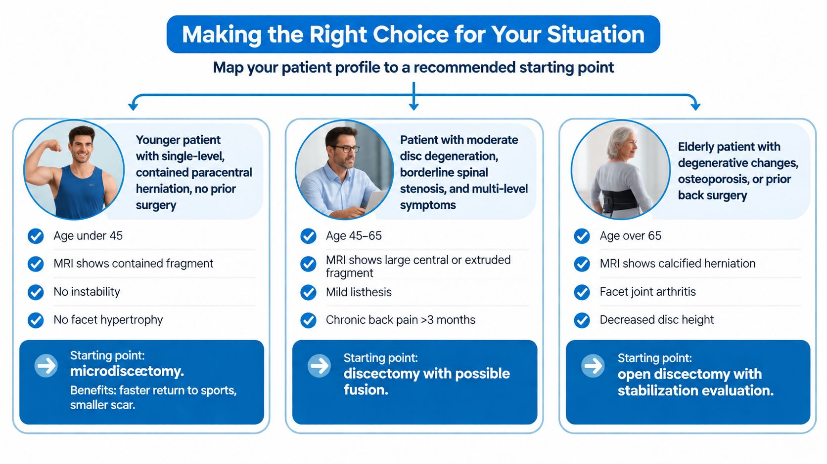
A recurrent herniation needs an experienced revision surgeon, regardless of the chosen label. Scar tissue changes the risk calculation, and a surgeon should explain whether a wider exposure, additional stabilization, or another treatment is necessary.
The consultation should produce direct answers to three questions:
- Which technique does the surgeon perform most often for this specific herniation morphology?
- What has the surgeon observed regarding reherniation and reoperation over the relevant follow-up period?
- How would recovery and complication management change if the initial corridor proved inadequate?
Interventional Pain Management evaluates herniated discs, radiculopathy, and spine conditions through coordinated pain-management, orthopedic, and spine-surgery services, including nonsurgical treatment and minimally invasive procedures. Patients comparing discectomy vs microdiscectomy can visit Interventional Pain Management to arrange an individualized review of symptoms, imaging, and treatment options before choosing an operation.