

You may feel pain just below the belt line, deep in one buttock, or across the back of the pelvis, yet still struggle to identify exactly where it starts. Sitting, standing, climbing stairs, or turning in bed can become difficult, and an MRI may not provide a clear answer. In that situation, a sacroiliac joint injection with fluoroscopy can serve two purposes: it may reduce pain, and it can test whether pain is coming from inside the SI joint.
The important distinction is that accurate needle placement doesn't equal a perfect diagnosis. Fluoroscopy helps the physician reach a difficult joint and confirm contrast spread, but it can't prove that the SI joint is the only pain generator. The most useful procedure combines careful patient selection, low-volume medication delivery, contrast confirmation, and a realistic plan for interpreting the response.
Table of Contents
- Understanding the Role of Fluoroscopy in SI Joint Pain
- The Clinical Evidence for Image Guided Accuracy
- Step by Step Walkthrough of the Procedure
- Diagnostic Limitations and Pain Generator Uncertainty
- Comparing Fluoroscopy to CT and Ultrasound Guidance
- Recovery Timeline and Post Procedure Care
- Evaluating Outcomes and Planning Next Steps
Understanding the Role of Fluoroscopy in SI Joint Pain
A patient with suspected SI joint pain often describes a broad, frustrating pattern rather than a pinpoint sensation. The discomfort may sit below the waist, spread into the buttock, or appear during weight-bearing movements. The SI joint lies deep within the pelvis and has an irregular shape, so surface landmarks alone don't reliably identify its joint space. Symptoms can also overlap with pain from the lumbar spine, hip, ligaments, and muscles around the pelvis. More background on this pattern appears in this guide to sacroiliac joint dysfunction.
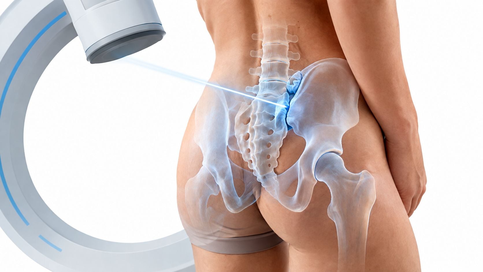
Fluoroscopy is real-time X-ray imaging. In the procedure room, the physician uses a C-arm to view the bony pelvis, adjust the angle, and guide the needle toward the target. The clinician isn't estimating the joint's position from the skin. The needle advances while its relationship to the sacrum and ilium remains visible.
Why the lower joint matters
The clinically useful target is generally the inferior synovial portion of the SI joint. This region is the part of the joint most suited to intra-articular access. Once the needle reaches the intended location, a small amount of contrast helps show whether the needle tip is inside the joint and whether the contrast follows the expected capsular pattern.
That confirmation changes the meaning of the injection. If a diagnostic anesthetic produces relief, the physician can interpret that response knowing where the medication went. If the needle position was only estimated, a lack of relief could reflect a missed joint rather than evidence against the SI joint as a pain source.
Practical rule: Fluoroscopy improves the technical question, “Did the needle reach the joint?” It doesn't answer the separate clinical question, “Is this joint responsible for all of the pain?”
The procedure therefore works best as one part of a broader evaluation. History, physical examination, competing diagnoses, and the patient's functional response all remain important.
The Clinical Evidence for Image Guided Accuracy
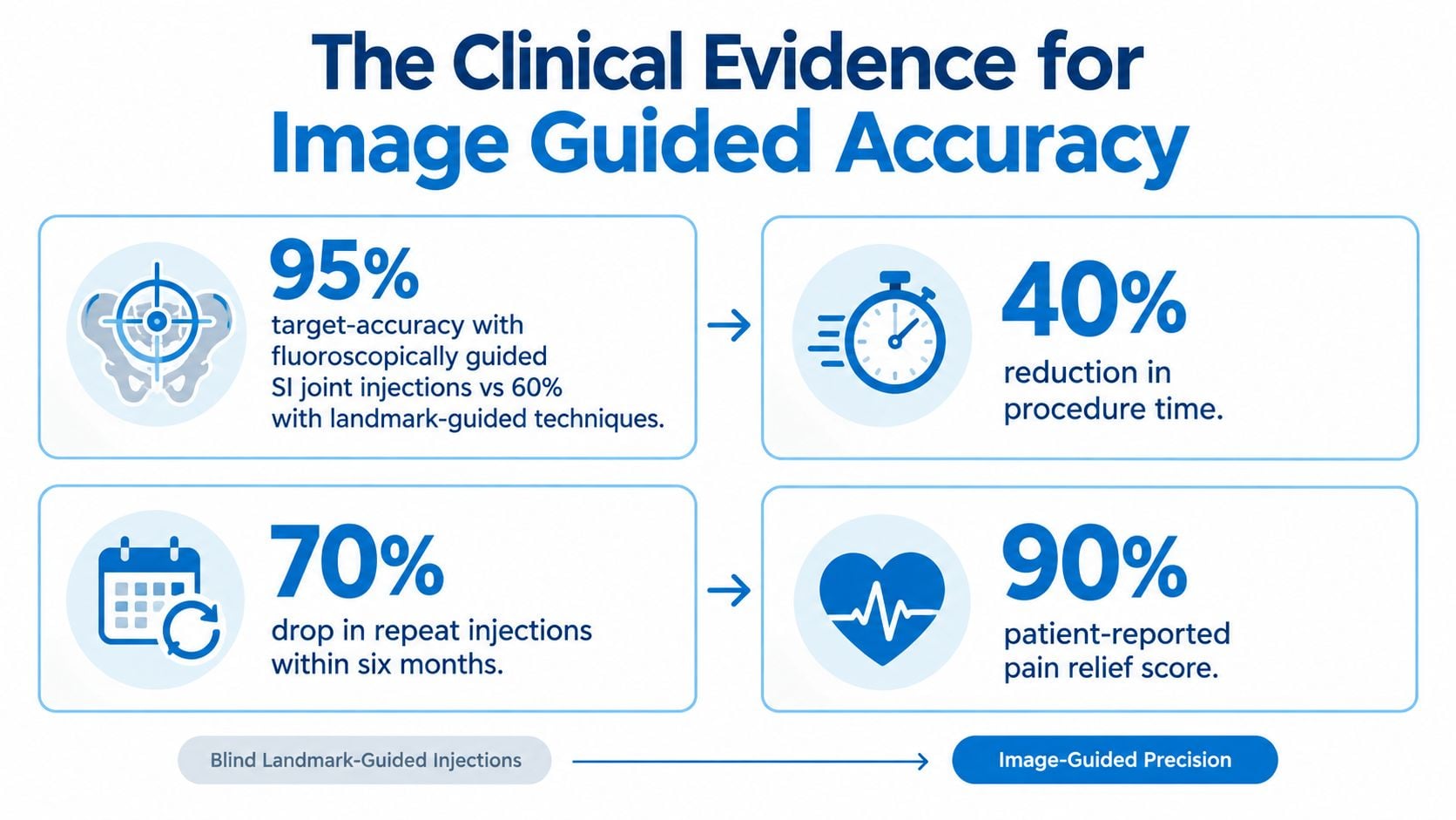
Fluoroscopy became central to SI joint injection practice because unguided needle placement is difficult to verify. An early Radiology report found that 30 of 31 injections were intra-articular, a 97% success rate, under fluoroscopic guidance. The authors described the technique as safe, rapid, and reproducible, supporting image confirmation for this anatomically difficult joint (historical fluoroscopic accuracy findings).
Unguided placement was accurate in only 12% of patients in comparative evidence, even when performed by experienced clinicians (evidence comparing guided and unguided SI injection placement). That limitation affects both treatment and diagnosis. Medication may miss the intended tissue, while a diagnostic block may appear negative because the anesthetic never reached the joint.

Accuracy isn't the same as guaranteed benefit
Technical precision improves the reliability of the injection, but it cannot guarantee lasting pain relief. In a later cadaveric experiment, dissection confirmed intra-articular placement in 10 of 20 fluoroscopy-guided injections, or 50%. Fluoroscopy performed significantly better than ultrasound in that experiment, yet the finding also shows that anatomy, needle position, and confirmation technique can still limit accuracy.
The practical implication is to use fluoroscopy with a small injectate volume and contrast confirmation. A low-volume injection limits spread into nearby structures, while contrast shows whether the needle is within the intended joint space. Without that confirmation, pain relief or lack of relief becomes harder to interpret.
Clinical outcomes require the same caution. A 2019 comparative trial found no significant difference between fluoroscopy-guided and landmark-guided blocks on its primary pain outcome. Positive blocks occurred in 61% versus 62%, and one-month categorical outcomes were 48% versus 40%, respectively. The reported intermediate-term advantage from fluoroscopy was modest and came with greater cost.
These findings separate two clinical questions:
- Placement accuracy: Fluoroscopy helps the physician target the joint and verify contrast distribution.
- Pain outcome: Relief depends on whether the joint is painful, whether extra-articular structures also contribute, and how the condition responds to treatment.
A well-performed injection makes the diagnostic result more trustworthy. It does not prove that the SI joint is the sole pain generator, nor does it promise improvement for every patient. History, examination, competing diagnoses, and functional response still determine what the result means.
Step by Step Walkthrough of the Procedure
A fluoroscopically guided SI injection is usually performed with the patient awake. The team reviews medications, allergies, relevant medical conditions, and the purpose of the injection before positioning the patient on the procedure table. The exact preparation can vary, particularly when blood-thinning medication, infection risk, pregnancy, or contrast concerns require a modified plan.
Before the needle enters
The patient lies face down so the physician can access the back of the pelvis. The skin is cleaned with antiseptic solution, sterile drapes are placed, and the injection site is identified using fluoroscopic views. A local anesthetic numbs the skin and deeper tissues. Patients may feel a brief sting from the numbing medicine, followed by pressure as the needle advances.
The physician then adjusts the C-arm to identify the lower synovial portion of the SI joint. Because the joint is narrow and its orientation varies, the imaging angle matters. A careful view helps distinguish the intended joint line from nearby bone and soft tissue.
Confirming the target
After the needle reaches the intended location, the physician injects contrast. This isn't decorative or optional imaging. The contrast pattern provides a visual check that the needle lies within the joint and that the injectate is spreading in the expected space.
The medication is then delivered in a deliberately small volume. Consensus guidance recommends a total volume under 2.5 mL, including contrast, because the inferior joint's capacity is less than 2.7 mL, and capsular distention can occur at volumes at or below 1.6 mL (consensus guidance on SI joint access and injectate volume).
Why low volume matters
A small joint doesn't need a large amount of fluid to produce pressure. Excessive volume can distend the capsule or spread beyond the intended target, reducing the specificity of a diagnostic block. Low-volume delivery is therefore a precision choice, not just a way to use less medication.
After the injection, the needle is withdrawn, the site is covered, and the patient is monitored according to the clinic's protocol. The procedure's value comes from the sequence: identify the joint, advance under live imaging, confirm with contrast, and inject only what the joint can reasonably accommodate.
Diagnostic Limitations and Pain Generator Uncertainty
A fluoroscope can show where the needle is. It can't show whether the SI joint is the only structure creating pain. This distinction matters because many patients understandably interpret immediate anesthetic relief as definitive proof that every symptom comes from the joint.
SI pain may arise from structures outside the joint, including surrounding ligaments and other pelvic tissues. Recent guidance emphasizes that extra-articular SI pain may be as common as intra-articular pain, so an injection into the joint doesn't test every possible SI-region pain generator (guidance on diagnostic uncertainty in SI joint pain).
What a diagnostic response can tell clinicians
A meaningful response during the anesthetic window can support the conclusion that the injected joint contributes to the patient's symptoms. The clinician still needs to compare the response with the patient's familiar pain, movement tolerance, and other examination findings. Relief that affects only one component of a complex pain pattern may indicate a partial contributor rather than a complete explanation.
Clinical examination and imaging have limitations here. Provocative maneuvers can reproduce familiar pain, but they have false-positive and false-negative limitations. Imaging can show structural changes without proving that those changes cause current symptoms. The block adds another piece of information, but it doesn't eliminate uncertainty.
Accurate placement makes a diagnostic block more interpretable, not infallible.
Recent guidance also notes that it remains unclear whether image-guided intra-articular local anesthetic injections reliably predict who will respond to later therapeutic injections. A patient can experience short-lived anesthetic relief without obtaining sustained benefit from anti-inflammatory medication. Conversely, an initially quiet response may reflect an incomplete pain map, technical factors, or a different dominant pain generator.
The responsible interpretation is therefore neither “the SI joint is definitely the cause” nor “the injection failed, so nothing can be learned.” The result should be integrated with the full clinical picture.
Comparing Fluoroscopy to CT and Ultrasound Guidance
Patients often ask why fluoroscopy is selected when CT and ultrasound also provide imaging. The answer depends on the clinical setting. Each modality balances visualization, radiation, access, equipment, and the physician's ability to guide the needle during the procedure.
A 2026 systematic review reported needle-placement accuracy ranges of 65% to 92% for fluoroscopy, 76% to 100% for CT, and 40% to 96% for ultrasound (systematic review comparing CT, ultrasound, and fluoroscopy guidance). These broad ranges reflect differences among studies, techniques, patient anatomy, and definitions of accurate placement.
| Modality | Accuracy range | Radiation exposure | Best use case |
|---|---|---|---|
| Fluoroscopy | 65% to 92% | Uses radiation | Routine image-guided access with live needle visualization and contrast confirmation |
| CT | 76% to 100% | Uses radiation | Selected cases where cross-sectional anatomical detail is especially valuable |
| Ultrasound | 40% to 96% | No radiation | Selected patients when radiation or contrast is a concern, including pregnancy or documented contrast allergy |
Why fluoroscopy remains common
Fluoroscopy provides live imaging while the needle advances, and it supports contrast confirmation during the same procedure. It is also broadly available in interventional procedure settings. That combination makes it a practical balance for many patients, even though CT may offer greater precision in selected anatomical circumstances.
CT can display anatomy in cross-section and may help when landmarks are unusually complex. Its limitations include radiation exposure and less convenient real-time procedural workflow in some settings. Ultrasound avoids ionizing radiation, which is an important advantage, but the SI joint's depth and irregular anatomy can make visualization and needle confirmation more variable.
Coverage policies generally require CT or fluoroscopy with contrast for most SI injections, while allowing ultrasound in selected situations such as pregnancy or documented contrast allergy. The choice should therefore be individualized. The best modality is the one that provides adequate target confirmation while respecting the patient's medical circumstances and the procedure's diagnostic purpose.
Recovery Timeline and Post Procedure Care
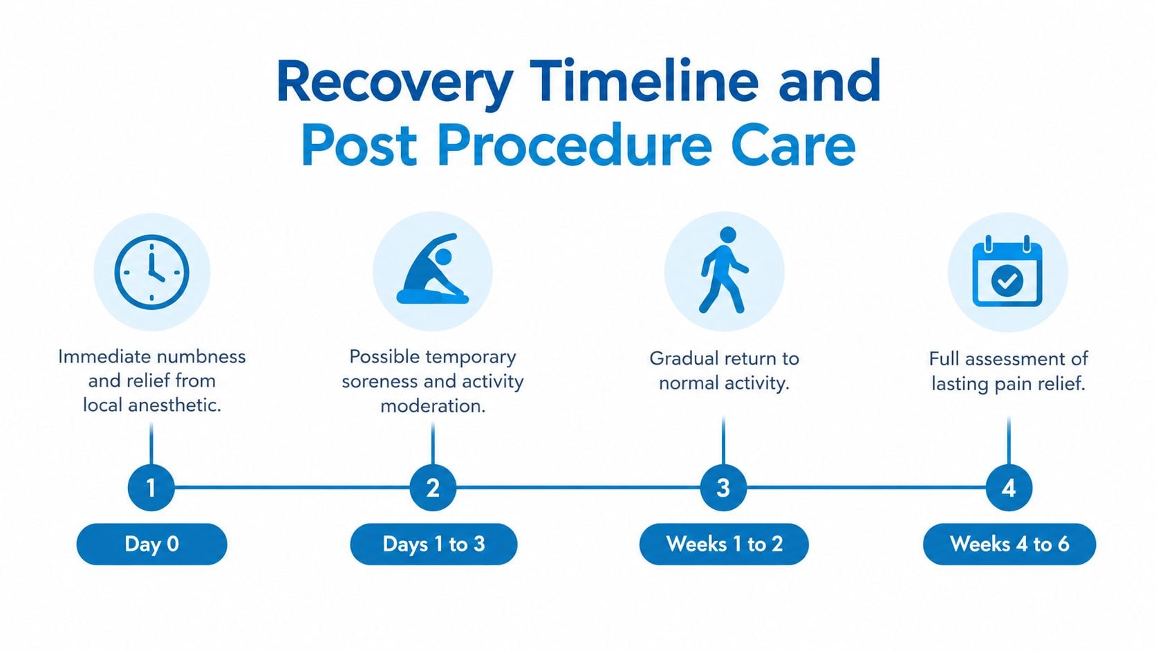
Most patients leave after a short observation period and can expect a manageable recovery. The local anesthetic may create temporary numbness or early relief, while the injection site can feel sore afterward. The first pain response shouldn't be treated as the final result because the anesthetic and anti-inflammatory components act on different timelines.
The early window
On the day of the procedure, patients should follow the clinic's instructions about driving, strenuous activity, and medication. A pain diary can record the familiar pain before the injection, any immediate change, and symptoms as the numbing medicine wears off. That record helps the clinician distinguish temporary anesthetic effects from later improvement.
For localized soreness, a wrapped ice pack may be helpful if the treating team permits it. Avoid pressing directly on the injection site, and contact the clinic if pain is severe, worsening, or accompanied by symptoms that seem unusual.
Returning to activity
The first day is generally a time for moderation rather than testing the joint. Normal movement can resume according to the physician's instructions, but heavy lifting, strenuous exercise, and activities that provoke the familiar pain should be postponed until the clinic says they're appropriate. Physical therapy can be resumed or adjusted once the treating physician and therapist coordinate the timing.
Patients looking for broader context on anti-inflammatory injections can review this overview of corticosteroid injections.
Call the clinic promptly for fever, increasing redness or drainage, progressive weakness, new bowel or bladder symptoms, or severe pain that doesn't follow the expected course. These symptoms aren't typical recovery sensations and require clinical assessment.
Evaluating Outcomes and Planning Next Steps
The most useful outcome measure is function, not just a lower number on a pain scale. The patient and clinician should review the pain diary, walking tolerance, sitting ability, sleep, transitions from sitting to standing, and the specific movement that usually triggers symptoms.
If the response supports an SI joint contribution, the next step may involve targeted rehabilitation to improve pelvic and hip stability. If the benefit is limited, the clinician should reassess other possible sources, including the lumbar spine, hip, nerves, and extra-articular pelvic tissues. A lack of relief can be diagnostically useful when the injection was properly targeted and interpreted in context.
Further treatment depends on the pain pattern and the patient's goals. Options may include rehabilitation, a different diagnostic approach, orthopedic assessment, neuromodulation, or carefully selected nerve treatment such as radiofrequency ablation. No single pathway fits every patient, and repeated injections shouldn't replace a search for a more durable strategy when relief is incomplete or short-lived.
Interventional Pain Management offers evaluation and image-guided interventional care for back, joint, and spine-related pain, including fluoroscopic procedures when clinically appropriate. Patients with persistent pain near the SI joint can visit Interventional Pain Management to request an assessment and discuss whether a low-volume, contrast-confirmed injection fits their diagnosis and treatment goals.