
A fracture of the sacroiliac joint is rarely an isolated “back injury.” Sacral fractures represent about 30% to 45% of pelvic ring injuries, and the sacroiliac region often determines whether the posterior pelvis remains stable, whether nerves are at risk, and whether surgery can restore normal mechanics (Orthobullets overview of sacral fractures). The initial bone healing matters, but it isn't the entire outcome. The reduction and fixation strategy can influence whether a patient later develops persistent sacroiliac joint dysfunction, altered load transfer, or chronic pelvic pain.
Table of Contents
- Why Sacroiliac Joint Fractures Demand Attention
- Anatomy and Mechanisms of Sacroiliac Joint Fractures
- Recognizing the Signs and Differentiating From Other Pain Sources
- Imaging and Diagnostic Workup for SI Joint Fractures
- Treatment Pathways From Conservative Care to Surgical Fixation
- Long-Term Outcomes and the Risk of Post-Traumatic SI Joint Dysfunction
- Red Flags and When to Seek Specialized Care
Why Sacroiliac Joint Fractures Demand Attention
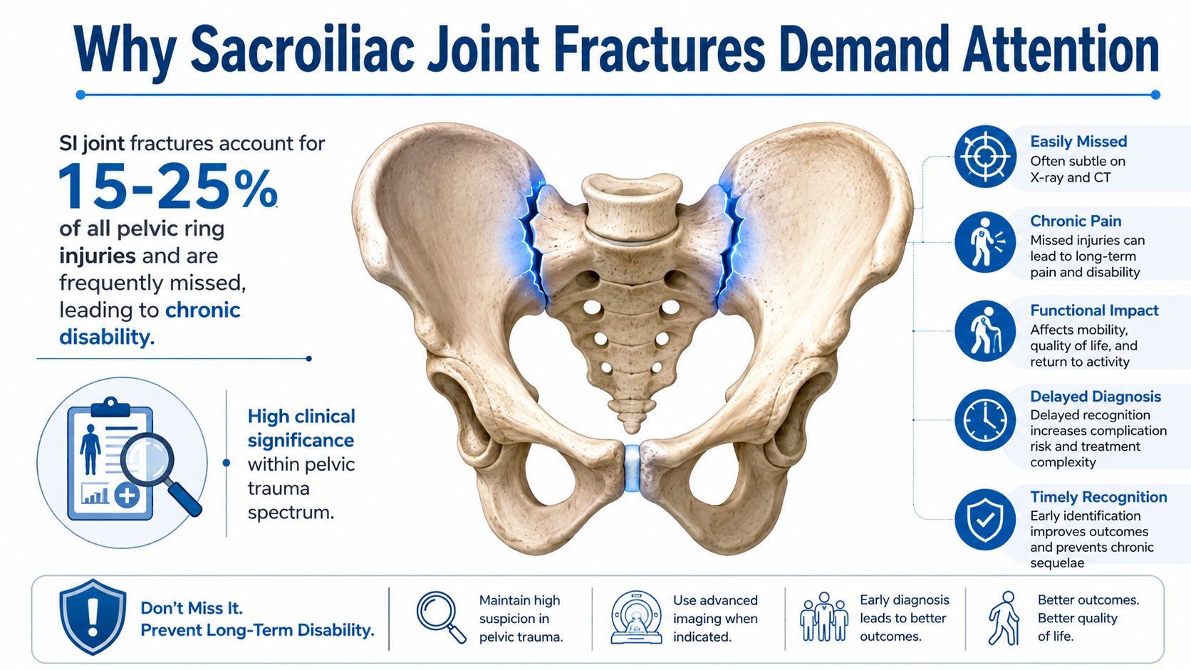
Sacroiliac joint fractures require specialist assessment because the joint lies within the posterior pelvic ring, the structural connection between the spine and lower limbs. A fracture extending into this region may occur with ligament disruption, displacement, nerve irritation, or mechanical instability that symptoms alone cannot reliably reveal. A clinical review reports that lateral compression involving the sacroiliac joint accounts for up to 80% of pelvic ring injuries, making this a common injury pattern rather than an unusual complication (NCBI Bookshelf clinical review).
The mechanism and the patient's age shape the clinical problem. Younger adults often sustain sacral fractures after high-energy trauma, including major collisions or falls from height. Older adults may fracture after a low-energy fall, particularly when bone strength and balance have declined. These situations differ, yet either can disrupt the posterior pelvic ring and cannot be managed like an uncomplicated lumbar strain.
Why anatomy changes the decision
Anatomic variation can affect both CT interpretation and fixation planning. A 2026 study reported an SI-joint anatomic variant in 74.1% of pelvic trauma patients, including a semi-circular defect in 24.4% and an ilio-sacral complex in 21.5% (Orthobullets summary). These variants do not prove that a fracture will occur, but they can alter the visible fracture path and reduce the safe corridor available for screws or other fixation.
Classification also has practical value. Medelman grouped sacral fractures in 1939, Bonnin proposed a six-type system in 1945 and 1947, and Denis and colleagues introduced the commonly used three-zone system in 1988 after reviewing 236 consecutive sacral fractures and dissecting 39 cadavers. Classification helps clinicians estimate stability, neurologic risk, and the technical demands of reduction.
Bone union is only one milestone. The reduction and fixation method can influence later load transfer, residual asymmetry, and post-traumatic SI joint dysfunction, so recovery expectations should extend beyond the initial healing period.
For patients arranging appointments, records, or insurance after an accident, a structured home service inquiry response system can help organizations respond consistently. It cannot replace urgent clinical triage when pelvic injury is suspected.
Anatomy and Mechanisms of Sacroiliac Joint Fractures
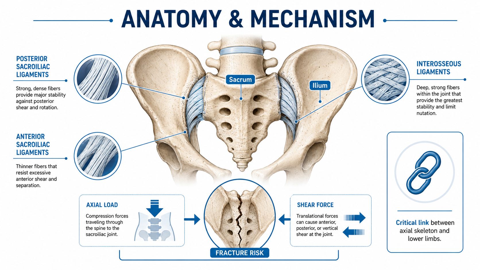
The sacroiliac joint connects the sacrum to the ilium and transfers load between the spine and lower limbs. Its limited normal motion allows the pelvic ring to absorb force without permitting large shifts. The posterior and interosseous ligaments provide much of this restraint, so disruption of the posterior ring changes the injury from a local fracture into a mechanically unstable pelvic problem.
How force creates the fracture
High-energy trauma can compress, rotate, or shear the pelvic ring. Lateral compression pushes the pelvis inward. Vertical shear can move one side upward, while an impact to the buttock transfers force through the ilium toward the joint. Axial loading travels upward from the legs through the pelvic ring.
The fracture line does not describe the full injury. Bone fragments may shift forward, backward, upward, or rotationally, and the supporting ligaments may be stretched, partly torn, or completely disrupted. Those differences affect stability, nerve risk, and whether protected weight-bearing can succeed. They also influence reduction and fixation, because residual displacement can alter load transfer even after the bone unites.
Why fixation planning affects long-term function
A posterior ligament disruption often requires stabilization rather than conservative care alone. Depending on the fracture pattern and safe bony corridor, fixation may include an iliosacral screw placed through the sacrum, or another posterior pelvic construct. The goal is to restore alignment while avoiding the sacral nerves, vascular structures, and adjacent joint surfaces.
Anatomic variants can narrow or redirect the available screw corridor. A semi-circular defect or an ilio-sacral complex may change the apparent joint shape and make a standard trajectory unsafe. CT-based planning helps distinguish normal anatomy from a fracture and determines whether the intended screw path has adequate bone around it. A corridor that is acceptable on one side may not be safe on the other.
The technique also creates trade-offs. More rigid fixation can protect a disrupted posterior ring during healing, yet malreduction, unrecognized instability, or altered joint loading may contribute to persistent pain and post-traumatic SI joint dysfunction. Bone union therefore marks only one stage of recovery. Alignment, pelvic symmetry, neurologic status, and tolerance of progressive loading matter as well.
Practical rule: Assess whether the posterior ring remains aligned and mechanically stable under load, then match the fixation path to the patient's three-dimensional anatomy.
Recognizing the Signs and Differentiating From Other Pain Sources
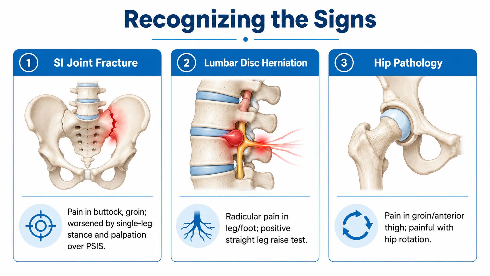
Pain from a fracture of the sacroiliac joint often sits deep in the buttock or low pelvis, usually near the posterior pelvic landmarks just below the belt line. Weight-bearing commonly aggravates it. Rolling in bed, standing from a chair, climbing steps, or shifting from one leg to the other can create sharp pain because these movements load the posterior ring unevenly.
Groin or thigh discomfort doesn't rule out an SI injury, but the pattern matters. A patient with a painful hip may have more prominent groin or anterior-thigh pain with hip rotation. A patient with lumbar nerve compression is more likely to describe radiating pain into the leg or foot with numbness, tingling, reflex changes, or weakness. SI-region pain can refer beyond the joint, but objective neurologic findings should prompt a careful spinal and sacral evaluation rather than an assumption of isolated SI dysfunction.
Comparing the common alternatives
| Possible source | Pattern that may point toward it | What still needs caution |
|---|---|---|
| SI joint fracture | Deep buttock or pelvic pain, worse with weight-bearing, transitions, or direct posterior tenderness | Pain location can't establish stability |
| Lumbar radiculopathy | Leg or foot radiation, numbness, weakness, or nerve tension signs | A pelvic fracture can coexist with spinal injury |
| Hip pathology | Groin or anterior-thigh pain, especially with hip rotation | Referred pain can overlap |
| Piriformis or myofascial pain | Buttock tenderness and movement-related muscle pain | Muscle guarding may be secondary to an unstable injury |
| Sacral insufficiency fracture | Deep sacral or pelvic pain after relatively minor trauma in a vulnerable patient | Early plain films may not define the injury |
Patients with suspected nerve-related leg pain can review the general clinical distinctions in this sciatica and pinched nerve resource, but a recent fall, collision, or inability to bear weight changes the priority. In that setting, pain should be evaluated for structural injury before being treated as routine sciatica.
A physical examination can identify tenderness and reproduce pain with controlled pelvic loading, but provocative maneuvers aren't a substitute for imaging after significant trauma. The key clinical clues are the mechanism, the ability to stand or walk, the presence of pelvic asymmetry, and any sensory or motor change.
Imaging and Diagnostic Workup for SI Joint Fractures
The mechanism often determines how urgently imaging is needed. Clinicians assess whether the patient could walk after the injury, whether pain is unilateral or central, and whether numbness, weakness, or bowel and bladder changes appeared. Examination adds information about pelvic alignment, posterior tenderness, hip motion, distal pulses, and neurologic function.
Plain pelvic radiographs can show obvious displacement or major pelvic ring disruption. They may miss injuries involving the posterior ring because the sacrum overlaps other pelvic structures, and a fracture crossing the SI joint can be difficult to follow on standard views. A normal or unclear radiograph therefore cannot reliably exclude a significant injury when the mechanism and symptoms remain concerning.
Why CT usually drives the decision
CT usually provides the clearest assessment of fracture morphology and posterior pelvic ring involvement, as described in this radiology guidance on sacroiliac joint injuries. It can show whether the fracture enters the joint, whether the sacrum is involved in more than one plane, and whether the ilium or nearby pelvic structures have shifted.
The review should address:
- Sacroiliac joint widening, which may indicate loss of ligament support.
- Vertical offset, suggesting upward movement of one pelvic component.
- Malalignment, which can reflect disruption of the posterior ring.
- Comminution or fragment rotation, findings that can make reduction and fixation more difficult.
- Neural foraminal or canal involvement, which may raise concern for nerve injury.
These details affect more than the initial fracture description. Widening, offset, and malalignment can indicate ligament failure and mechanical instability, factors that influence fixation planning and the later risk of persistent SI joint dysfunction even after the bone has healed.
When MRI adds information
MRI can show marrow injury, occult fractures, soft-tissue abnormalities, and nerve-related structures when CT does not explain ongoing symptoms. It may help when the suspected injury is subtle or when the differential includes a sacral insufficiency fracture, infection, tumor, or another nontraumatic condition.
Imaging interpretation also requires attention to anatomy. Normal joint variation can resemble traumatic separation, while that same anatomy affects safe screw placement if fixation is considered. The radiologist and treating surgeon should therefore review the images with the examination and injury mechanism rather than relying on an isolated report. Clear bone healing on follow-up imaging does not, by itself, prove that the joint has regained normal motion or that pain will resolve.
Treatment Pathways From Conservative Care to Surgical Fixation
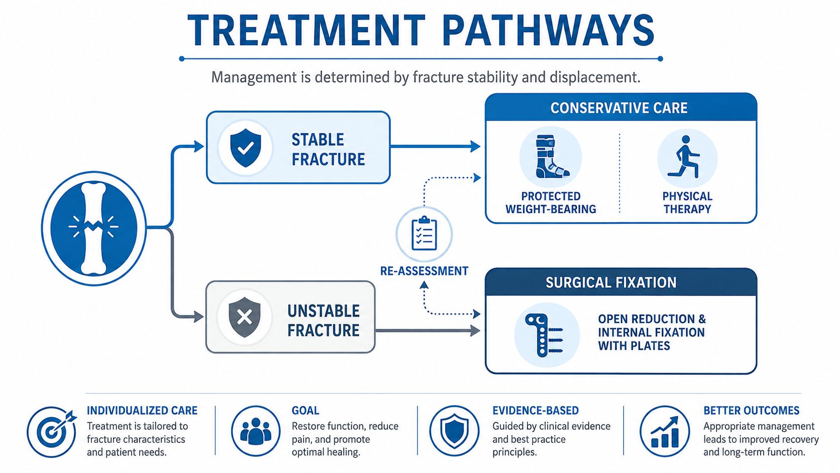
Treatment depends on stability, displacement, alignment, neurologic status, and functional demands. The presence of a fracture line alone doesn't determine whether surgery is necessary. A stable, minimally displaced injury may heal with protected loading, while a displaced fracture-dislocation can continue to deform if the posterior ring isn't stabilized.
Conservative care for stable injuries
For a stable fracture, the treating team may prescribe protected weight-bearing, temporary assistive devices, activity modification, medication, and physical therapy. The purpose isn't complete inactivity. It is controlled loading that protects the ring while preserving strength, circulation, and basic mobility.
Rehabilitation should progress according to pain, imaging, strength, and mechanical stability. Early therapy may focus on safe transfers, gait training, breathing, and maintaining motion in unaffected joints. Later work can address trunk control, hip strength, balance, and gradual return to meaningful activities.
An SI belt may provide external support for selected patients with painful pelvic motion, although it shouldn't be used as proof that the fracture is stable. A practical MedAmerica Rehab Center SI belt guide can help explain belt positioning and general support concepts, but the treating clinician must decide whether a belt is appropriate for a specific fracture.
When procedures become reasonable
Image-guided SI joint injection may help identify and treat persistent pain after the acute fracture has been assessed. It is generally considered only when the pain generator is sufficiently clear and the injection won't obscure an untreated instability or infection. Radiofrequency ablation may be considered for selected chronic SI-region pain after diagnostic evaluation, particularly when the pain persists after structural healing.
Surgery becomes more relevant when the posterior ring is unstable, displaced, malaligned, or associated with a fracture-dislocation. Iliosacral screws, transiliac fixation, plates, or combinations may be used depending on the fracture pattern and available bone corridors. The technical objective is not merely to insert hardware. It is to restore alignment and maintain stable fixation.
A broader overview of orthopedic evaluation and treatment for fractures can help patients understand why the same treatment isn't appropriate for every fracture. For complex SI injuries, orthopedic trauma, spine, radiology, and pain specialists may all contribute to the plan.
Long-Term Outcomes and the Risk of Post-Traumatic SI Joint Dysfunction
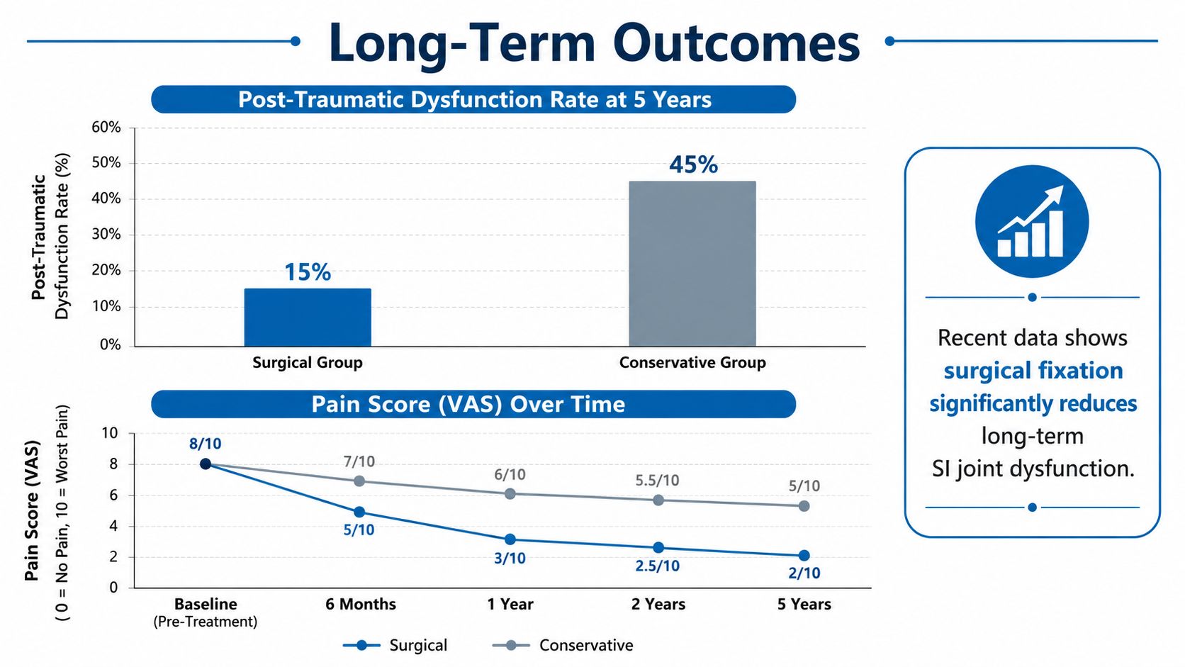
Radiographic union does not guarantee a symptom-free sacroiliac joint. Bone can heal while the joint and its supporting ligaments transmit load abnormally. Scar tissue, residual malalignment, muscle inhibition, hardware irritation, altered pelvic mechanics, and nerve sensitivity may each sustain pain.
The meaningful long-term question is whether the pelvis healed in a mechanically acceptable position and whether the patient can load it normally. Fracture-dislocations are difficult to reduce because fragments may shift in multiple planes. A review of SI joint fracture-dislocations associates accurate reduction and stable fixation with better functional results, while poor reduction is associated with poorer function.
What fixation technique may change
A 2025 to 2026 cohort of 80 patients found SI joint dysfunction in 25% after SI screw fixation for posterior pelvic ring injuries (Frontiers in Surgery cohort and review). Dysfunction occurred after unilateral fixation and not after bilateral fixation in that cohort. This does not establish bilateral fixation as the right choice for every fracture. Fixation must match the injury pattern, stability requirements, bone corridors, and expected loading demands.
If unilateral fixation was used, schedule a functional loading assessment at 12 weeks, provided healing and weight-bearing restrictions permit it. The assessment should examine gait, single-leg loading, pelvic control, symptom reproduction, and progression toward work or sport. A normal radiograph does not exclude emerging SI dysfunction.
The cohort also reported higher risk among younger patients. That finding may reflect injury mechanics, activity demands, or loading patterns rather than age-related degeneration alone. It should guide follow-up, not determine an individual prognosis without review of the fracture pattern and recovery course.
Setting realistic recovery expectations
Recovery usually progresses through protection and safe mobility, strength and coordination training, then endurance and return to work or sport. The pace depends on fixation stability, associated injuries, neurologic involvement, and the patient's loading demands.
Persistent pain does not automatically indicate failed surgery. It warrants a focused reassessment of alignment, hardware, neurologic status, hip and lumbar pain sources, and the SI joint itself. Patients with ongoing symptoms can review sacroiliac joint dysfunction evaluation and treatment as part of a broader discussion of targeted pain management after structural healing. Recovery should be judged by function and load tolerance, not bone healing alone.
Red Flags and When to Seek Specialized Care
A patient with a suspected fracture of the sacroiliac joint shouldn't wait for routine back-pain treatment when the pelvis may be unstable. Immediate evaluation is appropriate after significant trauma, especially when pain is severe, walking is difficult, or the pelvis appears asymmetric. A clinician needs to determine whether the injury is stable before recommending stretching, strengthening, or unsupervised weight-bearing.
Symptoms that require urgent attention
Seek emergency assessment for:
- Inability to bear weight, particularly after a fall, collision, or crush injury.
- New numbness or weakness in either leg, foot, buttock, or saddle region.
- Bowel or bladder dysfunction, including new retention or loss of control.
- Rapidly worsening pain, severe pain at rest, or pain out of proportion to the apparent injury.
- Faintness, pallor, or unusual sweating after pelvic trauma, which may indicate a broader emergency.
- Fever or systemic illness accompanying severe SI-region pain, especially without a clear mechanical explanation.
Neurologic symptoms may reflect nerve-root, sacral, or more extensive pelvic involvement. Bowel and bladder changes are particularly important because they can indicate serious neural compromise and shouldn't be monitored at home.
When specialist review is appropriate
A patient whose pain persists or worsens despite an initial conservative plan needs reassessment rather than repeated generic treatment. Specialist review may include a detailed neurologic examination, CT review, MRI when indicated, diagnostic injections after structural concerns are addressed, and orthopedic or spine consultation for unstable patterns.
In New Jersey and Staten Island, a multidisciplinary evaluation can coordinate interventional pain care, orthopedic assessment, spine surgery input, rehabilitation, and imaging review. This approach is valuable when the initial fracture has healed but the patient still has focal SI pain, impaired walking tolerance, pelvic asymmetry, or symptoms that don't match a simple lumbar diagnosis.
Patients should bring all imaging discs or digital files, radiology reports, operative notes, and a clear list of current symptoms. The most useful history identifies which movements provoke pain, whether symptoms are improving, and whether numbness, weakness, or bladder and bowel changes have appeared.
Interventional Pain Management evaluates sacroiliac-region pain with clinical examination, imaging review, and targeted treatment options when persistent symptoms remain after injury. Patients with a suspected or treated SI fracture can visit Interventional Pain Management to request an assessment that connects pelvic stability, fixation history, rehabilitation needs, and ongoing pain.