

A sharp electric shock at the base of the skull can be followed by a strange sense that the room is tilting, especially when the head turns. Many patients describe this combination after long periods of desk work, neck strain, whiplash, or an unexplained flare of head pain. They're often told the dizziness must be migraine, an inner-ear problem, sinus disease, or anxiety before anyone examines how the upper neck and occipital nerves might be contributing.
So, can occipital neuralgia cause dizziness? It can be associated with dizziness, but dizziness shouldn't automatically be blamed on occipital neuralgia. The more useful clinical question is whether the symptoms share a mechanical or neurologic source in the upper cervical spine, whether a vestibular disorder is present, or whether the apparent connection is coincidental. That distinction matters because a nerve block may help clarify the pain source, but it isn't a substitute for evaluating persistent or atypical dizziness.
Table of Contents
- When Neck Pain and Room Spinning Show Up Together
- What Occipital Neuralgia Actually Is
- How Occipital Nerve Irritation May Trigger Dizziness
- Occipital, Vestibular, and Cervicogenic Dizziness Compared
- When Dizziness Points to Something Other Than Occipital Neuralgia
- How Specialists Work Up the Dizzy Occipital Patient
- Treatment Options From Conservative to Interventional
- Practical Takeaways and Next Steps
When Neck Pain and Room Spinning Show Up Together
Consider a typical clinic presentation. A 38-year-old office worker develops brief, electrical pain from the base of the skull over one ear after a long day at a monitor. When she turns her head to reach for a coffee cup, the room seems to tilt. The dizziness lasts for several seconds after the pain spike and eases when she lies still.
That pattern is clinically important, but it doesn't identify the cause by itself. The pain may follow the path of an irritated occipital nerve, while the dizziness may arise from altered neck-position signals, a vestibular condition triggered by head movement, or another process entirely. Patients often describe “dizziness” for different sensations, including true spinning, rocking, visual instability, faintness, or general unsteadiness.
Why the combination gets mislabeled
In specialty practices, occipital pain with imbalance is a recurring diagnostic puzzle. The symptoms may be labeled migraine when the pain is severe, sinus disease when pressure spreads around the eye or ear, or anxiety when routine testing doesn't immediately reveal an explanation. Those labels can sometimes be correct, but they shouldn't end the evaluation.
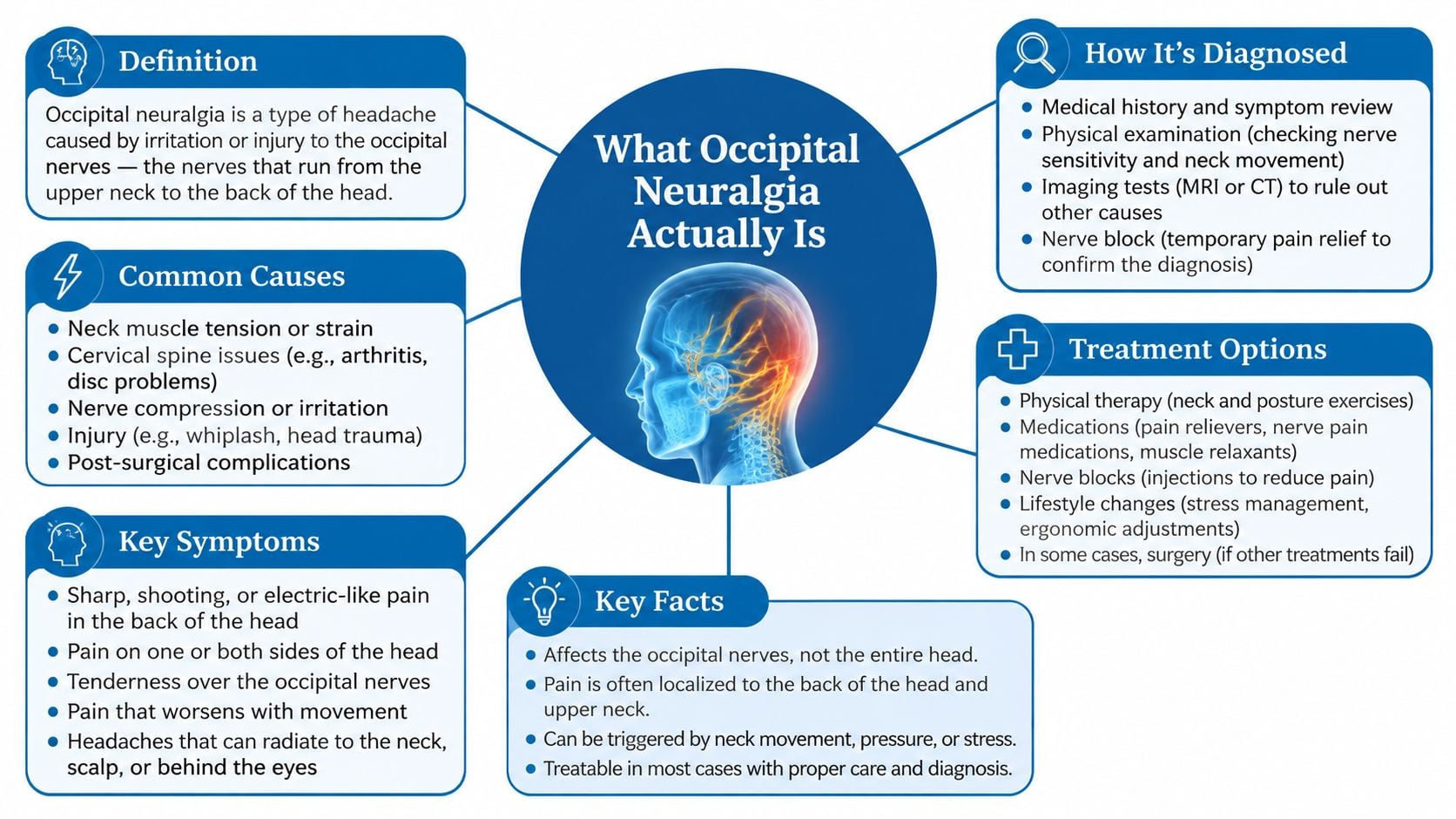
Occipital neuralgia generally involves sharp, shooting, or electric pain along the back of the scalp, often with tenderness or altered sensation over the nerve. A patient may also have restricted neck movement, muscle guarding, or symptoms that change with posture. Detailed information about neck pain evaluation and treatment can help patients understand why the cervical examination matters.
Practical rule: Dizziness that appears with occipital pain deserves a pattern-based assessment, not a yes-or-no answer.
The central issue is sequencing. A clinician must first determine whether the dizziness is plausibly cervico-occipital, primarily vestibular, cervicogenic without direct occipital nerve involvement, cardiovascular, or neurologic. Only after that reasoning should an occipital nerve block be considered.
What Occipital Neuralgia Actually Is
Occipital neuralgia is nerve pain arising from irritation of the greater, lesser, or third occipital nerves. Patients often describe sudden stabbing, burning, or electric shocks that begin near the upper neck or base of the skull and travel across the posterior scalp. Tenderness, tingling, numbness, and pain behind the ear or around the eye can occur because these nerves supply sensation to the back and side of the head.
The occipital nerves arise from branches of the upper cervical dorsal rami, particularly the C2 and C3 regions. They pass through muscles and connective-tissue planes near the skull base before traveling upward into the scalp. Tight muscles, irritated joints, scar tissue, trauma, or local inflammation can make these narrow pathways more sensitive.
The relay-wire analogy
A useful way to understand the anatomy is to think of the occipital nerves as relay wires. They carry sensory information from the upper neck and scalp into the nervous system, where that information is processed alongside signals from the face, eyes, inner ears, muscles, and joints.
The upper cervical sensory system communicates with the trigeminocervical complex, a network involved in head and facial pain processing. Nearby brainstem systems also integrate information related to head position and balance. This convergence makes it biologically plausible for irritation in the upper neck to produce symptoms felt beyond the exact location of the nerve, including the forehead, eye, ear, or a sense of disequilibrium.
That plausibility doesn't prove that occipital neuralgia directly causes rotational vertigo. It explains why a patient can experience head pain and balance symptoms together, while still requiring an examination for other causes. Occipital neuralgia is a pain diagnosis, whereas dizziness is a broad symptom category with neurologic, vestibular, cervical, and cardiovascular possibilities.
How Occipital Nerve Irritation May Trigger Dizziness
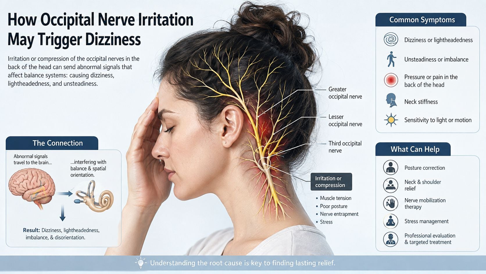
A patient may describe a sharp pain at the back of the head followed by rocking, tilting, or difficulty walking straight. That sequence deserves clinical attention. The relationship between occipital neuralgia and dizziness is clinically recognized but mechanistically unsettled, so dizziness should guide the diagnostic workup rather than serve as proof that the occipital nerve is responsible.
A clinical report indexed in PubMed found dizziness in 50% of patients with occipital neuralgia, alongside nausea in 42%, visual disturbances in 67%, scalp paresthesia in 33%, and tinnitus in 33%. The findings show a broader symptom pattern than localized pain, but they do not establish that nerve irritation directly produces every symptom. The historical clinical report remains useful because it documented this association.
Two mechanisms are most relevant in practice.
Direct neural cross-talk
Pain signals from the upper cervical region enter central pathways that interact with trigeminal and vestibular processing networks. A sudden increase in nociceptive input may change how the brain interprets head movement, visual information, and spatial orientation. Patients may feel rocking, tilting, imbalance, or motion sensitivity rather than classic inner-ear vertigo.
This mechanism is plausible, but clinicians cannot measure it directly in an individual patient. Timing provides a useful clue. Dizziness that consistently follows an occipital pain flare suggests a shared process more strongly than dizziness that appears independently throughout the day.
Cervicogenic disruption
The upper cervical joints and muscles provide proprioceptive information about head and neck position. Pain, muscle spasm, restricted motion, or joint dysfunction can distort those signals. The brain may then receive mismatched information from the neck, eyes, and vestibular organs, producing unsteadiness or visually induced dizziness.
Both mechanisms can operate together. Occipital pain may cause protective muscle tightening, while reduced neck movement further disrupts proprioceptive input. A nerve block may reduce pain without resolving the dizziness if cervical rehabilitation or vestibular assessment is also needed. For that reason, specialists assess the symptom sequence and examination findings before offering an intervention.
A separate clinical discussion also describes dizziness in 50% of patients with occipital neuralgia and emphasizes tinnitus, nausea, and visual disturbance. It proposes interactions among upper cervical afferents, cranial nerve pathways, and sympathetic connections, while acknowledging overlap with vestibular and cervical conditions. The clinical overview of occipital neuralgia and dizziness supports treating dizziness as a diagnostic signal, not as proof of causation.
Occipital, Vestibular, and Cervicogenic Dizziness Compared
Patients use the word dizziness to describe several different experiences. A room-spinning sensation suggests vertigo, while rocking, imbalance, visual discomfort, and near-fainting point toward different diagnostic pathways. The symptom's quality, trigger, duration, and examination findings matter more than the word itself.
Occipital vs. Vestibular vs. Cervicogenic Dizziness at a Glance
| Feature | Occipital-Related Dizziness | Vestibular Dizziness | Cervicogenic Dizziness |
|---|---|---|---|
| Symptom quality | Disequilibrium, tilting, motion sensitivity, or dizziness accompanying occipital pain | Often true rotational vertigo, motion illusion, nausea, or ear-related symptoms | Unsteadiness, rocking, visual disorientation, or a floating sensation |
| Common triggers | Occipital pain flares, head movement, pressure near the nerve | Position changes, visual motion, or inner-ear stimulation | Neck rotation, sustained posture, restricted cervical movement |
| Timing pattern | Often linked temporally to pain episodes, though patterns vary | May be brief or prolonged depending on the vestibular disorder | Often follows neck movement or persists while cervical dysfunction remains active |
| Examination focus | Occipital tenderness, sensory change, neurologic examination, symptom response to targeted treatment | Dix-Hallpike, head impulse test, smooth pursuit, vestibular and hearing assessment | Cervical range of motion, flexion-rotation test, posture, balance, and symptom provocation |
| What complicates diagnosis | Can mimic migraine or vestibular migraine | Can coexist with neck pain and headache | Can overlap with whiplash-associated and post-concussive symptoms |
A clinician may use the Dix-Hallpike maneuver to assess positional vertigo, the head impulse test to evaluate vestibulo-ocular reflex function, and smooth-pursuit testing to look for abnormalities in visual tracking. A HINTS examination may be relevant in selected patients with acute, continuous vertigo, but it requires appropriate training and should not be treated as a home test.
For patients with tinnitus, hearing fluctuation, ear pressure, or unexplained auditory symptoms, diagnostic hearing assessments in Boca Raton may be a useful part of the broader evaluation. Hearing findings can shift attention toward an inner-ear or auditory pathway disorder.
Cervical assessment adds another layer. The flexion-rotation test can provoke symptoms associated with upper cervical restriction, while palpation may reproduce familiar occipital pain. However, tenderness alone doesn't establish that the neck is causing the dizziness. Patients may have both myofascial pain and a separate vestibular disorder, which is why a focused myofascial pain evaluation may need to occur alongside vestibular screening.
When Dizziness Points to Something Other Than Occipital Neuralgia
The contrarian point is important: persistent or atypical dizziness often signals a different or additional diagnosis. A tender occipital nerve can be real without being responsible for the entire symptom pattern. Treating that tenderness first may delay evaluation of a vascular, structural, vestibular, or cardiac problem.
A clinician considers vertebrobasilar insufficiency, posterior circulation stroke, cerebellar disease, intracranial pressure disorders, vestibular migraine, Ménière disease, persistent postural-perceptual dizziness, and perilymphatic fistula when the history points away from a simple cervico-occipital pattern. Presyncope can also reflect a cardiac rhythm problem rather than vertigo. Patients trying to understand rhythm-related faintness may find a general SA node dysfunction patient guide useful, although it doesn't replace medical assessment.
Red flags that change the order of care
Urgent evaluation becomes more important when dizziness is:
- Sudden or severe: Abrupt onset, especially when unlike prior episodes, warrants prompt neurologic consideration.
- Neurologically accompanied: Weakness, numbness, double vision, speech difficulty, severe gait disturbance, or altered coordination should not be attributed to occipital nerve pain without evaluation.
- Trauma-related: Neck trauma, including whiplash, raises concern for cervical injury or vascular complications.
- Associated with unusual headache: A new or dramatically different headache pattern requires a broader workup.
- Linked to auditory change: New hearing loss, marked tinnitus, or ear pressure may point toward an otologic or vestibular disorder.
- Medication or risk-factor related: Anticoagulant use and vascular risk factors can change the threshold for emergency assessment.
The HINTS examination can help in the hands of a trained clinician when the presentation is appropriate, but imaging or specialist consultation may still be needed. Brain and vascular imaging can become relevant when symptoms suggest a posterior circulation process, while cardiac evaluation may be appropriate when episodes feel like near-fainting rather than spinning.
Suspecting an alternative diagnosis doesn't invalidate occipital neuralgia. It sequences the workup correctly. A nerve block should not be used to postpone emergency evaluation, and relief of scalp pain doesn't prove that a serious cause of dizziness has been excluded.
How Specialists Work Up the Dizzy Occipital Patient
A careful workup proceeds by decision points rather than by ordering every test at once. The first decision is whether the dizziness is episodic or continuous, movement-triggered or spontaneous, spinning or faint-like, and tightly linked to occipital pain or independent of it.
History first
The history should capture:
- The exact starting point of the pain and dizziness.
- Whether head turning, neck extension, standing, visual motion, or lying down changes symptoms.
- How long each episode lasts and whether pain precedes dizziness.
- Associated nausea, tinnitus, hearing change, visual symptoms, weakness, numbness, or speech difficulty.
- Recent trauma, infection, medication changes, or prior neurologic and vestibular diagnoses.
- What happened after medication, physical therapy, rest, or previous injections.
The physical examination then tests the working hypotheses. Cervical range of motion, posture, suboccipital palpation, the flexion-rotation test, gait, balance, eye movements, head impulse responses, and positional maneuvers can reveal whether neck movement or vestibular stimulation reproduces the familiar complaint. A complete neurologic examination remains essential.
Imaging and diagnostic blocks
MRI of the brain, with attention to the posterior fossa, may be considered when central causes are possible. MRA or CTA can be appropriate when vascular disease or arterial injury is a concern. Cervical MRI helps evaluate disc, joint, canal, cord, or other structural pathology when the examination supports a cervical source.
Imaging isn't automatically necessary for every brief episode of dizziness with familiar occipital pain. The decision depends on the clinical pattern, examination findings, trauma history, neurologic signs, and response to initial care.
A diagnostic occipital nerve block can provide useful information when the pain distribution and examination suggest nerve involvement. A meaningful reduction in familiar pain, and possibly the associated dizziness, after image-guided local anesthetic at the greater or lesser occipital nerve supports a cervico-occipital contribution. It doesn't prove causation, because anesthetic can reduce pain-driven guarding without correcting another source of dizziness. A negative response should redirect the workup rather than prompt repeated injections.
Neurology, otolaryngology, audiology, or vestibular therapy may be appropriate when the presentation doesn't fit a single pain diagnosis.
Treatment Options From Conservative to Interventional
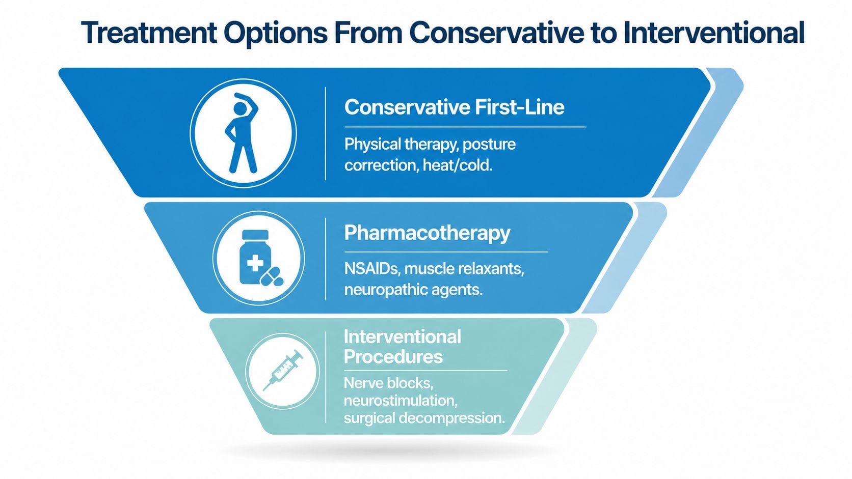
Treatment should follow the strongest working diagnosis. If cervical dysfunction and occipital nerve sensitivity appear central, conservative care usually comes before procedures. If the history suggests inner-ear disease, vestibular treatment takes priority. If red flags are present, urgent neurologic or vascular evaluation comes first.
Conservative care
Physical therapy may focus on the suboccipital muscles, upper cervical mobility, deep neck stabilization, posture, and movement retraining. Ergonomic changes can reduce prolonged forward-head positioning, but they shouldn't be treated as a complete solution when symptoms are severe or progressive. Graduated vestibular rehabilitation may help patients whose balance symptoms persist after pain and neck guarding improve.
Heat, activity modification, sleep positioning, and carefully selected home exercises can support recovery. Forceful neck manipulation isn't appropriate for every patient, especially when vascular or structural concerns haven't been evaluated.
Medication and injections
Medication choices depend on medical history, other prescriptions, kidney and gastrointestinal risks, mood, sleep, and the character of the pain. Clinicians may consider anti-inflammatory medication, a short course of a muscle relaxant, or nerve-targeted agents such as a tricyclic antidepressant or gabapentinoid. These medications can cause sedation, dry mouth, cognitive slowing, dizziness, or other adverse effects, so a patient already reporting imbalance needs careful monitoring.
An image-guided occipital nerve block can serve two purposes. It may reduce pain therapeutically, and it can help test whether the targeted nerve contributes to the symptom pattern. Information about nerve block procedures and their role in pain care can help patients prepare for a discussion with a treating clinician. Relief may be temporary, and a single injection shouldn't be presented as a guaranteed long-term answer.
Advanced options
For persistent, well-localized nerve pain after appropriate conservative treatment, clinicians may discuss pulsed radiofrequency or radiofrequency-based treatment in selected cases. Occipital nerve stimulation is generally reserved for carefully selected chronic neuropathic pain situations after less invasive approaches haven't provided adequate control. Surgical decompression is considered only when a specific compressive cause and appropriate surgical indication are established.
Referral may involve pain management, neurology, ENT or otolaryngology, vestibular therapy, physiotherapy, or spine surgery. Interventional Pain Management offers evaluation and procedures including nerve blocks within a broader pain and spine service, but the correct sequence still depends on the diagnostic findings.
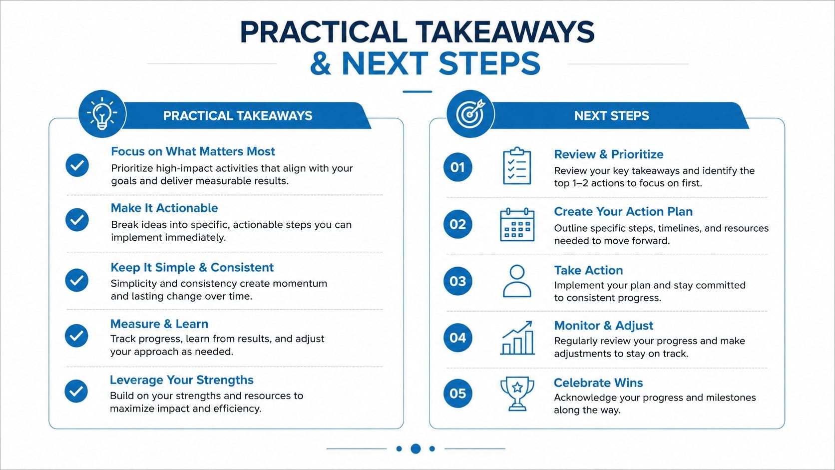
Practical Takeaways and Next Steps
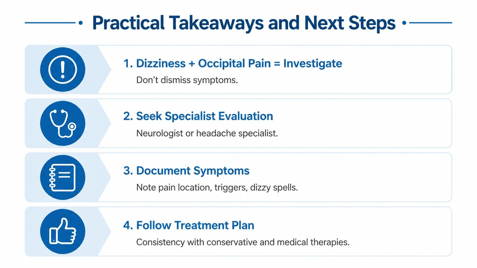
Dizziness with occipital pain is a signal to investigate, not a symptom to dismiss or self-manage indefinitely. Occipital neuralgia may contribute through pain-related neural processing or altered cervical position sense, but the same complaint can reflect vestibular disease, migraine, vascular pathology, structural disease, or presyncope.
A symptom log can make the first appointment more productive. Record:
- Episode timing: Note when pain and dizziness begin, whether one comes first, and whether episodes occur at rest or during activity.
- Sensation quality: Describe spinning, tilting, rocking, lightheadedness, visual instability, or near-fainting.
- Position triggers: Record neck rotation, looking up, standing, rolling in bed, walking, or screen use.
- Associated features: Include headache intensity, nausea, tinnitus, hearing changes, visual symptoms, weakness, numbness, and speech changes.
- Response to position: Note whether sitting, lying still, closing the eyes, or supporting the neck changes the episode.
- Treatment response: List medication, therapy, injections, and the specific symptom each intervention changed.
The likely referral depends on the pattern. A pain specialist or neurologist may be appropriate when electric occipital pain and nerve tenderness dominate. ENT, audiology, or vestibular therapy may be more suitable when vertigo, hearing changes, or positional symptoms lead the presentation. A vascular neurologist or emergency department is the safer route for sudden, persistent vertigo with neurologic signs or major vascular risk.
Patients should ask: When did the symptoms begin? What reliably triggers or relieves them? Does the dizziness occur without pain? What diagnoses have already been considered? Which imaging has been performed? Has a nerve block been offered, and what question is it intended to answer? If symptoms involve standing-related lightheadedness, the clinician should also assess autonomic and cardiovascular possibilities rather than assuming a cervical cause. General information about how salt helps POTS symptoms may provide context, but dietary changes shouldn't begin without appropriate medical guidance.
Interventional Pain Management evaluates occipital neuralgia, cervical pain, and related nerve conditions using history, examination, imaging review, and appropriately selected interventions. Patients with dizziness and occipital pain can visit Interventional Pain Management to request an assessment that separates pain-related symptoms from vestibular, neurologic, vascular, and cervical causes.