
If a mid-back or thoracolumbar injury has turned everyday movement into a negotiation, the next question usually isn't whether the pain is real. It's whether fusion is the right answer, and if so, what kind of fusion makes sense for this body, this bone quality, and this life. A careful decision starts long before the operating room, because thoracic lumbar fusion can stabilize a failing segment, but it also commits the spine to permanent trade-offs that deserve a clear explanation.
Table of Contents
- Understanding Thoracic and Thoracolumbar Fusion
- When Spine Surgeons Recommend Fusion
- Open vs Minimally Invasive Approaches
- Surgical Techniques Used in Thoracolumbar Fusion
- What Recovery Looks Like Week by Week
- Risks and the Bone Quality Question
- Making the Decision at a Multidisciplinary Center
Understanding Thoracic and Thoracolumbar Fusion
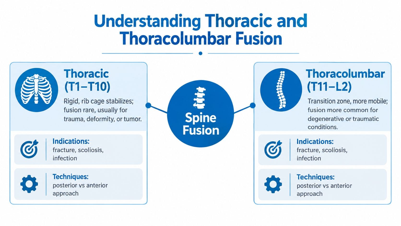
A patient with a burst fracture at the junction of the ribs and lower back often hears two words in the same conversation, thoracic and thoracolumbar. They sound close, but they don't behave the same way. The thoracic spine, roughly T1 through T10, is stiff by design because the rib cage helps stabilize it. The thoracolumbar junction, roughly T10 through L2, is the body's transition zone, where the rigid upper back meets the more mobile lumbar spine.
That transition matters because forces concentrate there. It's one reason surgeons see this area as mechanically demanding, especially when the problem is fracture, deformity, infection, or degenerative collapse. In published clinical series, thoracic and thoracolumbar fusion has shown high fusion success overall, with one study reporting a definite fusion rate of 88.81% across 278 levels and a regional difference between the thoracic spine and the thoracolumbar junction (NCBI study). The point isn't that fusion is guaranteed, it's that the region itself changes the odds.
How surgeons decide what to fuse
A real operation is rarely about one vertebra. The surgeon studies the damaged level, the segments above and below, the alignment on imaging, and the way the patient stands and walks. If the construct stops too short, the junction can fail under stress. If it extends too far, motion is sacrificed unnecessarily.
Practical rule: the smallest stable construct is often the right target, but only if it truly neutralizes the painful or unstable segment.
Fusion itself is straightforward in concept. Two or more vertebrae are joined with bone graft and instrumentation so they heal into one rigid segment. That rigidity can relieve painful motion and restore structural stability, but it also changes how nearby segments move. For many patients, the key question isn't “can fusion be done?” It's “which levels need help, and which levels should be left alone?”
What the imaging usually shows
On X-ray or CT, a typical construct looks like a row of screws and rods bridging the unstable segment. Sometimes the surgeon adds an interbody cage between vertebrae to support height and alignment. In severe collapse, the plan may include more than simple fixation, because the spine is not just painful, it's deformed. That distinction drives the rest of the decision-making, from candidacy to recovery expectations.
When Spine Surgeons Recommend Fusion
Most patients don't go straight to fusion. They first go through a structured nonoperative pathway, which often includes physical therapy, bracing, activity modification, and sometimes image-guided injections. That conservative sequence matters because pain alone is not enough to justify a fusion, and many mechanical complaints improve without surgery.
The situations that change the threshold
There are four broad reasons a surgeon may recommend thoracic lumbar fusion despite conservative care. The first is progressive neurologic deficit, such as new weakness or bowel and bladder changes. The second is mechanical instability, confirmed on imaging or CT, where the spinal segment moves in a way that it shouldn't. The third is fixed deformity, such as symptomatic scoliosis or post-traumatic kyphosis. The fourth is a fracture pattern that can't be safely managed without stabilization.
Red flags need fast triage, not a routine appointment. New weakness, saddle numbness, loss of bowel or bladder control, or rapidly worsening posture should be treated as urgent. Bone quality also enters the conversation early, especially in older women, because a fusion that looks solid on the table can still fail later if the vertebrae can't hold the hardware.
A fusion recommendation is strongest when the problem is not just pain, but instability, deformity, or neurologic risk.
What gets checked before a final recommendation
A multidisciplinary team usually looks at the same core questions. Is the spine mechanically unstable? Is there a progressive curve or collapse? Has enough conservative care already been tried? Is the bone dense enough to support screws, cages, and healing?
| Indications for Thoracic Lumbar Fusion at a Glance | Confirming Findings | Confirmatory Workup |
|---|---|---|
| Progressive neurologic deficit | New weakness, gait decline, bowel or bladder changes | MRI, full neurologic exam, urgent surgical review |
| Mechanical instability | Motion on flexion-extension imaging, fracture displacement | X-ray, CT, comparison with prior imaging |
| Fixed deformity | Symptomatic kyphosis or scoliosis, visible sagittal imbalance | Standing full-spine films, specialist assessment |
| Unstable fracture pattern | Burst injury or posterior tension-band failure | CT, trauma evaluation, neurologic status |
Open vs Minimally Invasive Approaches
The surgical approach should match the pathology, not the marketing language around it. Open fusion uses a longer incision and direct exposure. Minimally invasive surgery, or MIS, uses tubular retractors or percutaneous screws through smaller openings. Both can stabilize the spine, but they solve different problems.
When open surgery makes more sense
Open surgery is still preferred when the spine needs broad correction, when the injury involves multiple columns, when tumor resection is part of the plan, or when a revision must go through scar tissue. The exposure is larger, but the surgeon gets more room to decompress nerves, correct alignment, and place complex hardware. That matters when the anatomy is distorted or when the construct has to be rebuilt from scratch.
When minimally invasive surgery is reasonable
MIS tries to preserve muscle and soft tissue. That often means less blood loss, a shorter stay, fewer wound issues, and a quicker early recovery in selected patients. The trade-off is that the surgeon works through a narrower corridor, so severe deformity correction is more limited. In other words, MIS can reduce the footprint of the operation, but it doesn't make every case simpler.
| Open vs Minimally Invasive Thoracolumbar Fusion | Open Fusion | Minimally Invasive |
|---|---|---|
| Exposure | Larger incision, direct visualization | Small incisions, tubular or percutaneous access |
| Best fit | Severe deformity, complex trauma, revision surgery | Selected instability, limited decompression, smaller constructs |
| Early recovery | More soft-tissue disruption | Less tissue trauma, often easier early mobilization |
| Alignment correction | Broader corrective ability | More limited correction range |
| Bone and hardware planning | Flexible for complex reconstruction | Dependent on anatomy and trajectory accuracy |
An internal planning discussion often starts with the question of whether a less invasive route can still achieve reliable stabilization. For a practical overview of one minimally invasive lumbar fusion pathway, see the TLIF approach overview.
The best approach is usually the one that solves the problem with the fewest extra moving parts. Body habitus, prior surgery, and the exact pattern of collapse matter more than labels like “advanced” or “modern.”
Surgical Techniques Used in Thoracolumbar Fusion
A thoracolumbar fusion plan combines several technical decisions rather than one standard operation. Posterior instrumentation is often the foundation. Pedicle screws and rods placed from the back stabilize unstable levels, support long constructs, and help control deformity while correction is applied.
How access changes the job
Anterior and lateral approaches reach the spine through the chest wall or flank, sometimes with another surgical specialist involved. This route gives direct access to the front of the spine. It can help reconstruct a collapsed disc space, restore anterior column support, or correct kyphosis. It may also provide room for a larger interbody cage to rebuild height and alignment.
Interbody fusion adds support inside the disc space. TLIF, PLIF, and lateral interbody techniques use different paths to place a cage and bone graft. The graft may come from the patient, a donor source, or a biologic or synthetic substitute. The choice depends on the anatomy, bone quality, reconstruction goals, and the surgeon's judgment.
When the correction must be bigger
A fixed or severe deformity may require an osteotomy, such as a Ponte osteotomy or PSO. These procedures remove or reshape part of the bone so the spine can be brought into better alignment. They provide more correction than standard screw-and-rod fixation, but they also add surgical complexity and are reserved for cases that cannot be realigned safely by less aggressive methods.
Technique choice is not about finding one “best” operation. It means matching the surgical corridor, correction required, bone quality, and surgeon's training to the patient's specific problem.
| Comparison of Thoracolumbar Fusion Techniques | Access / Approach | Best Suited For |
|---|---|---|
| Posterior instrumentation | Back of the spine with screws and rods | Multilevel stabilization, deformity control |
| Anterior or lateral access | Through chest wall or flank | Direct reconstruction, height restoration, kyphosis correction |
| Interbody fusion | Disc space support with cage and graft | Segmental support and fusion surface creation |
| Osteotomy | Bone resection for realignment | Fixed severe deformity |
Hardware planning must account for bone strength. Weak bone may limit screw purchase and change the fixation strategy, so implant selection cannot rely on the scan alone. A practical overview of fixation options is available in this guide to spinal instrumentation. The final construct should be adapted to the patient's anatomy, bone quality, and correction needs.
What Recovery Looks Like Week by Week
Recovery after thoracic lumbar fusion is best understood as a sequence of milestones, not a promise of instant improvement. The first days are about pain control, safe mobility, and watching for early problems. The later months are about rebuilding function while the bone slowly matures.
The first six weeks
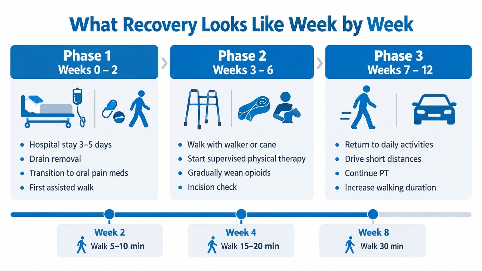
In the first 0 to 2 weeks, most patients are still dealing with the hospital phase, drains, wound care, and the shift from stronger pain medicine to oral medication. Early walking is encouraged with help, because movement protects the lungs, the circulation, and the gut. In weeks 2 to 6, the big rule is still simple, no bending, lifting, or twisting beyond what the surgeon allows.
Driving often returns somewhere around week four to six if pain is controlled and reaction time is safe. That window can't be rushed, and social support matters here because routine tasks, meal prep, and transport can all be difficult. Patients who live alone usually need a concrete plan before surgery, not after.
The later recovery window
From weeks 6 to 12, physical therapy usually starts focusing on gait and core stabilization. Desk work often returns in this period if the patient can sit safely and manage discomfort. From months 3 to 6, strengthening increases and brace use, if prescribed, starts to fall away.
By months 6 to 12, follow-up imaging helps confirm that the fusion is maturing as intended. Functional recovery often plateaus around a year, although nerve recovery can continue longer. That distinction matters, because bone healing and symptom healing don't finish on the same calendar.
Walking farther is not the same as healing faster. The goal is steady progress without provoking the construct.
The safest recovery plans are coordinated, specific, and realistic. A patient who knows what to expect week by week is less likely to panic when progress feels slow.
Risks and the Bone Quality Question
Every thoracic lumbar fusion has risks, and they should be named plainly. These include infection, hardware loosening, pseudarthrosis, adjacent segment disease, dural tear, and neurological injury. Thoracic cases also carry region-specific concerns such as pleural violation or, less commonly, chylothorax.
What drives risk
Some risks are tied to the patient, like diabetes, smoking, frailty, or poor bone quality. Others are tied to the operation itself, including the number of levels fused, the amount of correction needed, and whether the surgery is open or minimally invasive. In one large administrative analysis, serious complications were around 7.7% and any complication around 12.3%, while long-segment instrumented fusions reported wound complications at 4.24% (complication analysis).
Why bone quality changes the decision
Bone quality is the piece many patients never hear about until the plan is nearly final. A recent meta-analysis found that preoperative Hounsfield unit values under 118 were linked to a 2.8-fold higher risk of postoperative mechanical and junctional complications, and it proposed a risk tiering system of <120 HU high risk, 120 to 150 HU intermediate risk, and >150 HU low risk (2025 meta-analysis). That doesn't automatically rule out surgery, but it can change the fixation plan, the timing, and the conversation about alternatives.
A surgeon may order DEXA, review FRAX when appropriate, check vitamin D, and use CT-based bone assessment to understand whether screws are likely to hold. In lower-density bone, the plan may shift toward cement augmentation, larger or expandable screws, or a different fixation strategy. Some patients also need bone-building medication before and after surgery, especially if the goal is to improve the odds of a durable fusion.
For graft choices and fusion support, a practical overview of bone grafting is available through the bone grafting overview.
A technically successful fusion can still fail mechanically if the bone can't support the construct.
Before signing off, a patient should ask whether bone density has been checked, whether the construct has been adapted for bone quality, and whether the surgeon expects any special measures to reduce loosening or non-union.
Making the Decision at a Multidisciplinary Center
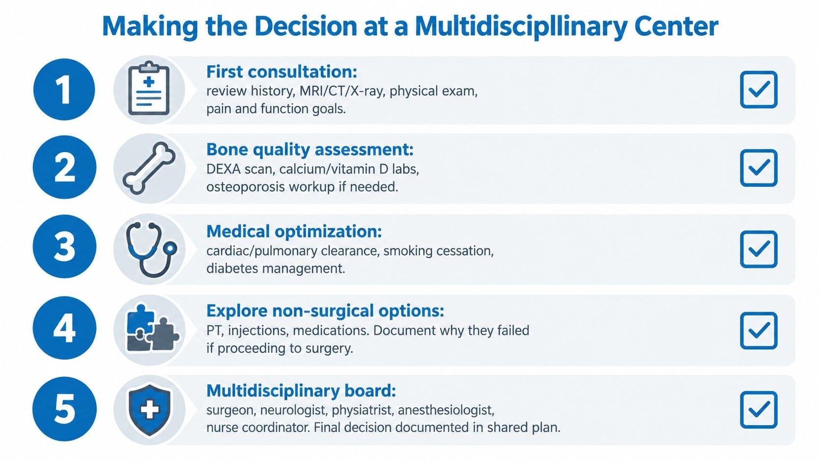
A good fusion decision is rarely made in isolation. It works best when the surgeon, physiatrist, pain specialist, anesthesiologist, and physical therapist are looking at the same problem from different angles. The first visit should cover the history, imaging, prior treatment, bone density results if they exist, and the patient's actual goals for walking, work, and daily life.
What the team is trying to answer
The question is not whether surgery is possible. It's whether fusion is the right trade-off compared with motion-preserving care, continued nonoperative treatment, or a different operation entirely. A patient with severe pain, major instability, and weak bone may need a very different plan from a patient whose symptoms are coming from one level and whose bone quality is strong.
Second opinions often change the surgical level or the approach. That happens because different surgeons weigh alignment, stability, and soft-tissue preservation differently, and because the long-term plan has to account for comorbidities, psychosocial stress, and recovery support at home. The best plan is explicit about re-operation risk, rehab, and who will help during the first weeks.
Questions that should be answered before consent
- Why fusion, and why now: The surgeon should explain why conservative care no longer fits.
- Which levels are included: The patient should know why the construct starts and stops where it does.
- How bone quality affects the plan: Bone density, vitamin D, and fixation strategy should be discussed clearly.
- What recovery support is needed: Transportation, help at home, and work restrictions should be planned in advance.
- What happens if symptoms persist: Re-operation risk and rehabilitation milestones should be part of the consent discussion.
At one multidisciplinary center, Interventional Pain Management, the spine team can coordinate diagnosis, conservative care, and surgical planning across pain management, orthopedics, and spine surgery when that mix of expertise is needed. That kind of coordination helps patients avoid a rushed decision and keeps the plan tied to the actual problem, not just the imaging report.
If thoracic lumbar fusion is on the table, Interventional Pain Management can help sort out whether surgery is the right next step, what bone quality means for the plan, and which non-surgical options still deserve a fair trial. A patient can learn more about coordinated spine evaluation and treatment at Interventional Pain Management, then bring those questions to a consultation that's built around clarity, not pressure.