
A planted foot, a sudden turn, and a knee that gives way can leave an active adult asking the same urgent question: Is this an ACL tear, an MCL tear, or both? A direct blow to the outside of the knee creates a different injury pattern, but pain, swelling, stiffness, and trouble walking can overlap enough to make self-diagnosis unreliable.
The distinction matters because the ACL and MCL have different jobs and different healing behavior. A stable, isolated MCL injury often responds to bracing and rehabilitation, while an ACL injury in a person who wants to return to pivoting sports may require reconstruction to restore dependable rotational stability. Combined injuries need an even more individualized plan.
The comparison below focuses on the decisions that affect recovery, including how symptoms differ, what an examination and MRI can establish, when bracing may be enough, and which objective tests should precede a return to sport. Adults in New Jersey and Staten Island, from recreational runners to competitive athletes and physically demanding workers, can use this information to prepare for an orthopedic evaluation without treating it as a substitute for one.
Table of Contents
- Introduction to ACL vs MCL Tears and Why the Difference Matters
- Understanding Knee Ligament Anatomy and Stability Roles
- How ACL and MCL Tears Happen and How Symptoms Differ
- How Clinicians Diagnose ACL and MCL Tears
- Treatment Options for ACL vs MCL Tears Including Combined Injuries
- Recovery Timelines and Return to Sport Criteria
- When to Seek Care and Which Option Fits Your Situation
Introduction to ACL vs MCL Tears and Why the Difference Matters
An ACL tear and an MCL tear are both knee ligament injuries, but they don't represent the same mechanical problem. The ACL sits inside the joint and controls forward and rotational movement. The MCL runs along the inner side of the knee and resists forces that push the knee inward.
ACL injuries are a major clinical concern. The reported annual incidence in the United States is about 1 in 3,500 people, and around 400,000 ACL reconstructions are performed each year, according to the NCBI clinical overview of ACL injury. The same source describes ACL injury as frequently occurring with other internal knee damage, so an MRI and careful examination may need to assess more than the ligament alone.
MCL injuries are also common in athletic settings. A review reports annual incidence ranging from 0.24 to 7.3 per 1,000 people, with MCL injuries accounting for 7.9% of athletic knee injuries in one review, as documented in this clinical review of MCL injuries.
A quick clinical comparison
| Feature | ACL tear | MCL tear |
|---|---|---|
| Location | Inside the knee joint | Inner side of the knee |
| Typical force | Pivoting, sudden deceleration, or twisting | Valgus force, often from a blow to the outside of the knee |
| Main symptom pattern | Giving way, rotational instability, rapid swelling | Localized inner-knee pain and tenderness |
| Healing tendency | Doesn't reliably heal spontaneously | Often has reliable healing potential |
| Common management question | Whether instability and activity goals justify reconstruction | Whether bracing and rehabilitation can restore stability |
| Combined injury issue | May require reconstruction for pivot stability | May be protected first, or reconstructed if severe instability persists |
A knee that feels unstable after a twist deserves prompt assessment, even if pain has eased. Delayed evaluation can make it harder to distinguish ligament laxity from pain-limited movement and can allow associated meniscal or cartilage problems to go unrecognized.
Understanding Knee Ligament Anatomy and Stability Roles
The easiest way to understand the ACL versus MCL difference is to locate each ligament in relation to the joint.
The anterior cruciate ligament crosses through the center of the knee, between the thighbone and shinbone. It helps limit forward movement of the shin relative to the thighbone and contributes to control during rotation, cutting, landing, and sudden deceleration.
The medial collateral ligament sits outside the joint on the inner side of the knee. It acts like a strong side support, resisting a valgus force, which is a force that drives the knee inward. Because the MCL is extra-articular, meaning outside the joint cavity, it generally has better healing potential than the intra-articular ACL.
A simple stability map
- Central pivot: The ACL helps control forward translation and rotation.
- Medial buttress: The MCL protects the inner side of the knee against inward angulation.
- Shared system: Both ligaments contribute to stability, but they respond to different loads.
- Combined failure: When both are injured, the knee can become unstable in more than one direction.
That biology explains a common treatment pattern. An isolated MCL tear can often heal as the knee is protected with a brace and gradually loaded through rehabilitation. The ACL's location inside the joint and its limited spontaneous healing capacity make reliable restoration more difficult, particularly for active adults who need to cut, pivot, or change direction.
The distinction doesn't mean every ACL tear requires surgery or every MCL tear can be managed without it. Tear completeness, associated injuries, examination findings, activity demands, and the patient's ability to regain functional stability all shape the recommendation.
Some patients also ask about general joint mobility products while recovering from a knee injury. Information about hyaluronic acid for joint mobility may be useful for understanding that topic, but it doesn't replace ligament protection, physical therapy, or an orthopedic diagnosis. General guidance on ligament and soft-tissue injuries is also available through this overview of sprains.
How ACL and MCL Tears Happen and How Symptoms Differ
Mechanism often provides the first clue, although it can't confirm the diagnosis on its own.
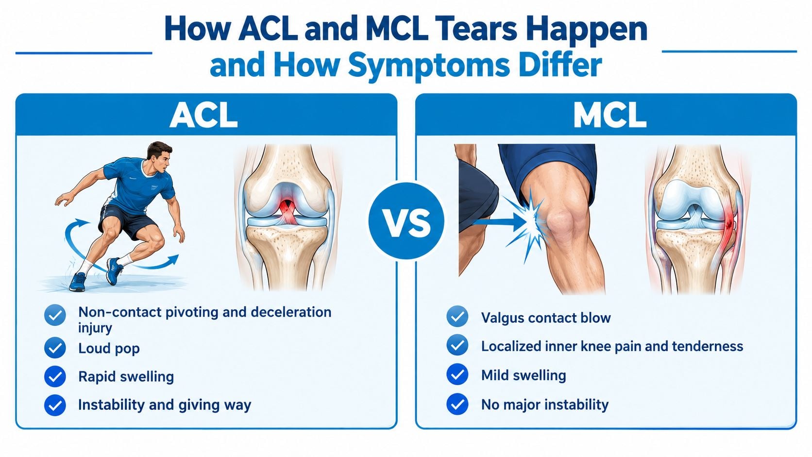
An ACL tear commonly follows a non-contact event. The athlete plants the foot, slows rapidly, lands awkwardly, or changes direction while the knee rotates. A pop, rapid swelling, and a sense that the knee won't hold during pivoting are classic warning signs.
An MCL tear more often follows a contact force. A blow to the outside of the knee can push the joint inward and strain the ligament on the medial side. Pain and tenderness usually concentrate along the inner knee, and swelling may be less dramatic than with an ACL injury.
Practical rule: A loud pop with rapid swelling and giving way raises concern for ACL injury, while focal inner-knee tenderness after a side impact raises concern for MCL injury. Neither pattern proves the diagnosis.
Symptoms that overlap
Both injuries can cause:
- Pain: Weight-bearing and bending may become uncomfortable.
- Swelling: The amount and timing vary with the injury pattern and associated damage.
- Stiffness: Swelling can limit full extension or flexion.
- Difficulty walking: Protective muscle guarding can make the leg feel weak.
- Reduced confidence: Fear of movement may persist even after pain improves.
An ACL tear tends to produce a deeper sense of instability, especially during turning or sudden direction changes. The knee may buckle even when the person can walk in a straight line. An MCL tear tends to hurt more directly over the inner ligament, particularly when the knee is stressed inward or when the medial tissues are touched.
A person with a twisting injury can also have a meniscal tear, bone bruise, cartilage injury, or more than one ligament injury. That's why pain location alone isn't enough. A patient resource on torn meniscus symptoms and care can help explain one source of overlapping symptoms, but a clinician still needs to determine which structures are injured.
For the first response, protecting the knee, limiting activities that provoke instability, and arranging an evaluation are safer than testing the joint repeatedly. Urgent evaluation is appropriate when the knee is visibly deformed, locked, rapidly enlarging, numb, unusually cold, or impossible to bear weight on.
How Clinicians Diagnose ACL and MCL Tears
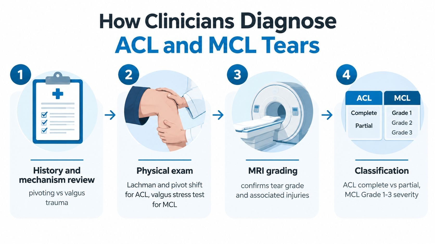
Diagnosis begins with the story of the injury. The clinician asks whether the knee twisted without contact, received a side impact, changed position during landing, or suffered a high-energy event. The timing of swelling, the presence of a pop, the location of tenderness, and any sensation of giving way help organize the examination.
The physical examination then tests the specific stability roles of each ligament. The Lachman test evaluates forward movement of the shin and is a key examination maneuver for ACL laxity. The pivot-shift test assesses abnormal rotational movement, although pain and swelling can limit its usefulness early after injury.
For the MCL, the clinician applies a controlled valgus stress test at selected knee positions. Medial gapping or pain can indicate injury, but the examiner also considers whether the looseness comes from the MCL, another ligament, or generalized swelling and guarding.
What MRI can and cannot tell
MRI helps show the location and extent of ligament damage and can identify associated injuries. MCL tears are commonly graded from 1 to 3:
- Grade 1: The ligament remains intact, with surrounding edema.
- Grade 2: The ligament is partially torn.
- Grade 3: The ligament is completely disrupted.
This grading system supports prognosis and rehabilitation planning, but location matters too. Proximal and midsubstance MCL tears often heal with increased stiffness and little residual laxity, while distal insertion injuries may heal less reliably and leave more valgus laxity, according to the Radiopaedia overview of MCL injury grading.
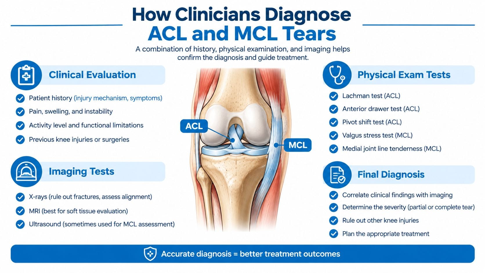
Partial ACL tears require a different interpretation. MRI may show abnormal internal signal with some intact fibers and preserved overall thickness, but an image can't determine by itself whether the knee is functionally stable. The clinician combines the scan with laxity testing, activity goals, symptoms, and the patient's ability to control the knee during movement.
A useful consultation includes questions about the tear location, whether the meniscus or cartilage is involved, how much laxity remains, and what findings would change the treatment plan. An academy comparison for beginners can help explain why training demands differ across activities, but the orthopedic evaluation must translate those demands into specific knee-stability requirements.
Treatment Options for ACL vs MCL Tears Including Combined Injuries
Treatment depends on the ligament, tear pattern, stability examination, and the person's goals. The simple rule that “ACL means surgery and MCL means bracing” is useful as a starting point, but it fails for partial tears, low-demand activities, persistent laxity, and combined injuries.
| Criteria | ACL Tear | MCL Tear | Combined ACL plus MCL |
|---|---|---|---|
| Healing biology | Intra-articular and not reliably healed spontaneously | Extra-articular with generally reliable healing potential | Each ligament must be assessed separately |
| Bracing role | May support selected nonoperative plans, but doesn't restore a torn ACL | Often protects the healing ligament during rehabilitation | Frequently used to protect the MCL before deciding on ACL surgery |
| Reconstruction indication | Considered when instability affects pivoting activity or function | Considered when severe or persistent medial laxity remains | Considered when the MCL remains unstable or the injury pattern threatens overall knee stability |
| Main risk to manage | Recurrent instability and associated internal damage | Residual valgus laxity or stiffness | Stiffness, graft stress, and instability in multiple directions |
| Rehabilitation focus | Motion, quadriceps control, strength, neuromuscular stability, and return-to-sport testing | Protection, motion restoration, progressive strengthening, and valgus control | Restore MCL stability while protecting motion, then address ACL deficiency when appropriate |
Isolated injuries
A stable, lower-grade MCL injury is commonly treated with a hinged brace, activity modification, progressive range of motion, and physical therapy. The rehabilitation team watches pain, medial tenderness, motion, and valgus stability rather than advancing activity because the calendar has moved forward.
An ACL-deficient knee may be managed without reconstruction in selected patients, especially when functional demands are modest and the knee remains stable during desired activities. Active adults who need repeated pivoting, cutting, or rapid deceleration may have a stronger reason to discuss reconstruction.
Combined ACL and MCL injury
The combined injury question is more complex. Expert guidance notes that ACL reconstruction outcomes are generally good when the MCL is treated nonoperatively, and operative MCL treatment can increase stiffness risk. Many protocols therefore protect the MCL for about 4 to 6 weeks before aggressive multiplanar loading, then reassess stability and motion, as described in this review of combined ACL and MCL injuries.
That approach isn't automatic. A severe grade 3 MCL injury, a distal avulsion pattern, or persistent valgus laxity may justify MCL repair or reconstruction. A recent systematic review found that MCL reconstruction may better restore biomechanics and reduce residual valgus laxity in grade 3 injuries, while broader evidence still reports conflicting findings and no single best strategy, as discussed in this systematic review of combined ACL and MCL treatment.
The practical decision is whether the medial side can become dependable enough to protect an ACL graft and support the patient's activity goals. Treatment planning may include conservative care, bracing, rehabilitation, minimally invasive procedures, or surgery. An overview of available orthopedic and interventional treatments can help patients understand the range of care, but the final plan must follow the examination and imaging findings.
Recovery Timelines and Return to Sport Criteria
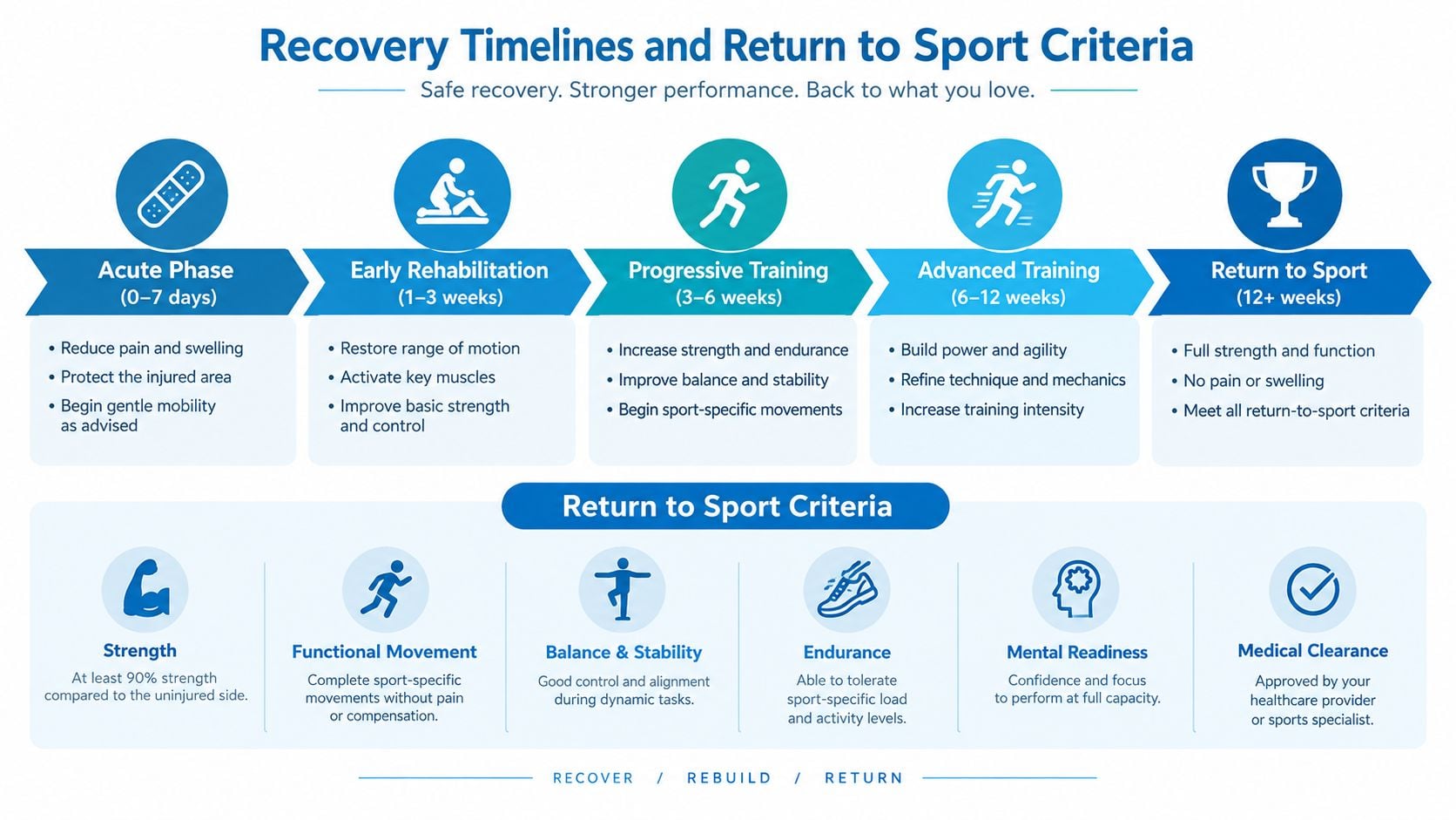
Recovery after an ACL or MCL injury shouldn't be reduced to a single number of weeks or months. The ligament's healing behavior, treatment pathway, motion, strength, associated injuries, and sport demands all influence progression.
MCL rehabilitation typically advances from protection and swelling control to restored motion, strength, balance, and controlled lateral loading. The clinician needs evidence that medial stability is returning before the athlete resumes cutting or contact. A painful or stiff knee isn't ready because walking has improved.
ACL reconstruction rehabilitation follows a longer performance pathway. Early work emphasizes swelling control, full extension, quadriceps activation, and a normal walking pattern. Later stages add progressive strengthening, running, landing mechanics, directional changes, and sport-specific drills.
Objective clearance matters
A 2025 return-to-play framework emphasizes several criteria, including:
- Time: At least 9 months after surgery, according to the published return-to-play framework.
- Strength: At least 90% quadriceps strength symmetry, using the same source.
- Hop performance: Hop-test symmetry at the stated threshold.
- Psychological readiness: Confidence and readiness to resume demanding activity.
These measures should be interpreted together. A strong quadriceps score can't compensate for swelling, restricted motion, poor landing control, or recurrent giving way. Conversely, psychological readiness matters because hesitation can alter movement mechanics and prevent a safe return even when physical tests look acceptable.
Sport level changes the expectation
Multiligament injuries create especially variable outcomes. A systematic review found return-to-sport rates ranging from 41.2% to 100%, with reported time to return ranging from 6.7 to 24.9 months, as reported in the same return-to-play evidence. Because that source URL should be used only once, those findings are cited here, where they directly inform clearance decisions.
A later study reported that only 25% resumed high-level pivoting sports after multiligament injury, also described in that source. The implication isn't that athletes must abandon their sport. It means the return plan should match the actual demands of the sport, and the care team should discuss whether the goal is recreational participation, full competitive performance, or a modified role.
When to Seek Care and Which Option Fits Your Situation
A knee injury deserves orthopedic evaluation when swelling appears rapidly, the knee gives way, full motion doesn't return, pain remains focused along the inner ligament, or weight-bearing is difficult. Immediate care is more appropriate for deformity, a locked knee, major swelling, numbness, a cold foot, or an inability to move the ankle and toes normally.
The right pathway depends on the person and the injury pattern:
- Competitive pivoting athlete: Evaluation should address rotational instability, associated injuries, graft planning, and objective return-to-sport testing.
- Recreational exerciser: A stable MCL injury may fit bracing and rehabilitation, while recurrent instability after an ACL injury requires a more detailed discussion.
- Physically demanding worker: The plan should account for lifting, climbing, kneeling, uneven surfaces, and the need for dependable medial and rotational stability.
- Person with a combined injury: The MCL's tear location and healing response should be reassessed before the ACL strategy is finalized.
Patients should bring MRI images and reports, list the injury mechanism, describe any pop or giving-way episodes, and identify the activities that must be regained. Interventional Pain Management evaluates joint and sports injuries, including ACL and MCL tears, through orthopedic assessment and coordinated treatment options ranging from non-invasive care to surgical planning.

Adults with an ACL tear, MCL tear, or combined knee injury can visit Interventional Pain Management for evaluation of symptoms, examination findings, and imaging, followed by a treatment plan matched to activity demands. The practice serves patients across New Jersey and Staten Island and can help clarify whether bracing, rehabilitation, or surgical consultation is the appropriate next step.