

Maria is 52, and the pain started while lifting groceries. A sharp line travels from her lower back into her right calf, making sleep difficult and turning every step into a calculation. After six weeks of activity changes, medication, and conservative care, her physician discusses an epidural steroid injection, not as a promise to erase the disc problem, but as a possible window in which the irritated nerve can settle enough for recovery work to begin.
That distinction matters. The benefits of epidural steroid injections are most meaningful when the diagnosis is accurate, the medication reaches the appropriate area, and the patient uses improved mobility for rehabilitation. An epidural steroid injection can reduce inflammation around a spinal nerve, but it can't rebuild a worn disc, widen a narrowed spinal canal, or guarantee permanent pain relief.
Table of Contents
- What an Epidural Steroid Injection Does for You
- How the Injection Targets Nerve Inflammation
- Proven Clinical Benefits and What the Evidence Shows
- Who Makes a Good Candidate for the Procedure
- Comparing Transforaminal, Interlaminar, and Caudal Approaches
- Realistic Expectations and the Limits of the Benefit
- Risks, Side Effects, and What the Day Looks Like
- Key Takeaways With Questions for Your Specialist
What an Epidural Steroid Injection Does for You
Pain that shoots down an arm or leg often reflects more than a mechanical pinch. A disc herniation or narrowed passage can irritate a spinal nerve, and the resulting inflammation may amplify otherwise ordinary signals into burning, tingling, or electric pain. An epidural steroid injection, or ESI, places corticosteroid and local anesthetic into the epidural space near that irritated nerve. This space sits outside the protective covering of the spinal cord and nerve roots.
An ESI has a narrower job than several treatments patients may confuse with it. Oral steroids circulate throughout the body. A trigger-point injection treats a painful muscle or taut band of muscle. Surgery changes or removes a structural source of compression. An ESI delivers medication near a suspected pain generator, aiming to reduce inflammation without repairing a disc, widening the spinal canal, or changing the underlying anatomy.
A patient guide to corticosteroid injections explains how these medicines are used. The planned injection still needs to fit the patient's symptoms, examination, imaging, and suspected nerve involvement.
What changes after the procedure
The local anesthetic may reduce pain or cause numbness during the first several hours. That early change can fade before the steroid has had time to work. The corticosteroid's anti-inflammatory effect often develops gradually over three to seven days, although timing varies.
A meaningful response may be practical rather than dramatic:
- Movement becomes easier: Walking, standing, or changing position may require less guarding.
- Sleep becomes less disrupted: Lower arm or leg pain can make comfortable positioning easier.
- Rehabilitation becomes possible: Physical therapy can address strength, flexibility, posture, and movement patterns when pain no longer dominates each exercise.
- Medication needs may fall: If function improves, some patients can discuss reducing pain medication with the prescribing clinician.
The injection needs a functional job. Walking farther, sleeping better, or participating in therapy is often a more useful measure than expecting every sensation to disappear.
Evidence supports a time-limited role. A systematic review found measurable pain improvement for sciatica from lumbar disc herniation within three months, with a mean difference of 0.44, and within six months, with a mean difference of 0.66. Long-term pain relief remained limited (National Library of Medicine evidence summary). The goal is a temporary anti-inflammatory bridge that creates room for rehabilitation and recovery, not a claim that the structural condition has been cured.
How the Injection Targets Nerve Inflammation
An irritated nerve root can be compared with a frayed electrical wire surrounded by swollen insulation. The wire hasn't necessarily been severed, but the surrounding irritation makes normal signals feel like burning, tingling, or electric pain. A corticosteroid acts less like a repair crew and more like a cooling treatment that reduces the inflammatory environment around the nerve.
The process usually involves several parts:
- The clinician identifies the target. Symptoms, neurological findings, and imaging help determine which nerve root may be responsible.
- The skin is numbed. A small amount of local anesthetic reduces discomfort at the entry point.
- The needle is guided into the epidural space. This fat-filled sleeve lies outside the dura, the protective membrane around the spinal cord and nerve roots.
- Medication is deposited near the inflamed area. The steroid reduces inflammatory activity, while the anesthetic temporarily quiets pain transmission.
The steroid may be dexamethasone or methylprednisolone. The anesthetic may be lidocaine or bupivacaine. These medicines serve different purposes. The anesthetic can provide short-lived confirmation that the treated region is relevant, while the corticosteroid is intended to produce the more gradual anti-inflammatory effect.
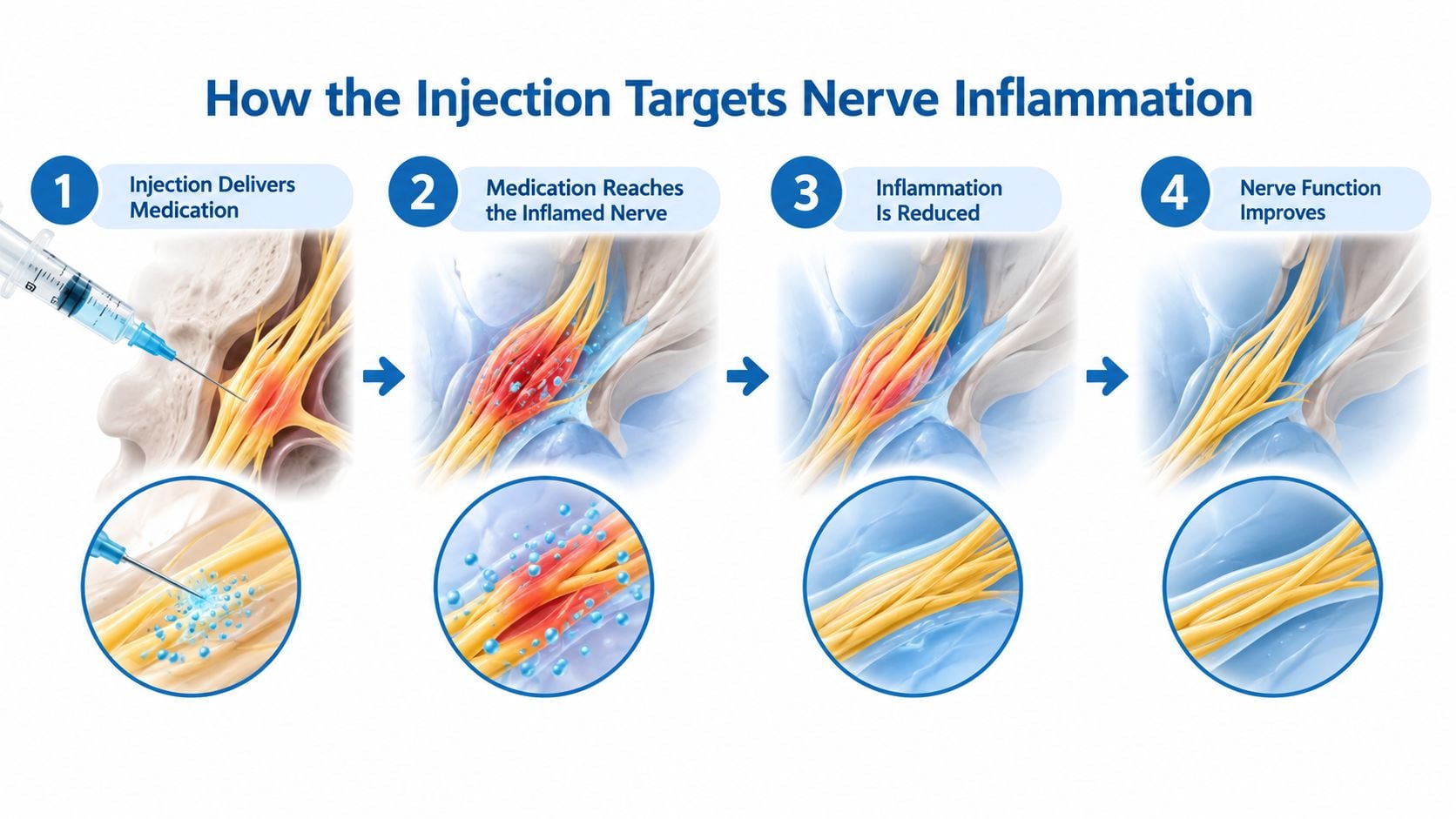
Why image guidance matters
Fluoroscopy or CT guidance lets the physician see the needle's position rather than relying on surface landmarks alone. Contrast helps confirm that the medication is spreading in the intended pattern and helps the operator avoid unwanted vascular or intrathecal placement. For a transforaminal injection, imaging also supports close deposition near the selected nerve-root sleeve.
A typical sequence of sensations is straightforward. There may be brief pressure during needle placement, temporary numbness or heaviness from the anesthetic, and possible soreness afterward. The early anesthetic effect may wear off the same day, while the steroid's benefit, if it occurs, usually develops over the following days. Worsening pain, new weakness, or another concerning neurological change isn't part of the expected recovery and should prompt a call to the treating clinic.
Proven Clinical Benefits and What the Evidence Shows
The clearest evidence supports radicular pain, which comes from irritation of a spinal nerve root. Pain may travel from the neck into an arm, or from the lower back through the buttock and leg. ESIs are less dependable for isolated axial low back pain, where symptoms remain mainly in the back without a recognizable nerve-root pattern.
For sciatica related to lumbar disc herniation, a systematic review and meta-analysis summary found pain relief in the short to medium term. Improvement was seen within three months and six months, but the analysis did not establish durable long-term pain control. The injection can calm an inflamed nerve long enough to support walking, sleep, and rehabilitation. It does not remove a disc herniation or permanently correct spinal narrowing.
Pain relief is only one outcome
The evidence is supportive but modest. A meta-analysis of randomized trials found that epidural corticosteroids increased the odds of short-term pain relief compared with placebo, with an odds ratio of 2.61. Benefit remained detectable through as long as twelve months, with an odds ratio of 1.87 (primary care evidence review). These figures describe outcomes in study groups, not a promise for one patient.
Function may improve too. A review summarized in neurological guidance reported a number needed to treat of 4 for pain and 6 for disability in cervical and lumbar radiculopathy, while finding no strong long-term pain effect (neurology guideline review). In practical terms, a carefully selected patient may move, sleep, or exercise more comfortably, even if some pain remains.
Medication use is another possible benefit. The evidence summary reported reduced opioid use after injection, with a mean difference of −14.45. That result suggests a medication-sparing effect for some patients, but medication changes still require clinical supervision.
ESI Benefit Outcomes by Time and Condition
| Outcome | 2-4 Weeks | 6-12 Weeks | Beyond 12 Weeks |
|---|---|---|---|
| Radicular pain | Short-term relief is supported | Relief may continue for some patients | Durable pain relief is uncertain |
| Disability | Modest improvement may appear | Functional gains can persist in some cases | Long-term benefit is inconsistent |
| Opioid use | Reduction may occur after injection | Medication needs should be reassessed clinically | No reliable long-term medication effect is established |
| Axial back pain | Evidence is weaker | Benefits are less consistent | No dependable lasting benefit is established |
For sciatica from lumbar disc herniation, reviews found greater leg-pain relief and functional improvement at about six weeks than placebo, although the gains often did not reach a minimal clinically important difference (spine evidence review). The meaningful goal is therefore improved function during a limited window for rehabilitation. Diagnosis, injection technique, and patient selection determine whether that window becomes useful, rather than serving as a definitive treatment for structural spine disease.
Who Makes a Good Candidate for the Procedure
The clearest candidate has a recognizable nerve-root pattern. A person may have pain following a particular route through the arm or leg, altered sensation, or weakness that fits a cervical or lumbar nerve distribution. A disc herniation or symptomatic narrowing on MRI can strengthen the case when it matches the clinical findings.
The decision usually follows a period of conservative care. Activity modification, physical therapy, and oral analgesics may be tried first, unless the pain or neurological symptoms require a different pace of evaluation. The important question isn't whether an MRI shows an abnormal disc. The question is whether the abnormality explains the symptoms the patient has.
Selection depends on correlation
A focused evaluation typically includes:
- A symptom map: The clinician notes where pain, tingling, or numbness travels and what activities provoke it.
- A neurological examination: Strength, reflexes, sensation, gait, and functional movement help identify nerve involvement.
- Imaging review: MRI findings are compared with the examination, rather than treated as a diagnosis by themselves.
- A recovery plan: The patient identifies what improved function would make the procedure worthwhile.
Patients seeking an explanation for sciatica and a pinched nerve should expect the consultation to address both the pain pattern and the suspected source.
When the evidence is weaker or the timing is wrong
An injection is less compelling for nonspecific back discomfort without leg or arm symptoms, or for pain that appears primarily muscular or mechanical. Post-surgical pain can require a more individualized assessment because scar tissue, altered anatomy, hardware, and several possible pain generators may be involved.
Certain health conditions can make an injection unsafe or require postponement. Active infection, uncontrolled diabetes, bleeding disorders, and pregnancy need specific medical discussion before proceeding. Anticoagulant use also requires careful medication planning. A patient shouldn't stop a blood thinner independently.
A good candidate isn't defined by an MRI alone. The diagnosis, examination, imaging, safety profile, and rehabilitation goal must point in the same direction.
Shared decision-making matters because an ESI is optional care for many patients, not an automatic step. The clinician should explain expected benefit, alternatives, possible risks, and what happens if the injection doesn't help.
Comparing Transforaminal, Interlaminar, and Caudal Approaches
The approach determines where the needle enters and how medication spreads. None is universally superior. The physician chooses based on the suspected nerve root, the number of levels involved, prior surgery, anatomy, and the desired coverage.
ESI Technique Comparison
| Technique | Needle Entry | Best Indication | Coverage |
|---|---|---|---|
| Transforaminal | Near the exiting nerve-root sleeve | Single-level radicular pain | Focused coverage |
| Interlaminar | Between the laminae through the midline epidural space | Bilateral or multilevel symptoms | Broader spread |
| Caudal | Through the sacral hiatus | Selected cases after lumbar surgery or altered anatomy | Spread upward through the epidural space |
A transforaminal injection is the most targeted option of the three. It places medication near the exiting nerve root and is often considered when one level clearly matches the patient's symptoms. Because the medication is concentrated near the suspected source, the approach can be useful when the clinical picture is focal.
An interlaminar injection enters the central epidural space. Medication can spread across more than one level, which may suit bilateral symptoms or multilevel narrowing. A caudal injection enters through the sacral hiatus. That route can be useful when previous lumbar surgery, scar tissue, or hardware makes a higher entry more difficult.
Each technique should use fluoroscopic or CT guidance and contrast to verify placement and reduce the chance of intravascular or intrathecal injection. Recent guideline summaries indicate that transforaminal injections tend to perform better for radicular pain, while interlaminar and caudal effects can be shorter or less consistent, with no meaningful long-term superiority across approaches (recent guideline summary).
Patients can review general types of injections in pain management, but the final choice belongs to the treating specialist. Repeat injections should follow a clearly positive prior response and a defined functional objective, not a routine schedule.
Realistic Expectations and the Limits of the Benefit
An ESI doesn't dissolve a disc herniation or rebuild a narrowed spinal canal. It doesn't restore disc height, reverse degenerative changes, or guarantee that surgery will be avoided. The structural finding may remain on imaging even when the patient feels substantially better.
The most useful analogy is a bridge over a difficult stretch of road. The steroid may reduce nerve-root swelling long enough for the patient to walk, sleep, exercise, and participate in rehabilitation. Physical therapy and activity changes then address strength, movement confidence, and daily mechanics that medication alone can't provide.
What success should look like
Complete pain elimination isn't the only meaningful result. A patient may judge the procedure successful if sitting through a workday becomes possible, walking no longer stops at the end of the driveway, or sleep improves enough to support daytime activity.
Evidence summarized in recent reviews describes mainly short-term benefit, with modest radiculopathy improvement for about three months and disability improvement sometimes extending toward six months, but no reliable proof of durable long-term pain relief or consistent surgery avoidance (evidence discussion of ESI duration). The benefit can be real without being permanent.
Before the injection, patients should write down a small number of functional goals:
- Mobility: Walk, stand, or climb stairs with less interruption.
- Rest: Sleep in a more comfortable position.
- Rehabilitation: Complete prescribed exercises without a severe flare.
- Medication: Discuss a safe reduction if symptoms improve.
Steroid benefit often develops over several days. Injection-site soreness or a temporary flare can occur. Durable improvement is more plausible when the patient uses the improved period for guided rehabilitation instead of returning immediately to the movements that aggravated the condition.
Risks, Side Effects, and What the Day Looks Like
Most image-guided ESIs are outpatient procedures, and serious complications are uncommon. Still, a careful consent discussion is essential because the needle enters a sensitive area near spinal nerves and the medication can have effects beyond the injection site.
Common short-term effects include local soreness, flushing, brief numbness, and temporary blood-sugar elevation in people with diabetes. Less common complications include a dural puncture headache, infection, bleeding, or nerve injury. The exact risk depends on the spinal region, approach, medical history, medications, and operator technique, so patients should ask the treating clinician for procedure-specific estimates rather than relying on a universal figure.
What happens on procedure day
The visit generally follows a predictable sequence:
- Arrival and review: The team checks medications, allergies, medical conditions, and the planned target.
- Consent: The physician explains expected benefits, alternatives, and risks.
- Positioning: The patient lies in a position that gives the imaging equipment access to the spine.
- Skin preparation: The area is cleaned carefully, and local anesthetic numbs the entry site.
- Image-guided placement: Fluoroscopy or CT guides the needle, and contrast confirms the intended spread.
- Medication delivery: The steroid and anesthetic are injected, often with pressure or brief reproduction of familiar symptoms.
- Recovery: The patient rests while the team checks sensation, strength, blood pressure, and overall comfort.
Light sedation may be offered in selected cases, but it isn't always necessary. Patients receiving sedation usually need a driver. The treating clinic gives individualized instructions about regular medications, activity, diabetes monitoring, and anticoagulants.
When to call
Same-day discharge is typical after observation, but a responsible adult may be needed if sedation was used. Patients should contact the clinic promptly for fever, increasing redness or drainage, severe worsening headache, new or progressive weakness, loss of bladder or bowel control, or numbness that doesn't resolve as instructed.
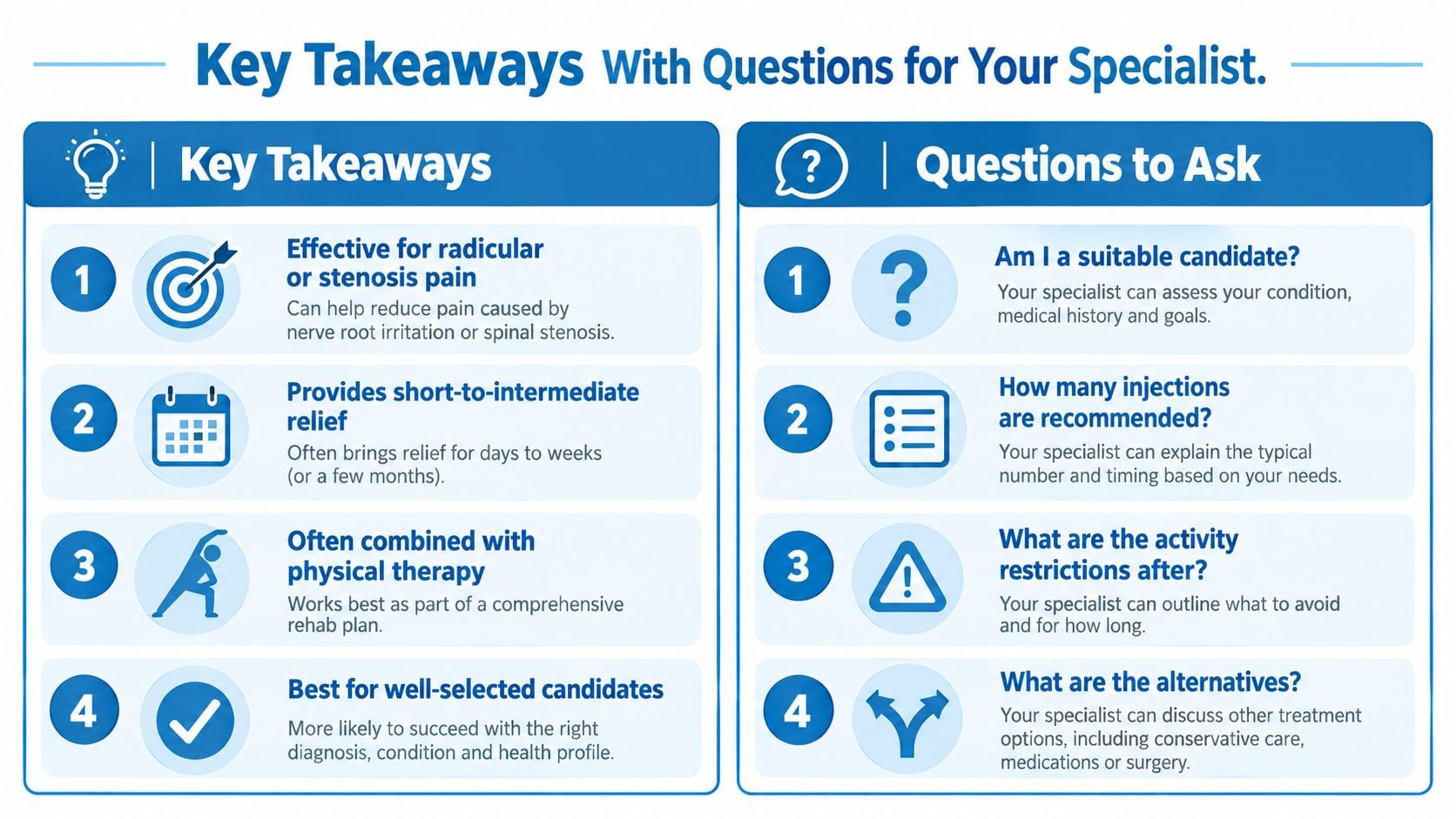
Key Takeaways With Questions for Your Specialist
The central message is simple. Epidural steroid injections can provide meaningful short-to-intermediate anti-inflammatory relief for well-selected radicular pain, but their value depends on what happens during that window. The injection is most useful when it helps a patient move, sleep, reduce medication reliance under supervision, and participate in a broader recovery plan.
The consultation should produce more than a procedure date. It should produce a diagnosis, a target, a technique, a safety plan, and a functional goal.

Questions worth bringing
- Diagnosis: Which nerve root or structural problem is being treated?
- Evidence: Does the symptom pattern match the imaging?
- Approach: Why is transforaminal, interlaminar, or caudal access appropriate?
- Goal: What specific activity should improve if the injection works?
- Timing: When should pain relief begin, and when should the patient call if it doesn't?
- Rehabilitation: What physical therapy or activity plan should follow?
- Medication safety: How should diabetes medicines, anticoagulants, or pain medicines be managed?
- Repeat treatment: What response would justify another injection, and what alternatives exist?
Short FAQ
Can an ESI cure a herniated disc?
No. It can calm inflammation around an irritated nerve, but it doesn't repair the disc or remove spinal narrowing.
Is temporary relief still useful?
Yes, if it enables meaningful rehabilitation or improves function. Relief without a recovery plan may not produce durable change.
Do all patients with sciatica benefit?
No. Diagnosis, symptom pattern, imaging correlation, approach, and medical safety all influence the outcome.
What if the injection doesn't help?
The clinician may reassess the diagnosis, review the imaging and technique, continue rehabilitation, or discuss another treatment. New weakness or serious neurological symptoms require prompt medical attention.
Patients in New Jersey and Staten Island can discuss diagnosis, image-guided epidural injections, rehabilitation planning, and alternatives with the multidisciplinary team at Interventional Pain Management. A consultation can clarify whether an ESI fits the patient's symptoms, health history, and functional goals before any procedure is scheduled.