
Facet joint syndrome is arthritis-like degeneration, inflammation, or injury of the small joints connecting neighboring vertebrae, and lumbar facet joints account for about 15% to 45% of chronic low back pain cases. These joints also contribute substantially to chronic neck pain, but no single symptom, examination maneuver, or scan can prove that a facet joint is the pain source.
A patient may feel this problem after lifting a grocery bag, turning to check a blind spot, or standing after a long meeting. The pain often settles as a deep ache or stiffness rather than the burning, electric sensation associated with nerve irritation. That pattern can point toward the facet joints, but it can also overlap with disc, muscle, sacroiliac, or nerve-related pain.
That uncertainty matters. A scan may show arthritis in a facet joint without explaining the pain, while a person with significant symptoms may have only modest imaging changes. Doctors usually build a working diagnosis from the history and examination, then use an image-guided diagnostic block when the result will change treatment.
Table of Contents
- What Facet Joint Syndrome Looks Like Day to Day
- The Anatomy of a Facet Joint and Why It Hurts
- What Causes Facet Joint Syndrome and Where the Pain Shows Up
- How Doctors Actually Confirm a Facet Joint Diagnosis
- Treatment Options From Conservative Care to Procedures
- When to See a Specialist and What to Expect
- Common Questions and the Bottom Line on Facet Pain
What Facet Joint Syndrome Looks Like Day to Day
A person with facet joint syndrome may bend toward a grocery bag and feel a sudden, localized pain in the lower back. Someone with cervical facet pain may turn the head to check traffic and notice a deep ache near the neck, shoulder, or upper back. Standing after prolonged sitting can also bring on stiffness because the joints have remained in one position and then face a sudden change in load.
Facet joint syndrome describes arthritis-like wear, inflammation, or injury affecting the small spinal joints that connect adjacent vertebrae. These joints help guide movement, and they're a significant contributor to chronic low back and neck pain. An acute flare can follow an awkward movement, while chronic symptoms often reflect progressive joint degeneration or repeated mechanical stress.

Common sensations and movements
The discomfort is often dull, aching, or stiff. Extension, twisting, or leaning backward may compress the affected joint and worsen symptoms. Rest may reduce the intensity, although remaining still for too long can make stiffness more noticeable when movement resumes.
Pain can spread into nearby areas, such as the buttock, shoulder, upper back, or thigh. Lumbar facet pain usually doesn't travel below the knee, which helps distinguish it from some nerve-root patterns, but location alone cannot establish the diagnosis. Morning stiffness and pain after prolonged sitting can occur as well.
Practical rule: A familiar pain pattern can raise suspicion for facet pain, but it cannot confirm the diagnosis.
No specific history, physical examination finding, or imaging result reliably identifies a painful facet joint. The symptoms help a clinician decide whether further testing, including a diagnostic block, makes sense. The rest of the evaluation must also consider discs, muscles, sacroiliac joints, spinal stenosis, and nerve compression.
The Anatomy of a Facet Joint and Why It Hurts
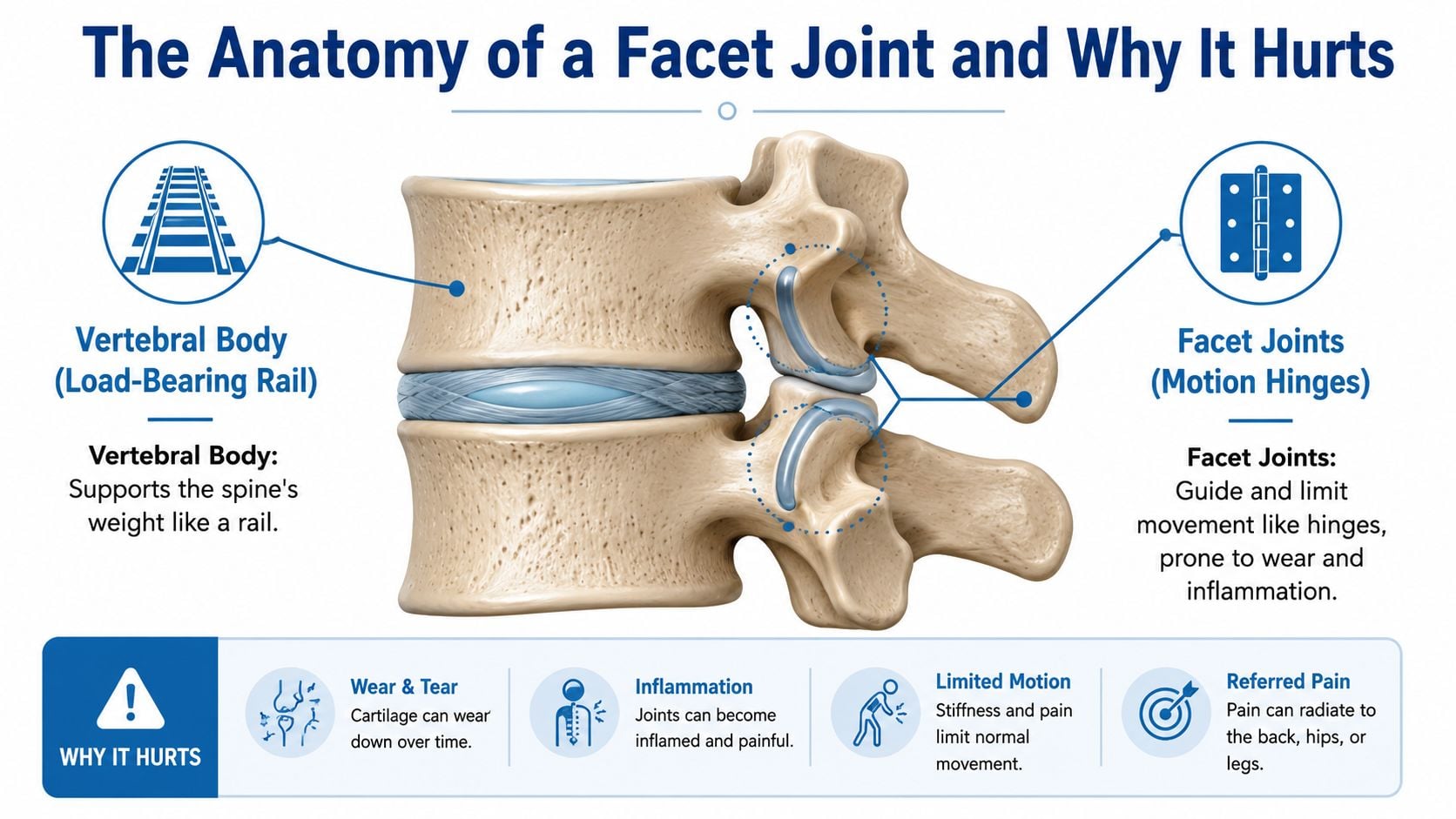
The spine can be understood as a rail-and-hinge system. Each vertebral body at the front acts like a strong, load-bearing rail, while paired facet joints sit toward the back and guide how neighboring vertebrae move. The facets are angled differently in the neck, mid-back, and lower back, so each spinal region balances bending, rotation, and stability in its own way.
The joint surfaces are covered with cartilage, and ligaments around the motion segment help control movement. A healthy facet behaves somewhat like a well-oiled door hinge. It permits motion while limiting excessive movement that could strain the spine.
What changes inside the joint
Cartilage can become less smooth with age, repeated loading, or injury. As the protective surface wears, the joint may develop roughened areas, inflammation, narrowing, and bone spurs. Those changes can increase pressure during extension or rotation and irritate pain-sensitive tissues around the joint.
Facet joints receive sensory input mainly through the medial branches of spinal nerves above and below the joint. Irritation inside the joint capsule or around these small nerve endings can create local back or neck pain, along with referred pain in nearby regions. Referred pain is different from nerve-root pain, because it doesn't necessarily follow a narrow strip down an arm or leg.
Aging commonly affects the facet joints. A broader guide to ageing muscles at Understanding Ageing can help explain how changes in muscles, bones, and joints influence mobility over time.
Why an MRI isn't a diagnosis
Facet degeneration is only one possible explanation for spinal pain. A scan can show an irregular or enlarged joint, but that image doesn't reveal whether the joint is actively generating symptoms. Several structures may look abnormal in the same spine, and imaging can't identify which one matters without clinical correlation.
That's why a pain physician treats the scan as a map, not a verdict. The patient's movement pattern, functional limitations, neurological findings, and response to targeted diagnostic testing all influence the final decision.
What Causes Facet Joint Syndrome and Where the Pain Shows Up
The most common pathway is age-related osteoarthritis. Cartilage gradually loses its smooth surface, the joint may narrow, and bony overgrowth can develop. Repeated bending, twisting, lifting, prolonged extension, poor conditioning, excess body load, prior spinal injury, and previous surgery can add mechanical stress.
Younger adults may develop facet symptoms after trauma or because of inflammatory arthritis or an inherited tendency toward joint problems. Infection is uncommon, but a hot, swollen, severely tender joint with fever requires a different medical evaluation from routine degeneration.
The affected spinal region shapes the symptoms:
- Lumbar facets can cause lower-back stiffness with aching into the buttock, groin, or back of the thigh, usually stopping above the knee.
- Thoracic facets can create mid-back pain that may wrap around the ribs.
- Cervical facets may cause neck stiffness with referral toward the shoulder, upper back, or head.
Extension and rotation often aggravate facet pain because those movements bring the joint surfaces closer together. Sitting, standing, or lifting can also provoke symptoms depending on posture and load. Because discs, muscles, sacroiliac joints, and compressed nerves can produce overlapping patterns, people with persistent symptoms may also benefit from reviewing lower-back pain from sitting all day while arranging a proper clinical assessment.
| Spinal Region | Typical Pain Pattern | Common Aggravators | Important Caution |
|---|---|---|---|
| Lumbar | Low-back ache with possible buttock, groin, or posterior-thigh referral | Backward bending, rotation, prolonged standing, lifting | Pain location overlaps with disc and sacroiliac disorders |
| Thoracic | Mid-back pain that may wrap around the ribs | Rotation, extension, sustained posture | Rib, muscular, and disc conditions can feel similar |
| Cervical | Neck ache and stiffness with shoulder, upper-back, or head referral | Turning, looking upward, prolonged neck posture | Headache or arm symptoms require broader evaluation |
Facet-mediated pain is common but not uniform across spinal regions or diagnostic methods. Reviews estimate lumbar facet involvement at about 15% to 45% of chronic low back pain cases, with one review placing facetogenic chronic low back pain at 15% to 41%. Broader controlled diagnostic studies have estimated lumbar facet pain at 27% to 40%, thoracic facet pain around 34% to 48%, and cervical facet-mediated pain around 27% to 67%, as summarized in this clinical review of facet joint syndrome.
How Doctors Actually Confirm a Facet Joint Diagnosis
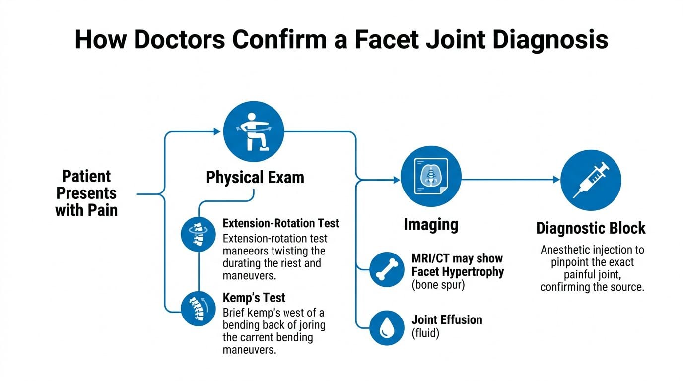
Facet diagnosis looks straightforward until a clinician has to identify the exact painful joint. Extension-rotation maneuvers, including Kemp's test, may reproduce a patient's familiar pain, but they don't reliably distinguish facet pain from other spinal sources. MRI or CT may show facet hypertrophy, arthritic change, or fluid in the joint, yet those findings can exist without symptoms.
The diagnostic pathway therefore begins with a clinical suspicion, not certainty. The physician reviews the pain location, aggravating movements, neurological symptoms, prior treatments, and competing diagnoses. Imaging helps assess anatomy and rule in or rule out other problems, but it doesn't independently identify the pain generator.
Why diagnostic blocks matter
The accepted technical standard is an image-guided medial branch block or an intra-articular facet block. A small amount of local anesthetic is placed near the medial branch nerves supplying the suspected joint, or directly into the joint, while the patient tracks pain during the anesthetic window.
Guidance from clinical and coverage sources identifies diagnostic injections as the gold standard and commonly calls for a second confirmatory block before proceeding to radiofrequency treatment. The diagnostic process is complicated by false-positive responses. One evidence review reported false-positive rates of 25% to 44%, even while estimating facet pain prevalence at 27% to 41% in low back pain populations, as detailed in this evidence review of facet interventions.
A second block can reduce the chance that the first result reflects placebo response, medication spread, or an unrelated temporary improvement. The procedure also has practical considerations, including needle-related discomfort and radiation exposure from image guidance. Patients considering a diagnostic injection can review what a nerve block procedure involves and discuss the specific target with the treating physician.
The reported prevalence of facet pain ranges from 5% to 90% when clinicians rely on history, examination, and imaging alone, which illustrates why a confident diagnosis requires more than a scan or a painful movement. The relevant consensus guidance is summarized in this review of cervical facet pain diagnosis.
Treatment Options From Conservative Care to Procedures
Treatment should match the strength of the diagnosis, the severity of disability, and the patient's goals. Conservative care usually comes first because it can improve movement and reduce mechanical stress without exposing the patient to procedural risks.
A physical therapist may emphasize trunk stabilization, hip mobility, posture, and controlled loading. Activity modification doesn't mean avoiding all movement. It means reducing the specific positions that repeatedly provoke symptoms while preserving walking, daily activity, and progressive conditioning. Nonsteroidal anti-inflammatory drugs may help selected patients, but medication safety depends on kidney function, stomach health, cardiovascular risk, and other medicines.
Weight management may reduce mechanical demand on the spine, although it doesn't reverse established cartilage loss. Patients recovering after pregnancy or caring for a newborn may also find practical movement ideas in guidance on postpartum back pain exercises, while recognizing that an individualized assessment remains important.
Comparing procedural options
When conservative care doesn't restore function, a clinician may discuss diagnostic and therapeutic procedures. Medial branch blocks can provide short-term relief and help determine whether the targeted joints are responsible. Intra-articular steroid injections may reduce symptoms for a limited period, but the evidence is not equally strong across studies and techniques.
Radiofrequency ablation, also called medial branch neurotomy, uses thermal energy to interrupt pain signaling from confirmed facet joints. It has moderate or level II evidence for sustained benefit after diagnostic confirmation, and guidelines commonly place it among the more evidence-supported procedures for appropriately selected patients. Details about radiofrequency ablation should be reviewed with a physician who can explain candidacy, alternatives, and expected recovery.
| Treatment | Mechanism | Evidence Level | Typical Duration of Benefit | Best For |
|---|---|---|---|---|
| Physical therapy | Improves stabilization, mobility, and load control | Foundational conservative care | Variable and dependent on continued participation | Patients seeking functional improvement without procedures |
| Medication and activity modification | Reduces symptoms and limits provoking stress | Symptom-focused care | Variable | Flares and early management |
| Medial branch block | Temporarily anesthetizes nerves supplying suspected facets | Diagnostic standard, possible short-term relief | Short term | Confirming the pain generator |
| Intra-articular steroid injection | Places anti-inflammatory medication in the joint | Evidence is limited and uneven | Short to intermediate term when helpful | Selected patients with suspected joint inflammation |
| Radiofrequency ablation | Interrupts medial branch pain signaling | Moderate to level II evidence after confirmation | Sustained benefit varies | Confirmed facet-mediated pain that persists despite conservative care |
| PRP, hyaluronic acid, or MSC-based care | Attempts to alter the joint environment or inflammation | Emerging and uncertain | Not established | Carefully selected patients considering experimental options |
| Surgery | Addresses instability, stenosis, or another structural problem | Depends on the coexisting condition | Depends on the operation and diagnosis | Structural disease not controlled with nonoperative care |
Evidence deserves particular caution. A 2025 systematic review found very low-quality evidence for fluoroscopically guided lumbar facet steroid injections, with sham-controlled trials not demonstrating efficacy. The same review described moderate evidence for PRP and weak evidence for MSCs, so regenerative procedures shouldn't be presented as established replacements for proven diagnostic pathways. The findings are reported in this 2025 review of facet injections and regenerative treatments.
Surgery is rarely directed at facet pain alone. Referral becomes more relevant when instability, severe stenosis, or another structural problem coexists and conservative and interventional care haven't addressed the underlying limitation.
When to See a Specialist and What to Expect
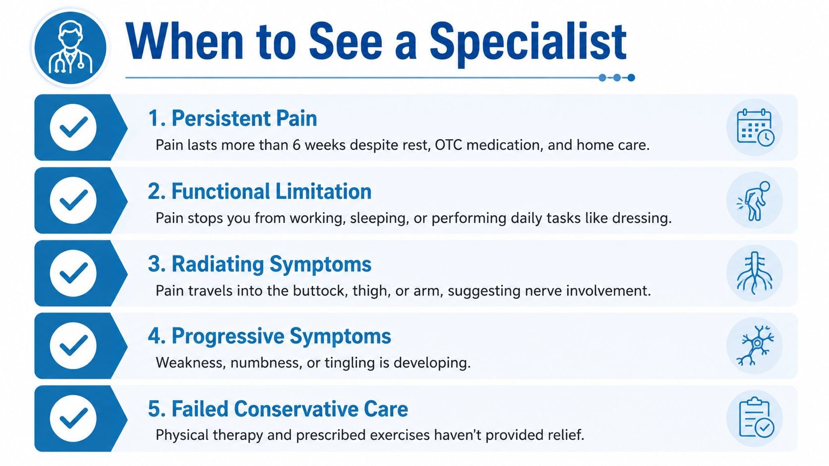
A specialist evaluation makes sense when pain persists despite home care, limits work or sleep, or continues after prior treatment. Persistent symptoms can have several causes, so the goal isn't to label the pain as facet syndrome. The goal is to identify the structure most likely responsible and choose testing that can change management.
Urgent assessment is needed for saddle numbness, progressive leg weakness, bowel or bladder changes, fever, unexplained weight loss, or a history of cancer. These features can signal conditions that require prompt evaluation rather than routine facet treatment.
At the first visit, the physician typically reviews symptom timing, movement triggers, previous treatments, medications, neurological complaints, and functional goals. A focused examination may be followed by review of existing imaging. New imaging or a diagnostic block should be timed according to the clinical question, not ordered automatically.
Patients can bring:
- Prior procedure records: Include injection names, targets, dates, and responses.
- Imaging reports: Bring reports and available images, not just a verbal summary.
- A symptom diary: Record triggers, relief, sleep disruption, and activity limits.
- Functional goals: State what improvement would matter, such as walking, working, or turning the head.
Common Questions and the Bottom Line on Facet Pain
Can facet syndrome resolve on its own? An acute flare may settle, especially when triggered by a strain or temporary irritation. Degenerative changes usually remain, so recurring symptoms may require ongoing exercise, activity adjustment, and targeted care.
How long do medial branch blocks or radiofrequency ablation last? A medial branch block is primarily a short-term diagnostic test, although some patients experience temporary therapeutic relief. Radiofrequency ablation can provide more sustained relief in appropriately selected patients, but the duration varies and the nerve signaling pathway can recover.
Can facet syndrome spread or damage nerves? Facet pain can refer into nearby areas, but the joint condition itself isn't the same as nerve-root compression. New weakness, progressive numbness, or bowel and bladder changes require urgent evaluation.
How does it differ from a herniated disc? Facet pain often feels localized and worsens with extension or rotation. Disc-related nerve pain may produce burning or electric symptoms that follow a nerve distribution, but overlap is common and examination alone may not settle the issue.
The central point is simple: imaging can show anatomy, but diagnostic blocks help test whether a facet joint is causing the pain. Treatment should target a confirmed pain generator and usually works best when a procedure, if needed, is paired with active rehabilitation rather than used alone.
Interventional Pain Management evaluates back and neck pain through clinical examination, imaging review, diagnostic nerve blocks, and procedure-based care when appropriate. Patients can visit Interventional Pain Management to request an evaluation and discuss whether facet pain, disc disease, nerve irritation, or another condition best explains their symptoms.