

You're on the sideline after a planted foot twisted, the knee buckled inward, and a pop was followed by swelling. Now the MRI mentions both the ACL and MCL, and the word “surgery” makes it sound as if both ligaments automatically need to be fixed. That isn't usually the decision.
The ACL and MCL do different jobs, heal differently, and create different surgical risks. ACL MCL surgery is therefore not one all-or-nothing choice. The central question is whether the MCL can regain useful stability with protection and rehabilitation, or whether its injury pattern makes repair or reconstruction necessary, either at the same operation as the ACL or in a staged plan.
Table of Contents
- When Two Knee Ligaments Fail at Once
- How the ACL and MCL Actually Work
- Surgery or Conservative Care for Each Ligament
- Repair Versus Reconstruction and Graft Choices
- What Happens on the Day of Surgery
- Rehabilitation Phases From Week One to Month Six
- Return to Sport and Long-Term Knee Health
- Making Your Decision and Questions to Ask
When Two Knee Ligaments Fail at Once
A soccer player plants one foot to change direction. The shoe stays fixed, the body rotates, and the knee caves inward. The force can overload the ACL through rotation while simultaneously stretching the MCL along the inner side of the joint. A tackle, ski fall, or awkward landing can create the same combination.
The immediate experience often feels more dramatic than a routine knee sprain. A pop may occur, swelling can develop rapidly, the inner knee may be tender, and the joint may give way when the athlete tries to stand. Those symptoms don't prove that both ligaments are torn, but they justify prompt examination and imaging.
Why the combination changes the mechanics
The ACL is the knee's internal guide against forward movement of the shinbone and unwanted rotation. The MCL is the medial-side restraint against valgus stress, meaning a force that pushes the knee inward. When both are injured, the knee may lose control in more than one direction.
That matters during the examination. A clinician assesses the ACL with tests such as the Lachman and pivot-shift examinations, while the MCL is stressed at different degrees of knee flexion. MRI helps show the tear's location and associated meniscus, cartilage, or bone injuries, but the physical examination still determines how the knee behaves under load.
Practical rule: Pain describes the injury, but instability helps determine the treatment plan.
ACL reconstruction has become increasingly common in large health systems. In England, age-sex standardized rates rose from 2.0 to 24.2 per 100,000 population between 1997-1998 and 2016-2017, while a U.S. population study recorded 86,837 reconstructions in 1994 and 134,421 in 2006, a 37% increase in the population-adjusted rate. A broader U.S. analysis identified 283,810 ACL reconstructions from 2002-2014, with the annual rate rising from 61.4 to 74.6 per 100,000 person-years, a 22% increase (British Journal of Sports Medicine review).
The MCL decision is less automatic. Its extra-articular location gives many tears a better opportunity to heal, so the key issue is not whether the MRI says “MCL tear.” The surgeon must determine whether the medial side remains unstable, where the ligament has torn, whether the injury is acute or chronic, and how much valgus stability the athlete needs.
How the ACL and MCL Actually Work
A useful analogy is a door hinge. The ACL acts like a strong rope inside the hinge mechanism. It helps stop the shinbone from sliding too far forward and controls rotational movement when the foot is planted. The MCL acts more like a strap on the inner edge of the door, resisting a force that tries to fold the knee inward.
Why the MCL often heals differently
The ACL sits inside the knee joint, where the environment and blood supply are unfavorable for the original torn ligament ends to reunite in a dependable way. A complete ACL tear can leave an active person with repeated buckling, especially during cutting, jumping, or sudden deceleration. More information about symptoms and diagnosis appears in this guide to an ACL tear.
The MCL lies outside the joint and generally has a more favorable healing environment. That's why bracing and progressive rehabilitation can restore useful function for many MCL injuries. This doesn't mean every MCL tear heals perfectly, particularly when the tear is displaced, located on the tibial side, or combined with broader medial and posteromedial damage.
Reading the injury grade
The grade describes the amount of structural damage and laxity, but it isn't the entire treatment decision.
| Injury grade | Typical meaning | What the examination may show |
|---|---|---|
| Grade I | Fibers are stretched but the ligament remains functionally stable | Tenderness with little or no laxity |
| Grade II | A partial tear has developed | Some opening or looseness under valgus stress |
| Grade III | The ligament is completely torn | Clear medial instability, sometimes in more than one knee position |
The same grading language applies broadly to both ligaments, but the consequences differ. A Grade III ACL tear in a pivoting athlete often produces a mechanical problem that rehabilitation alone can't correct. A Grade III MCL tear may still heal with protection, depending on its location and the stability pattern.
MCL injuries range from a mild sprain to a complete disruption involving nearby structures. A patient should ask the surgeon whether the MRI and examination show isolated medial injury, persistent valgus opening, instability in extension, or a tear pattern that may not heal in its normal position.
Surgery or Conservative Care for Each Ligament
The treatment conversation should begin with two separate questions:
- Does the ACL need reconstruction?
- Does the MCL need repair or reconstruction, or can it be protected while it heals?
For the MCL, isolated Grade I and Grade II injuries are commonly managed with a hinged brace, activity modification, and rehabilitation. Some Grade III injuries can also be treated without surgery when the ligament has a favorable tear pattern and the knee becomes stable during healing. Surgery may add scar tissue and stiffness without providing a meaningful advantage if the MCL can restore functional stability on its own.
The ACL follows a different logic. A complete tear in a person who wants to return to pivoting sports, or a knee that repeatedly gives way during ordinary activity, often leads to reconstruction. Reconstruction replaces the damaged ligament with a tendon graft, rather than expecting the original ACL to reconnect reliably.
The timing problem in combined injuries
When the MCL is significantly torn, the knee can be swollen and difficult to move. Performing major surgery before the joint regains motion may increase the chance of postoperative stiffness. Conversely, allowing an unstable ACL to keep shifting the tibia can place stress on a healing MCL.
Evidence supports careful timing rather than a universal calendar rule. In one retrospective study of combined ACL and MCL injury, patients who underwent ACL reconstruction at least 10 weeks after initial non-operative MCL bracing had fewer postoperative motion losses and fewer re-arthroscopies for loss of extension than patients treated with acute ACL reconstruction (study abstract). That finding supports delayed ACL reconstruction in that specific injury pattern, but it doesn't mean every combined injury should wait.
| Injury grade | ACL approach | MCL approach |
|---|---|---|
| Grade I | Reconstruction depends on instability, activity goals, and associated injuries | Usually bracing and rehabilitation |
| Grade II | Complete or functionally unstable tears may require reconstruction | Often non-surgical care, followed by repeat stability examination |
| Grade III | Reconstruction is commonly considered for persistent functional instability | Bracing may work for selected patterns, while displaced or persistently unstable tears may need repair or reconstruction |
A 2024 systematic review found that acute ACL reconstruction with acute MCL reconstruction produced the highest reported valgus stability at 30 degrees of flexion, 81% to 100%, while non-operative MCL treatment with delayed or non-operative ACL strategies showed lower stability, 27% to 68%. The review found no clear Lysholm score advantage across MCL strategies, so stability findings and patient function still have to be weighed against stiffness risk (2024 systematic review).
The MCL choice, not the ACL label alone, often determines whether surgery is simultaneous, staged, or limited to the ACL. A detailed explanation of medial-side symptoms and treatment options is available in this resource on an MCL tear.
Repair Versus Reconstruction and Graft Choices
The ACL and MCL are different surgical problems. Repair reattaches the patient's own ligament, while reconstruction replaces damaged tissue with a tendon graft. The decision depends on the tear's location, tissue quality, timing, residual instability, activity goals, and the condition of the other ligaments. In a combined injury, the MCL decision often determines whether treatment stays limited to ACL reconstruction or includes a medial procedure, either during the same operation or in stages.
ACL surgery usually involves reconstruction because torn ends often retract and do not heal predictably inside the joint. Common autografts include the patellar tendon, hamstring tendons, and quadriceps tendon. Each has a different donor-site profile, fixation method, and rehabilitation effect. Allograft tissue avoids harvesting the patient's own tendon, but age, activity level, tissue quality, and the possibility of later revision still influence that choice.
How MCL procedures differ
An MCL repair can work when the ligament has pulled away from its attachment and the remaining tissue is strong enough to hold sutures. Anchors may secure the ligament back to bone. Suture tape or an internal brace can provide added protection during biological healing, although positioning must be accurate to avoid making the medial side too tight.
MCL reconstruction becomes more appropriate when the ligament is chronically stretched, the tissue is poor, the tear cannot be returned to its normal attachment, or several medial structures remain unstable. A hamstring tendon may be used as the graft. The surgeon must also consider whether removing the hamstrings takes away a secondary stabilizer from a knee that already lacks medial support.
A 2025 systematic review reported lower arthrofibrosis with MCL reconstruction than with MCL repair, 5.4% versus 11.6%, and lower failure rates, 2.9% versus 5.7%. ACL graft failure was also lower in reconstruction cohorts, 0.2% versus 2.3%. These results do not make reconstruction automatically preferable. They combine different injury patterns, techniques, and patient groups, so your examination and tissue quality remain central to the decision.
| Procedure | Best candidate | Common graft or technique | Key trade-off |
|---|---|---|---|
| ACL reconstruction | Complete, symptomatic instability in an active patient | Patellar tendon, hamstring, quadriceps tendon, or selected allograft | Restores a central restraint but creates graft-harvest or graft-related considerations |
| MCL repair | Acute tear with repairable tissue and a favorable attachment pattern | Sutures and bone anchors | Preserves native tissue but may fail if the tear is displaced or tissue is poor |
| MCL repair with augmentation | Repairable injury needing additional protection | Suture tape or internal brace | Adds support but can contribute to overtightening if positioned incorrectly |
| MCL reconstruction | Chronic, irreparable, or high-grade multi-ligament instability | Tendon graft, often hamstring-based | Provides a new restraint but may increase stiffness and rehabilitation demands |
The goal is stable motion, not maximum tightness. An accurately positioned reconstruction should control inward angulation while allowing the knee to bend and straighten. Too tight can restrict motion. Too loose can leave valgus instability.
What Happens on the Day of Surgery
Arrival usually includes a medical review, consent confirmation, examination of the operative knee, and marking of the surgical side. Anesthesia may include general anesthesia with a regional nerve block to reduce pain during the first part of recovery.
For ACL reconstruction, the surgeon makes small arthroscopic portal incisions and examines the joint. The graft is harvested or prepared, tunnels are created in the thighbone and shinbone, the graft is passed into position, and fixation is secured with a button, screw, or another fixation construct.
When the MCL is addressed
If the MCL requires treatment at the same sitting, the surgeon may add an open medial incision to repair or reconstruct the ligament while completing the ACL arthroscopy. If the knee is markedly swollen or motion is restricted, the MCL and ACL procedures may be staged instead. Staging allows the first phase of care to focus on swelling, motion, and medial-side healing before the next operation.
The surgical team discusses the expected duration beforehand. Combined cases may take roughly 90 to 150 minutes, although the actual time depends on the tear pattern, graft choice, meniscus treatment, and whether additional stabilizing procedures are needed.
After surgery, the recovery team monitors pain, circulation, sensation, and movement. A nerve block may leave the leg numb for approximately 12 to 24 hours, and patients receive instructions for protecting the limb as sensation returns. Before discharge, the team fits the brace, explains crutch use, reviews medication and wound care, and clarifies whether weight-bearing is allowed immediately or must be limited.
The patient shouldn't assume that a standard ACL protocol applies. The MCL repair or reconstruction may require stricter control of valgus stress, flexion, rotation, or loading.
Rehabilitation Phases From Week One to Month Six
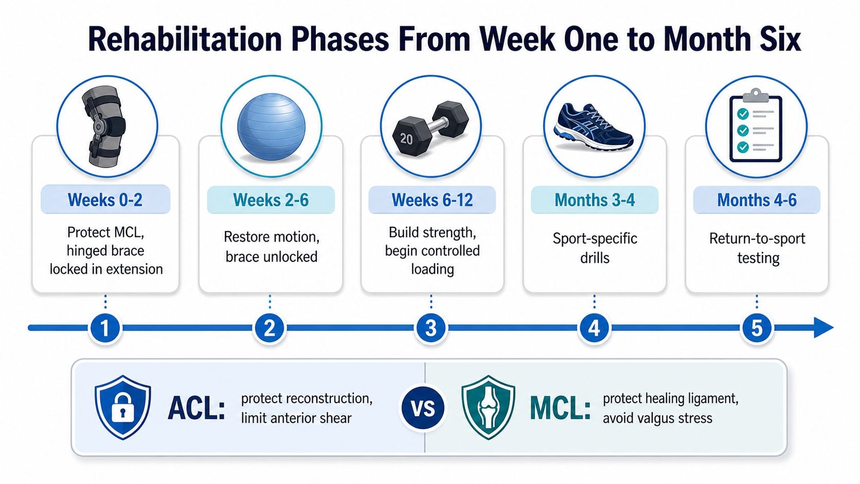
Combined ACL and MCL rehabilitation balances two demands. The healing MCL must be shielded from valgus stress and excessive rotation, while the knee needs enough movement to prevent stiffness, especially loss of extension. The brace and exercise plan act like guardrails: they limit dangerous force without keeping the joint still longer than necessary.
The early protection window
During weeks 0 to 2, treatment centers on swelling control, quadriceps activation, safe transfers, and gradual motion recovery. A hinged brace may stay locked in extension while walking. Heel slides, ankle movement, and prescribed exercises support circulation and begin restoring knee control. Early progress is measured by safe movement and a stable knee, not by forcing range.
During weeks 2 to 6, the brace may open for selected activities, and weight-bearing can increase according to the surgeon's instructions. A stationary bicycle may be added once motion allows. Hamstring loading needs particular care when the medial structures are healing or hamstring tendons supplied the graft.
Loading without losing motion
From weeks 6 to 12, therapy commonly advances to controlled closed-chain strengthening, balance work, and recovery of full range of motion. The therapist checks how the knee responds after each increase. Persistent loss of extension deserves prompt attention because a stiff knee can become harder to correct later.
From months 3 to 5, selected patients may start a jogging progression on stable surfaces, lateral-control exercises, and light plyometric work. Strength, landing mechanics, swelling, graft choice, and the stability examination determine the sequence. During months 5 to 6, sport-specific drills may begin for patients who meet the required milestones. Cutting and contact work still require objective assessment, not enthusiasm alone.
Your therapist may adjust brace use, exercise range, or loading from one visit to the next. A repair that needs protection may progress more slowly than a reconstruction, while swelling or limited motion can require a temporary change in emphasis. The practical goal is steady progress without trading ligament protection for avoidable stiffness. The guide to injury triage and rehab pathways explains how clinicians organize these decisions.
Motion matters: The safest protocol protects the healing ligament while preventing avoidable stiffness from becoming a second problem.
Contact the surgical team if swelling rises sharply, extension worsens, the brace causes pressure injury, or pain prevents the prescribed exercises. These changes do not automatically mean the operation has failed, but they warrant timely review before the problem affects the next rehabilitation phase.
Return to Sport and Long-Term Knee Health
A calendar date can open the door to return-to-sport testing, but it shouldn't make the final decision. The knee must demonstrate strength, control, confidence, and stability under tasks that resemble the athlete's sport.
The clearance checklist
- Strength: The surgical leg should approach the performance of the opposite leg on quadriceps and relevant lower-limb testing.
- Hopping: Single-leg hop tasks should show controlled landings, quiet alignment, and no protective hesitation.
- Agility: Cutting, deceleration, and directional-change drills should be completed without valgus collapse or swelling afterward.
- Patient-reported function: Instruments such as the IKDC can help document symptoms and perceived knee function.
- Psychological readiness: Fear of reinjury and uncertainty during a single-leg landing can limit performance even when strength looks acceptable.
Some clinicians use a limb symmetry index above 90% for strength and hop testing, but the threshold must be interpreted alongside movement quality and the demands of the sport. A linear runner may progress differently from an athlete whose sport requires repeated cutting, contact, and pivoting. Calendar estimates should therefore remain approximate, and a cutting athlete may need substantially longer than a person returning to straight-line activity.
Protecting the knee beyond clearance
ACL and MCL injuries can coexist with meniscus and cartilage damage. Persistent swelling, catching, or joint-line pain may indicate another structure needs evaluation, including a possible torn meniscus. Continued neuromuscular training, landing practice, hip and trunk control, and progressive strength work help support long-term knee function.
Patients also benefit from broader joint-health habits, particularly once rehabilitation shifts from protected recovery to lifelong maintenance. Practical arthritis care tips can complement, but not replace, an individualized orthopedic plan.
Return should happen when the knee is quiet, stable, strong, and trusted by the athlete. A cleared athlete who still avoids loading the surgical leg may need more graded exposure and psychological support before full competition.
Making Your Decision and Questions to Ask
The decision becomes clearer when the patient separates the two ligaments. The ACL plan reflects instability, activity goals, and associated injuries. The MCL plan reflects tear location, grade, tissue quality, valgus laxity, chronicity, and the risk that surgery could produce stiffness.
The MCL can heal without surgery in many cases, but a favorable MRI alone isn't enough. The surgeon should compare the injured knee with the opposite side, test stability in extension and flexion, and explain whether the medial side is becoming stable during rehabilitation.
Common questions deserve direct answers:
- Can the MCL heal on its own? Often, particularly with selected isolated injuries, but displaced or persistently unstable tears may not.
- How should the ACL graft be chosen? The decision should account for age, sport, prior surgery, tendon quality, donor-site effects, and the need to preserve medial stabilizers.
- What happens if surgery is delayed? Delay may allow motion and swelling to improve, but ongoing instability can affect the treatment strategy.
- Is repeat imaging always needed? Not necessarily. Repeat imaging is usually guided by symptoms, examination findings, surgical planning, or an unexpected recovery course.
A consultation checklist can include:
- How unstable is the MCL in extension and flexion?
- Where exactly has the MCL torn?
- Can the MCL heal with bracing?
- Why is simultaneous or staged surgery recommended?
- Which ACL graft fits the patient's sport and anatomy?
- What brace, motion, and weight-bearing restrictions will apply?
- What findings would change the plan?
- Which milestones determine return to work and sport?
The right plan matches the knee's instability pattern and the patient's goals. The fastest operation isn't always the fastest recovery, and “fixing both” isn't always better than protecting the MCL while reconstructing the ACL.
Interventional Pain Management evaluates joint and sports injuries, including ACL and MCL tears, through coordinated orthopedic assessment, imaging review, and individualized treatment planning. Patients in New Jersey and Staten Island can visit Interventional Pain Management to discuss diagnosis, surgical options, rehabilitation needs, and pain management with the appropriate clinical team.