
A person with spinal stenosis often notices the problem before learning its name. A familiar walk becomes shorter, standing in line becomes difficult, and leaning over a shopping cart brings surprising relief. The change can seem like ordinary aging, but a shrinking walking distance, leg heaviness, numbness, or position-dependent pain deserves a closer look.
Effective spinal stenosis pain management isn't one treatment. It's a sequence of decisions based on the dominant symptoms, the anatomy on imaging, walking ability, medical risks, and how well non-surgical care has worked. Epidural injections can create a useful window for movement, but they don't widen a narrowed canal. Surgery can relieve pressure, but it isn't automatically the next step for every MRI finding. The practical question is what each option can accomplish, for whom, and for how long.
Table of Contents
- Why Stenosis Pain Is Different From Ordinary Back Pain
- What Spinal Stenosis Actually Is and How It Shows Up
- Conservative Care That Actually Helps
- Injections, Nerve Blocks, and Other Interventional Options
- Minimally Invasive Decompression and Surgical Treatments
- Building a Stepwise Pain Management Plan
- Common Misconceptions Worth Putting Aside
- When to See a Specialist and What to Ask
Why Stenosis Pain Is Different From Ordinary Back Pain
A person may begin by walking a mile without difficulty. Gradually, the distance falls to a few blocks. At the grocery store, leaning forward on the cart makes the legs feel better, while standing upright brings back burning, heaviness, or aching. Many people blame age or weak muscles, especially when sitting still feels relatively comfortable.
That pattern differs from ordinary muscular back pain. Muscle pain is often localized and linked to lifting, twisting, or a particular movement. Stenosis-related symptoms may be triggered less by activity itself than by the position of the spine during activity. Standing and walking extend the lower back, which can reduce available space around nerve roots. Sitting or bending forward tends to flex the spine and may give those nerves more room.
The walking clue
This position-dependent pattern is called neurogenic claudication. The legs may feel heavy, cramped, burning, weak, or numb after standing or walking. A person may stop at a storefront, sit on a bench, or bend forward over a counter. The relief isn't imaginary, and it isn't just a matter of poor conditioning. The posture changes the mechanical environment around the nerves.
The coping behaviors can become so routine that patients don't mention them unless asked. They may avoid museums, shopping trips, cooking at the counter, or neighborhood walks. A cane or cart becomes less a mobility aid than a way to keep the back flexed.
Practical rule: A marked difference between walking upright and walking while leaning forward is a useful clinical clue, not a minor quirk.
Stenosis can overlap with sciatica or a pinched nerve, but the pattern matters. A helpful explanation of how nerve irritation can produce leg symptoms appears in this guide to sciatica and pinched nerve. When symptoms repeatedly follow a predictable distance or standing time, rest alone may not solve the problem. Earlier clinical assessment and appropriate imaging can clarify whether narrowing is contributing to the limitation.
What Spinal Stenosis Actually Is and How It Shows Up
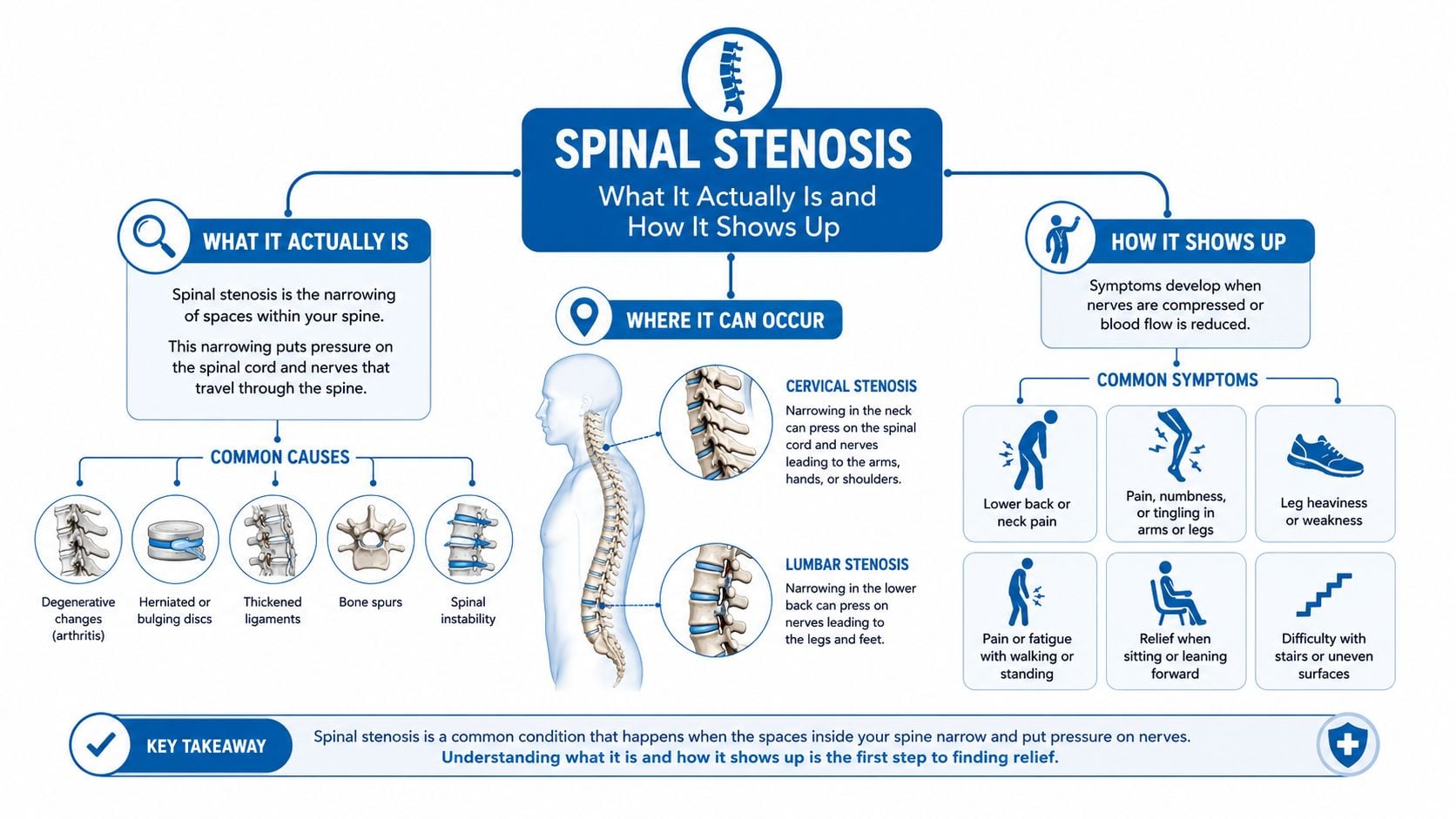
The spinal canal is a protected passageway for the spinal cord and nerve roots. Spinal stenosis means that bone, disc material, thickened ligaments, or shifted spinal structures reduce the available space. Common contributors include enlarged facet joints, thickening of the ligamentum flavum, disc bulging, and spondylolisthesis, where one vertebra shifts in relation to another.
The location of narrowing helps explain the symptom pattern:
- Central canal stenosis affects the main passageway and can produce symptoms in both legs, especially with walking or standing.
- Lateral recess stenosis narrows the area where a nerve root travels before leaving the canal.
- Foraminal stenosis affects the opening through which an individual nerve exits, often producing symptoms along one nerve-root distribution.
Two patterns patients should recognize
Neurogenic claudication usually involves bilateral leg heaviness, aching, burning, cramping, or fatigue during walking and standing. Sitting or leaning forward often helps. The back may hurt, but the walking limitation can be more disabling than the back pain.
Radiculopathy is more often sharp, electric, or burning pain that follows a single nerve root. Numbness, tingling, or weakness may travel down one leg in a recognizable distribution. A patient can have both patterns at once, which is why the history and examination remain important even when an MRI shows several areas of narrowing.
Most lumbar stenosis is acquired and degenerative. Cervical stenosis requires a different level of caution because compression in the neck can affect the spinal cord itself. Hand clumsiness, worsening balance, unsteadiness, or changes in coordination can indicate myelopathy and warrant prompt evaluation rather than routine self-care.
Conservative Care That Actually Helps
Conservative treatment works best when it has an active purpose. The aim isn't to keep a patient resting until symptoms disappear. The aim is to improve walking tolerance, strength, balance, confidence, and the ability to manage flare-ups while the clinical team watches for signs that a stronger intervention is needed.
Physical therapy should be functional
A structured program may include flexion-tolerant conditioning, trunk stabilization, hip mobility, gait work, balance training, and graded walking. Passive heat or hands-on treatment can make movement easier, but exercise and self-management should remain central. A therapist can also teach modifications for standing tasks, lifting, stairs, and community walking.
Hip and calf mobility can influence stride and posture. Patients looking for plain-language mobility guidance may find this resource on how to improve hip and calf flexibility useful as general education, while an individualized program should account for neurologic symptoms and balance risk.
A graded walking plan might begin with tolerable intervals, supported cycling, or repeated short walks rather than one long effort that provokes a flare. The checkpoint isn't zero pain. It's whether the person can stand longer, walk farther, sleep more consistently, or recover more quickly after activity.
Medication requires trade-offs
Medication choices depend on kidney function, gastrointestinal risk, blood pressure, other prescriptions, age, and the type of pain present.
- Acetaminophen may have a role for mild baseline discomfort, within safe dosing limits.
- Nonsteroidal anti-inflammatory drugs can help during selected flares, but prolonged or frequent use may be inappropriate for some patients.
- Gabapentinoids or duloxetine may be considered when burning, tingling, or nerve pain dominates. Drowsiness, dizziness, interactions, and other adverse effects matter.
- Muscle relaxants are generally used cautiously because sedation can increase fall risk.
Long-term opioids, extended bed rest, unproven supplements, and expensive passive treatments shouldn't replace active rehabilitation without a clear reason. Manual therapy, acupuncture, and weight management may serve as adjuncts, particularly when they help someone participate in exercise, but they don't correct mechanical narrowing.
A conservative trial is reasonable when symptoms are stable and neurologic function isn't declining. Reassessment after a defined period, often six to twelve weeks, should examine function rather than pain alone. If walking continues to deteriorate or weakness appears, the plan needs to change sooner.
Injections, Nerve Blocks, and Other Interventional Options
Interventional procedures can answer two different questions. A diagnostic injection tests whether a particular nerve, facet joint, or pain pathway contributes to symptoms. A therapeutic injection aims to reduce irritation long enough for rehabilitation, sleep, or a larger treatment decision to become possible.
Matching the procedure to the symptom
An epidural injection places medication in the epidural space around irritated nerve structures. The approach matters:
- Interlaminar injections enter the epidural space from the back and may suit symptoms involving a broader central or multilevel region.
- Caudal injections enter through the sacral opening and can be useful when medication needs to spread upward through the epidural space.
- Transforaminal injections approach a selected nerve root and may fit a more focal radicular pattern, although evidence for stenosis is weaker than for some other approaches.
Research published in the mid-2010s found significant pain and functional improvement with fluoroscopy-guided caudal and lumbar interlaminar epidural injections using local anesthetic, with or without steroid, in lumbar central spinal stenosis. The same evidence did not show that adding steroid clearly outperformed local anesthetic alone, while transforaminal treatment had weaker support for short-term benefit, as described in this systematic review of epidural injections for lumbar spinal stenosis.
A later randomized trial also found minimal or no added analgesic benefit from steroid in many patients with lumbar stenosis, with limited effects on pain and walking ability in the trial record. That doesn't make injections useless. It means the expected benefit should be framed as symptom modulation, not anatomical repair.
A practical comparison
| Procedure | Best For | Typical Relief Duration | Key Caveat |
|---|---|---|---|
| Caudal epidural injection | Broader or multilevel lumbar symptoms | Temporary and variable | Doesn't remove narrowing |
| Interlaminar epidural injection | Central canal symptoms and broader irritation | Temporary and variable | Steroid may add limited benefit |
| Transforaminal injection | Selected nerve-root symptoms | Temporary and variable | Evidence is weaker for stenosis |
| Facet or medial branch block | Suspected facet-mediated back pain | Diagnostic or temporary | Doesn't treat nerve compression |
| Selective nerve-root block | A focal radicular pattern | Temporary and variable | Requires accurate level selection |
| Radiofrequency ablation | Confirmed facet-mediated pain | Variable | Not a treatment for central stenosis |
Steroid exposure also deserves individualized discussion for people with diabetes, low bone density, infection risk, or concerns about repeated procedures. The decision should consider whether a short functional window justifies the medication risk. Further patient education about indications and limitations is available through epidural steroid injections.
Other interventions may fit different diagnoses. Radiofrequency ablation targets selected facet pain, while basivertebral nerve ablation addresses vertebrogenic pain rather than canal narrowing. Interspinous spacers and adhesiolysis may be considered in selected circumstances. These options are usually bridges, targeted solutions, or alternatives for carefully chosen patients, not universal replacements for decompression.
Minimally Invasive Decompression and Surgical Treatments
Surgery becomes more relevant when leg symptoms restrict daily life, conservative care hasn't restored function, or neurologic deficits progress. Guidance recommends reassessment and surgical referral when symptoms persist beyond three to six months, with decompression considered when non-surgical care is inadequate. Fusion is generally reserved for selected cases because its added benefit isn't certain in every stenosis presentation according to this clinical guideline.
Less invasive options
Minimally invasive lumbar decompression, often considered when ligamentum flavum thickening is a major contributor, removes tissue through a small access point while aiming to preserve stability. Endoscopic decompression and tubular techniques use specialized corridors to limit muscle disruption. Their suitability depends on the exact anatomy, the number of levels involved, the presence of instability, and the surgeon's assessment.
Interspinous process spacers are motion-preserving devices intended for selected patients whose symptoms improve with flexion and worsen with extension. They aren't appropriate for every pattern of stenosis, especially when instability, deformity, or broad compression requires more direct treatment. Patient education about a minimally invasive decompression approach is available through Midwest Pain & Wellness spinal care.
Traditional decompression and fusion
A laminectomy removes part of the lamina to create space for compressed nerves. A laminotomy removes a smaller portion, while a foraminotomy enlarges a nerve's exit opening. More information about the procedure is available in this overview of laminectomy.
Fusion may be added when instability, deformity, or a structural problem makes decompression alone inadequate. It shouldn't be added just because stenosis appears on an MRI. The operation should match the mechanical problem.
Surgery often offers greater functional improvement than continued conservative care for appropriately selected patients with moderate-to-severe symptoms after a failed non-surgical trial. Recovery varies by procedure and health status. Patients may walk soon after surgery, but returning to unrestricted activity takes longer, and risks include infection, nerve injury, recurrent narrowing, reoperation, and adjacent-level problems. A consultation should focus on walking, standing, sleep, and independence, not only the scan.
Building a Stepwise Pain Management Plan
A useful plan has checkpoints, but it isn't a rigid ladder. A patient with stable symptoms and preserved strength may move gradually. Someone with progressive weakness may need specialist review before completing every conservative step.
The first phase focuses on capacity
During the initial six to twelve weeks, education, activity modification, targeted medication, and active physical therapy usually form the foundation. The patient and clinician should define measurable functional goals, such as standing through meal preparation, walking to a nearby store, or sleeping without repeated interruption.
A procedure becomes more reasonable when symptoms continue to limit walking, sleep, or rehabilitation despite a well-designed conservative program. The injection should match the dominant pattern. Broad bilateral claudication may lead to discussion of a caudal or interlaminar approach, while a focal nerve-root syndrome may prompt consideration of a selective or transforaminal technique, recognizing the evidence differences.
Reassessment determines the next move
At three to six months, persistent disabling symptoms, declining walking capacity, or progressive neurologic findings should prompt a surgical discussion. Referral doesn't commit a patient to an operation. It gives the patient a chance to compare continued non-surgical care with decompression before disability becomes more entrenched.
Patients who aren't surgical candidates, or who continue to have neuropathic pain after surgery, may discuss neuromodulation. Spinal cord stimulation and dorsal root ganglion stimulation are considered for selected persistent pain syndromes, particularly after failed back surgery. The evidence and indications differ from treatment of untreated mechanical stenosis, so the diagnosis must remain clear.
The strongest goal is usually not pain elimination. It is a safer, longer walk, more stable standing, better sleep, or enough relief to complete rehabilitation.
Shared decision-making should include the expected duration of benefit, risks, alternatives, and what happens if the treatment only helps partially. That last question, “What next if this helps a little?”, often produces a more realistic plan than promising a single definitive fix.
Common Misconceptions Worth Putting Aside
A severe-looking MRI doesn't automatically mean severe symptoms. Imaging shows narrowing, but the clinical impact depends on which nerves are affected, whether they are irritated, how the body responds to loading, and whether weakness or functional loss is present. A person with dramatic imaging may have manageable symptoms, while another with less impressive narrowing may struggle to walk.
An injection doesn't physically widen the spinal canal. Local anesthetic can temporarily interrupt pain signaling, and steroid can reduce inflammation around an irritated nerve, but neither reverses bony overgrowth, disc height loss, or ligament thickening. That is why an injection may help someone walk enough to exercise or make a decision without becoming a permanent solution.
Rest can calm an acute flare, but prolonged inactivity often reduces strength, balance, and confidence. A safer approach usually combines symptom-guided movement with positions that are better tolerated. Forward-flexed exercise may suit many lumbar stenosis patients, but a clinician or therapist should adapt the plan to the individual.
Not everyone with stenosis needs surgery. Conversely, surgery shouldn't be postponed indefinitely when weakness, falls, or severe functional decline are developing. A patient also doesn't have to accept persistent numbness as an unavoidable part of aging. Numbness can have several causes, and a changing neurologic examination deserves assessment.
The most useful question isn't whether the MRI looks bad. It's whether the symptoms, examination, imaging, and functional decline tell the same story.
When to See a Specialist and What to Ask
Some symptoms require urgent evaluation rather than a routine appointment. These include rapidly worsening leg weakness, new bowel or bladder changes, saddle numbness, progressive foot drop, or increasing unsteadiness that leads to falls. New or worsening balance and coordination problems are especially important when cervical stenosis may affect the spinal cord.
Routine specialist evaluation also makes sense when walking distance continues to decline despite a structured conservative program, sleep is repeatedly disrupted, or previously helpful medication no longer controls symptoms. A consultation can clarify whether the main problem is central claudication, focal radiculopathy, facet pain, another neurologic condition, or a combination.
What to bring
- Imaging records: Bring the actual MRI or CT images when available, not only the written report.
- Symptom diary: Record walking distance, standing time, sitting relief, weakness, numbness, and sleep disruption.
- Medication list: Include prescriptions, over-the-counter products, supplements, allergies, and prior side effects.
- Treatment history: List physical therapy, injections, their results, and any previous operations.
- Functional goals: Describe the activities that need to return, such as shopping, cooking, work, or outdoor walking.
Questions that improve the visit
Patients can ask which anatomical finding matches the symptoms, whether the proposed injection is diagnostic or therapeutic, how long benefit might reasonably last, and what alternatives exist if relief is incomplete. If surgery is discussed, the key questions concern the specific nerve being decompressed, whether fusion is necessary, expected functional recovery, and the plan if symptoms persist.
The purpose of specialist care isn't to choose the most aggressive treatment. It's to identify the least burdensome option that can restore meaningful function while preserving a safe path to the next decision.
Interventional Pain Management evaluates spinal stenosis through examination, imaging review, rehabilitation planning, image-guided injections, and surgical coordination when appropriate. Patients can visit Interventional Pain Management to request an evaluation focused on walking ability, neurologic symptoms, treatment goals, and the next practical step.