
A person can go to bed with a mildly stiff shoulder and wake at 3 a.m. with a deep, hot ache that no position relieves. For many adults, that sudden change raises an understandable fear: does the shoulder need surgery, or can the calcium be removed another way?
The answer depends on what the deposit is, where it sits, and whether it is soft and inflammatory or mature and bone-like. Removal of calcium deposits isn't a single procedure or a binary choice between enduring pain and undergoing an operation. The right pathway may involve observation, physical therapy, shockwave treatment, ultrasound-guided lavage, or surgery in selected cases.
Table of Contents
- What Calcium Deposits Are and Why They Form
- Types of Calcium Deposits You Should Know About
- Recognizing Symptoms and Getting the Right Diagnosis
- Conservative and Noninvasive Treatment Options
- Minimally Invasive Procedures That Target the Deposit
- Surgical Removal of Calcium Deposits
- Choosing the Right Treatment for Your Situation
- Recovery, Follow-Up, and When to See a Specialist
What Calcium Deposits Are and Why They Form
A shoulder deposit is not “extra calcium” circulating through the body. Calcium deposits are collections of hydroxyapatite crystals inside or around soft tissue, most often within a tendon. In the shoulder, they commonly involve the rotator cuff and may irritate the tendon, the nearby bursa, or both.
The distinction matters because a calcific deposit differs from an osteophyte, or bone spur. A bone spur is an outgrowth of bone connected to an existing bone surface, often associated with joint wear. Calcific material is a mineral collection within soft tissue. These lesions can look similar on an X-ray, yet their texture, stage, and response to treatment may differ.
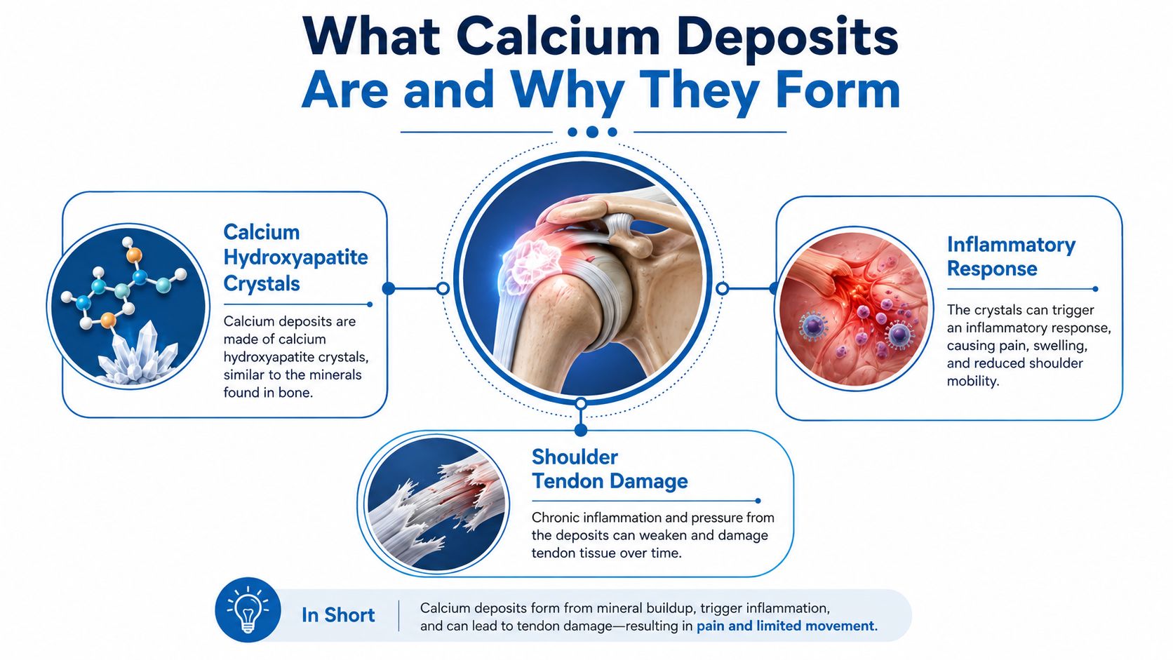
Blood and tissue fluids normally keep calcium dissolved and regulated. Local changes can disrupt that balance. Reduced oxygen within a tendon, repeated small injuries, altered tendon cells, and conditions such as diabetes or thyroid disorders may contribute. The process usually begins locally, rather than from eating too much calcium.
How the deposit changes over time
A sluggish pipe offers a useful comparison. As flow and tissue conditions change, mineral material can settle along the inner surface. It may remain quiet, then soften or fragment as the body starts clearing it. That clearing response can produce substantial inflammation.
Clinicians commonly describe a formative phase, a resting phase, a resorptive phase, and a later repair phase. During formation, the material becomes more organized. During rest, symptoms may be mild or absent. During resorption, it may become paste-like or fragmented, causing pain that seems disproportionate to the deposit's size.
Calcific tendinitis has been reported at a prevalence of 2.7% to 20% in adults, with about 35% to 45% of cases becoming symptomatic and roughly 50% of patients eventually developing shoulder pain in a clinical review of calcific tendinitis. It most often affects adults aged 30 to 50, occurs about twice as often in women as in men, and involves both shoulders in about 10% of patients.
The visible mineral is only part of the picture. A soft, inflamed deposit may behave differently from a dense, mature lesion, while an ossified structure may require a different assessment altogether. Imaging should therefore clarify the lesion's tissue type, size, location, and phase before removal is considered.
Types of Calcium Deposits You Should Know About
“Calcium deposit” describes several different findings. A radiology report may use similar wording for lesions with very different behavior, so the tissue type should be identified before removal is considered.
Calcific tendinitis
Calcific tendinitis contains hydroxyapatite within a tendon, often in the rotator cuff. Its texture can range from a firm, well-defined mass to softer, fragmented material, much like dried toothpaste that has hardened in place and later begun to break apart.
The deposit may be quiet during its resting phase, then become intensely painful as the body tries to clear it. Severe shoulder pain, night discomfort, painful movement, and restricted function are common during this inflammatory stage. Some deposits are found incidentally, however. Earlier studies described deposits in asymptomatic workers and shoulders, showing that the presence of calcium does not automatically identify the source of pain, as noted in the earlier discussion.
Periarticular and soft-tissue calcification
Some deposits develop beside a joint capsule, ligament, bursa, or another soft-tissue structure rather than clearly inside a tendon. Their appearance and behavior depend heavily on location. Trauma, systemic disease, and changes in mineral metabolism may influence how they form, while nearby inflammation can determine which symptoms a patient feels.
The deposit may be only part of the problem. Pain near a tendon or bursa can also reflect tendinitis and bursitis, so treatment should address the irritated structure instead of assuming that every visible deposit needs to be removed.
Ossified or heterotopic lesions
An ossified lesion has organized bone formation within soft tissue. Imaging may show a mature outer cortex or internal bone architecture, features that separate it from hydroxyapatite deposits. A mature bony mass is more like a small piece of bone fixed within the surrounding tissue than a soft deposit that can be disrupted.
Needling is generally intended for accessible, softer calcific material. It is usually a poor match for a mature ossified lesion, particularly when the lesion causes persistent mechanical restriction. Surgical assessment may become more relevant in that setting.
| Feature | Calcific Tendinitis | Periarticular Calcification | Ossified / Heterotopic Deposit |
|---|---|---|---|
| Material | Hydroxyapatite crystals | Mineral deposits near soft tissue structures | Organized bone within soft tissue |
| Typical feel on imaging | Dense, paste-like, or fragmentary | Variable and location-dependent | Bone-like architecture |
| Common behavior | May enter a painful resorptive phase | Often depends on the surrounding bursa or capsule | Usually more mechanically fixed |
| Response to needling | Often favorable when accessible and soft | Selective, depending on location | Usually limited |
| Main treatment question | Can the deposit be disrupted or aspirated? | What structure is inflamed? | Does the lesion require excision? |
The practical distinction is clear: focused shockwave therapy and ultrasound-guided procedures generally fit accessible intratendinous deposits, especially when they are soft or in an inflammatory or resorptive phase. A dense, mature lesion may need a different approach, while an ossified structure may call for surgical consideration. A narrative review of deposit phenotypes also supports matching treatment to the deposit's material, location, size, and stage rather than treating all calcium findings as one condition.
Recognizing Symptoms and Getting the Right Diagnosis
A patient may arrive with severe night pain and almost no shoulder movement, yet the deposit on imaging looks modest. That mismatch can occur during the resorptive phase, when the surrounding tendon and bursa are highly inflamed. Swelling, marked tenderness, and pain with ordinary movements may develop. A low-grade fever can occur occasionally, so infection must be considered rather than automatically attributed to calcific tendinitis.
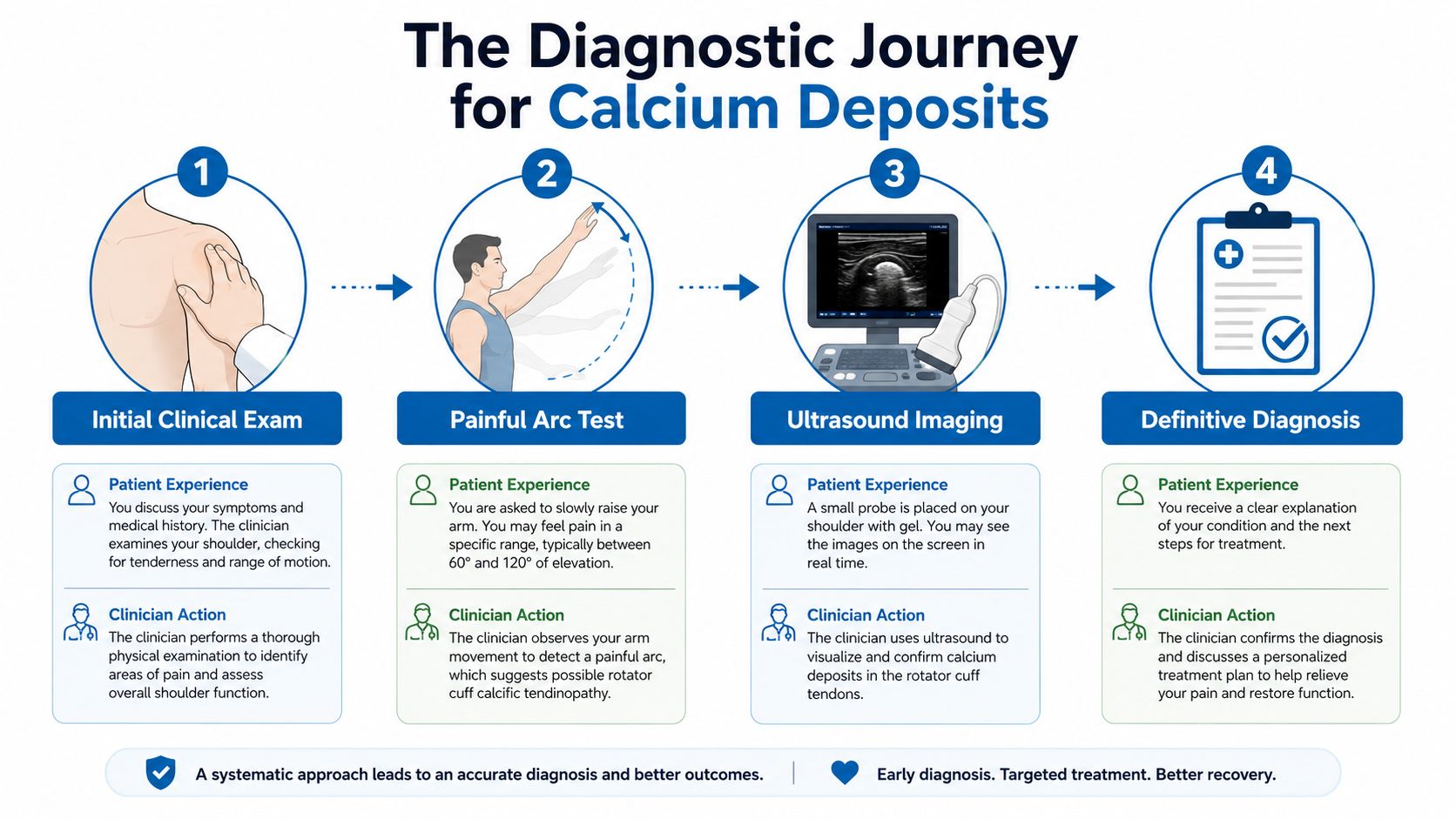
Diagnosis begins with the shoulder examination, not the scan alone. A clinician checks rotator cuff strength, tests resisted abduction, identifies painful arcs of elevation, and observes how the arm moves. Pain between about 60 and 120 degrees may suggest irritation beneath the acromion, but it does not establish that a calcium deposit is the cause.
The examination also helps separate a calcific deposit from an ossified lesion. Calcific material may be soft, fragmented, or undergoing resorption, which can make ultrasound-guided disruption or lavage technically suitable. A mature, bone-like lesion is more fixed and may require a different assessment. Symptoms, lesion phenotype, size, and phase must be considered together.
Why ultrasound often comes first
Musculoskeletal ultrasound shows the deposit while the shoulder moves. It uses no radiation, evaluates the tendon and bursa in real time, and can show whether the material looks dense, arc-like, fragmented, or more fluid. Those features help determine whether a minimally invasive procedure is reasonable.
Ultrasound can also reveal a tendon tear, bursal fluid, or a deposit near a nerve. Material safely positioned within an accessible tendon may be suitable for lavage. A deposit embedded in damaged tissue or lying close to a nerve may call for another plan.
When X-ray or MRI adds information
An X-ray provides a baseline view of the deposit's size, density, and position. It can also describe its appearance with systems such as the Gärtner classification, and it helps when ultrasound findings are uncertain or need comparison over time.
MRI is usually chosen to answer a specific question. It can assess a suspected rotator cuff tear, mass effect, unusual pain pattern, or atypical deposit location. The final diagnosis should connect the deposit type, its phase, its relationship to the tendon, and the patient's functional problem.
Conservative and Noninvasive Treatment Options
A painful shoulder does not automatically need the deposit removed. For many people with calcific tendinitis or periarticular calcification, conservative care is the appropriate starting point. The aim is to settle irritation, preserve movement, and give the tendon time to recover while the deposit stays stable or begins natural resorption.
Match treatment to the tissue state
A small, stable deposit with manageable symptoms may improve through activity changes and rehabilitation. A softer, resorptive deposit can provoke more inflammation, so short-term symptom control may be needed while the tissue settles. The deposit's consistency and phase matter because soft calcific material differs from organized, bone-like tissue.
- Activity adjustment: Reduce repetitive overhead work, heavy pressing, and positions that reproduce the painful arc. This lowers compression on the tendon without unnecessarily stopping all shoulder movement. Complete immobilization can increase stiffness.
- Ice and local care: Cold therapy may temporarily ease pain around an inflamed tendon or bursa. A brief sling can help during an acute flare, but prolonged use should be avoided unless a clinician has a specific reason.
- Medication review: Nonsteroidal anti-inflammatory drugs may help for a short course when medically appropriate. Kidney disease, gastrointestinal bleeding risk, cardiovascular disease, anticoagulant use, and other medicines can change their safety, so dosing should be individualized.
- Physical therapy: Stretching works on lost motion, while progressive strengthening improves rotator cuff and scapular control. Eccentric loading may be added as pain allows. An aggressive program during a highly reactive phase can worsen symptoms.
A supervised plan often combines mobility exercises with gradual loading. Therapeutic ultrasound or shockwave treatment may be offered as an adjunct in some clinics, but the choice should follow the lesion's imaging appearance, size, and phase rather than the simple presence of calcium. A calcific deposit that remains soft may later suit lavage or another deposit-targeting procedure, while an ossified lesion generally requires a different discussion.
Recovery is measured by function, not by pain on one isolated day. A short flare does not necessarily mean treatment has failed. Look for gradual improvement in motion, sleep, and daily activities.
Corticosteroid treatment can calm inflammation in the subacromial bursa or another painful space. It manages symptoms rather than mechanically removing the deposit. Patients can review the role, benefits, and limitations of corticosteroid injections with a treating clinician before choosing an injection.
Minimally Invasive Procedures That Target the Deposit
A patient may have severe pain from a deposit that is still soft and fragmentary, while another patient has a firm, organized lesion that behaves more like bone. Both may be described casually as “calcium,” but they are not the same treatment problem. After conservative care has been tried appropriately, the procedure should match the deposit's composition, size, location, and phase.
Shockwave therapy
High-energy extracorporeal shockwave therapy sends focused acoustic energy through the skin. No needle enters the shoulder. The energy can break up susceptible material and may encourage the body to resorb the fragments over time.
A network meta-analysis summarizing extracorporeal shockwave therapy outcomes found that high-energy shockwave therapy significantly reduces pain, promotes complete deposit resolution, and reduces the need for surgery. It may suit a visible intratendinous deposit when a patient wants to avoid needle treatment or when the material is not suitable for aspiration.
Treatment commonly requires multiple visits. The shoulder can feel temporarily more irritated afterward, so improvement is usually gradual rather than immediate. Shockwave therapy is less suitable for a mature ossified lesion. Organized bone does not respond like soft hydroxyapatite material, just as a compact pebble does not break apart like wet sand.
Ultrasound-guided needling and barbotage
Barbotage, also called ultrasound-guided percutaneous needle lavage, uses live ultrasound to guide a needle into the deposit. Sterile fluid helps fragment the material, and the clinician removes what can be aspirated. This approach targets the physical source of irritation rather than only reducing the surrounding inflammation.
A review reported a mean radiographic calcium reduction of 39.9 mm², with 80% of patients achieving more than 60% deposit resolution, and a very low reported complication rate in the reviewed series, according to a review of ultrasound-guided percutaneous needle lavage outcomes. In a prospective study of 121 patients, pain disappeared in 89.26% and calcification disappeared completely in 86.78%. Vasovagal reactions occurred in 5%, as reported in the same review.
The strongest candidate is usually a well-localized, reachable intratendinous deposit, especially when imaging suggests soft or fragmentary material. Size and access still matter. A large lesion may require more than one treatment discussion, while a small deposit in a risky location may not justify needle entry. A steroid injection can be added for bursal inflammation, but it does not make an otherwise fixed deposit removable.
Steroid injections alone
A steroid injection can calm inflammation and improve sleep or movement when bursitis is the main pain generator. It does not mechanically fragment or aspirate calcium. Pain relief and radiographic removal are therefore separate outcomes.
| Treatment | Mechanism | Best Deposit Phase | Key Evidence | Recovery Time |
|---|---|---|---|---|
| Shockwave therapy | External acoustic energy disrupts susceptible material | Intratendinous inflammatory or resorptive deposits | High-energy treatment reduces pain and supports complete resolution, based on the linked network meta-analysis | Gradual improvement |
| Needle lavage | Direct fragmentation and aspiration under ultrasound | Accessible, softer, or fragmentary deposits | Clinical evidence reports substantial deposit and pain improvement, as described in the linked barbotage review | Usually outpatient recovery |
| Steroid injection | Suppresses local inflammation | Inflamed bursa or surrounding tissue | Symptom control, not physical removal | Often brief activity modification |
| Surgery | Debridement or excision | Refractory or mechanically fixed lesions | Reserved for selected cases | Structured rehabilitation |
Patients considering an operative pathway can review endoscopic surgery for deposit removal. Imaging should first establish that the lesion is mechanically contributing to symptoms and that the proposed procedure matches its phenotype.
Surgical Removal of Calcium Deposits
A person may still have pain and limited shoulder movement after months of appropriate nonoperative care. If imaging shows a deposit that is mechanically irritating the tendon or blocking motion, surgery may become part of the discussion. Reviews commonly place it after at least 6 months of non-surgical care, although timing depends on the lesion's size, material, location, tendon condition, disability, and response to earlier procedures systematic review of treatment options.
The lesion's structure matters. Soft or fragmentary calcific material may be removable through a small arthroscopic opening. Dense, mature, bone-like tissue is more organized and may require a different surgical plan. Surgery is not a larger version of needle lavage. It is selected when the deposit, its phase, or associated tendon damage makes less invasive treatment unsuitable or unsuccessful.
What arthroscopy involves
Arthroscopic removal uses small portals and a camera to inspect the shoulder. The surgeon identifies the deposit, opens the involved tendon when needed, removes accessible material, and clears unhealthy tissue. An acromioplasty may be added when the anatomy creates meaningful compression, but it is not automatically required.
The goal is safe decompression, not excavation at any cost. A technical review notes that complete removal of every residual fragment isn't necessary if aggressive excavation would damage the tendon. Debridement and removal of loose residual material may provide a better balance between removing the irritant and preserving tendon strength surgical review.
Open excision is less common. It may be considered for deep, recurrent, unusually positioned, or difficult-to-access lesions. Possible complications include infection, postoperative stiffness, tendon re-tear, and incomplete pain relief. Removing the deposit can address one mechanical source, while bursitis, tendon injury, or joint disease may still require care.
Recovery requires rehabilitation
Rehabilitation protects the debrided or repaired tendon, restores passive motion, and then builds strength gradually. Sling use and exercise timing depend on whether the surgeon also repaired the tendon. Overhead activity may take several months to resume when tendon healing is part of the operation.
A meta-analysis of six studies involving 294 shoulders found low complication rates and no reoperations, with no significant outcome differences among acromioplasty plus removal, acromioplasty alone, or removal alone over 12 months to 5 years surgical evidence review. These findings support matching the operation to the lesion and tendon, rather than choosing the most extensive procedure automatically.
Choosing the Right Treatment for Your Situation
A patient may have an obvious deposit on an X-ray yet little pain, or severe symptoms from a smaller lesion. Treatment should follow the deposit's behavior and the tissue around it, rather than the scan alone.
A practical decision framework
A clinician weighs several factors:
- Material: Soft hydroxyapatite can respond differently from organized ossified tissue.
- Phase: A resorptive deposit may be inflamed and suitable for lavage, while a stable deposit may be observed.
- Size and access: A larger, accessible lesion offers a clearer target for ultrasound-guided treatment. A small lesion near vulnerable structures may not.
- Pain generator: Bursal inflammation, tendon injury, joint disease, and the deposit itself can coexist.
- Functional goal: A manual worker, overhead athlete, and person seeking better sleep may choose different timelines.
- Prior care: The response to activity changes, medication, therapy, shockwave treatment, or injection helps determine the next step.
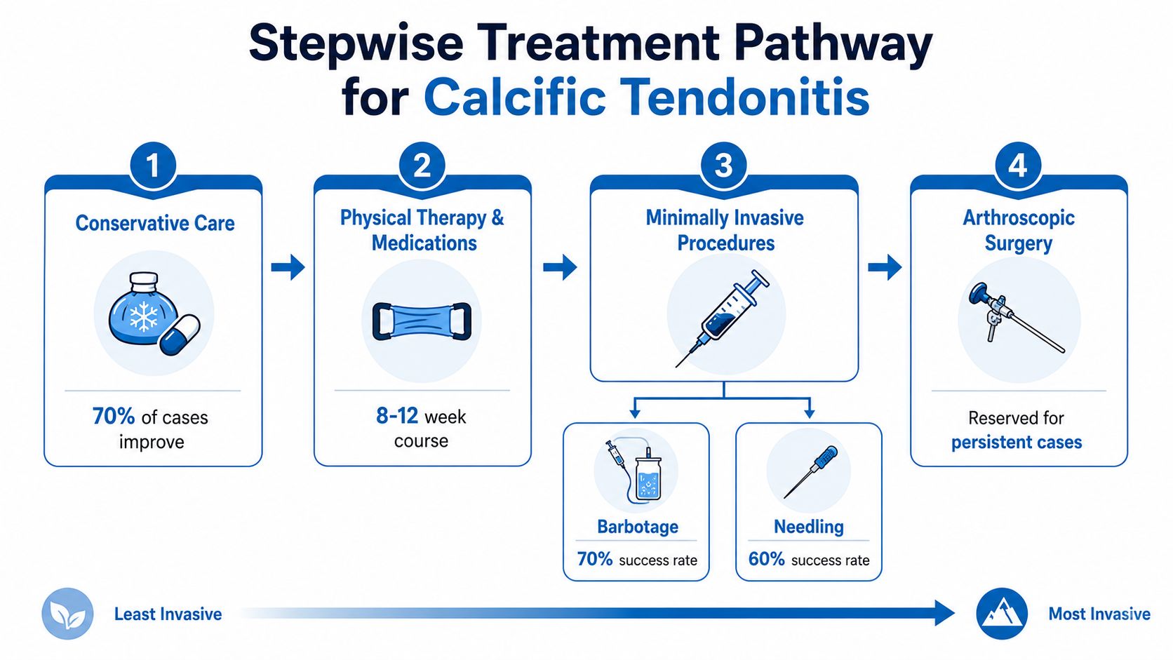
A small, stable lesion with mild symptoms often fits rehabilitation and observation. A painful, accessible, resorptive deposit that continues to limit sleep and movement after conservative care may justify discussion of shockwave treatment or barbotage. A mature ossified lesion, tendon tear, or persistent mechanical block may instead call for an orthopedic opinion.
The evidence supports matching the option to the phenotype. A 2025 systematic review of 33 randomized trials found that physical therapy ranked best for function, while ultrasound-guided needling combined with subacromial corticosteroid injection showed promise for deposit resolution, as described in a 2025 systematic review ranking physical therapy and needling approaches. These findings do not make needling the right choice for every lesion. They show why function, deposit composition, access, and treatment goals must be considered together.
The best option matches the lesion, not the fear attached to the scan.
Shared decision-making should identify the dominant symptom, review the images, and set a realistic time horizon before escalation. The right choice is the least invasive treatment that fits the deposit's type, phase, location, and the patient's goals.
Recovery, Follow-Up, and When to See a Specialist
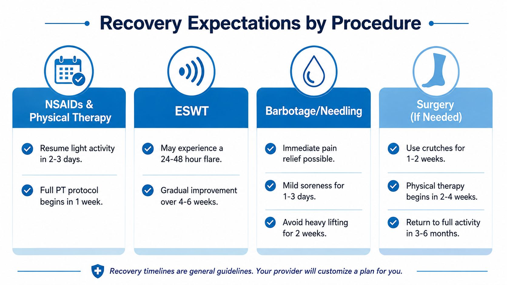
Recovery depends on the procedure and the deposit being treated. Medication and physical therapy usually build comfort, motion, and tendon capacity over time. Shockwave therapy can briefly increase soreness before symptoms improve. Barbotage may reduce a soft, accessible deposit more directly, while the surrounding tendon and bursa still need time to settle. An ossified lesion behaves more like formed bone, so its recovery plan may differ from that of a resorptive calcific deposit.
Follow-up should measure function alongside imaging. Repeat ultrasound or X-ray can help when symptoms stop improving, the clinician needs to assess deposit change, or the original diagnosis remains uncertain. A smaller deposit is only one possible result. Better sleep, improved range of motion, and a return to normal tasks may matter more than complete radiographic disappearance.
Signs that need prompt evaluation
Seek urgent medical assessment for:
- Sudden swelling: Rapid enlargement or severe new swelling needs evaluation.
- Fever with worsening pain: Infection can resemble an inflammatory deposit and should not be dismissed.
- Neurovascular changes: New numbness, weakness, color change, or unusual coldness requires prompt attention.
- Escalating night pain: Rapidly worsening pain, especially with systemic symptoms, deserves reassessment.
- Loss of function: Inability to lift the arm or use the joint may signal tendon injury or another problem.
A specialist visit is more useful when you bring prior imaging, a complete medication list, a brief pain diary, and written functional goals. These details help distinguish an abnormal scan from a shoulder that still cannot perform an important task.
After surgery, plan the home environment before the procedure. Arranging mobility support, a safe sleeping setup, medication organization, and household assistance can reduce avoidable strain. The DME Superstore surgery recovery guide covering post-procedure mobility and household setup discusses these practical preparations.
An interventional pain and orthopedic evaluation can combine diagnostic ultrasound, image-guided treatment, surgical co-management, and coordinated rehabilitation. Patients in New Jersey and Staten Island with plateaued shoulder pain, disrupted sleep, or failed conservative care can request an assessment to clarify whether the lesion is calcific, periarticular, or ossified before selecting removal of calcium deposits.
Interventional Pain Management offers diagnostic evaluation, image-guided minimally invasive procedures, orthopedic consultation, and coordinated rehabilitation for persistent joint and tendon pain. Patients can visit Interventional Pain Management to request an appointment and discuss a plan matched to the deposit's type, phase, location, and functional impact.