

A person can spend weeks doing physical therapy, changing pillows, taking medication, and trying to work around persistent neck pain, only to hear that an MRI looks “normal for age.” That experience is frustrating, but it doesn't mean the pain is imaginary or untreatable. A cervical facet joint can generate substantial pain even when imaging doesn't clearly identify it as the source.
The diagnostic challenge is that facet pain often overlaps with muscle strain, disc-related pain, and ordinary stiffness. The scan may show arthritis without proving that the arthritic joint hurts, while a painful joint may look unremarkable. A careful history, examination, and properly controlled diagnostic blocks provide a more reliable path toward treatment. For people seeking additional background on persistent neck symptoms, this advanced neck care in Sandy Utah resource offers another patient-focused perspective.
Table of Contents
- Recognizing Cervical Facet Joint Pain
- Understanding Cervical Facet Joint Anatomy and Function
- Symptoms and Warning Signs of Facet-Mediated Neck Pain
- Why Imaging Alone Cannot Diagnose Facet Joint Pain
- Treatment Options From Conservative Care to Advanced Procedures
- Expected Outcomes and Recovery Timelines
- When to See an Interventional Pain Specialist
Recognizing Cervical Facet Joint Pain
Consider a patient whose neck has hurt for months. Physical therapy helped briefly, chiropractic treatment produced inconsistent results, and medication made daily activities easier without resolving the underlying pain. The MRI report mentions degenerative changes, but no obvious surgical problem. The patient may leave the appointment believing that nothing specific can be done.
That conclusion is often premature. Cervical facet joints are small paired joints at the back of the neck, and they're a major, well-established source of chronic neck pain. In a controlled diagnostic-block study, facet joint pain accounted for 60% of chronic neck pain cases, with a 95% confidence interval of 50% to 70%. The same study reported a 40% false-positive rate for controlled diagnostic blocks, which shows why a single injection can overestimate the likelihood that a facet joint is the true pain generator (controlled diagnostic-block study).
Why patients are often misdirected
Facet-mediated pain may feel deep, dull, and difficult to pinpoint. It can spread toward the base of the skull, shoulder, or shoulder blade without following the predictable line of a compressed spinal nerve. Those features overlap with muscular pain and disc-related pain, so symptoms alone can suggest a diagnosis without confirming it.
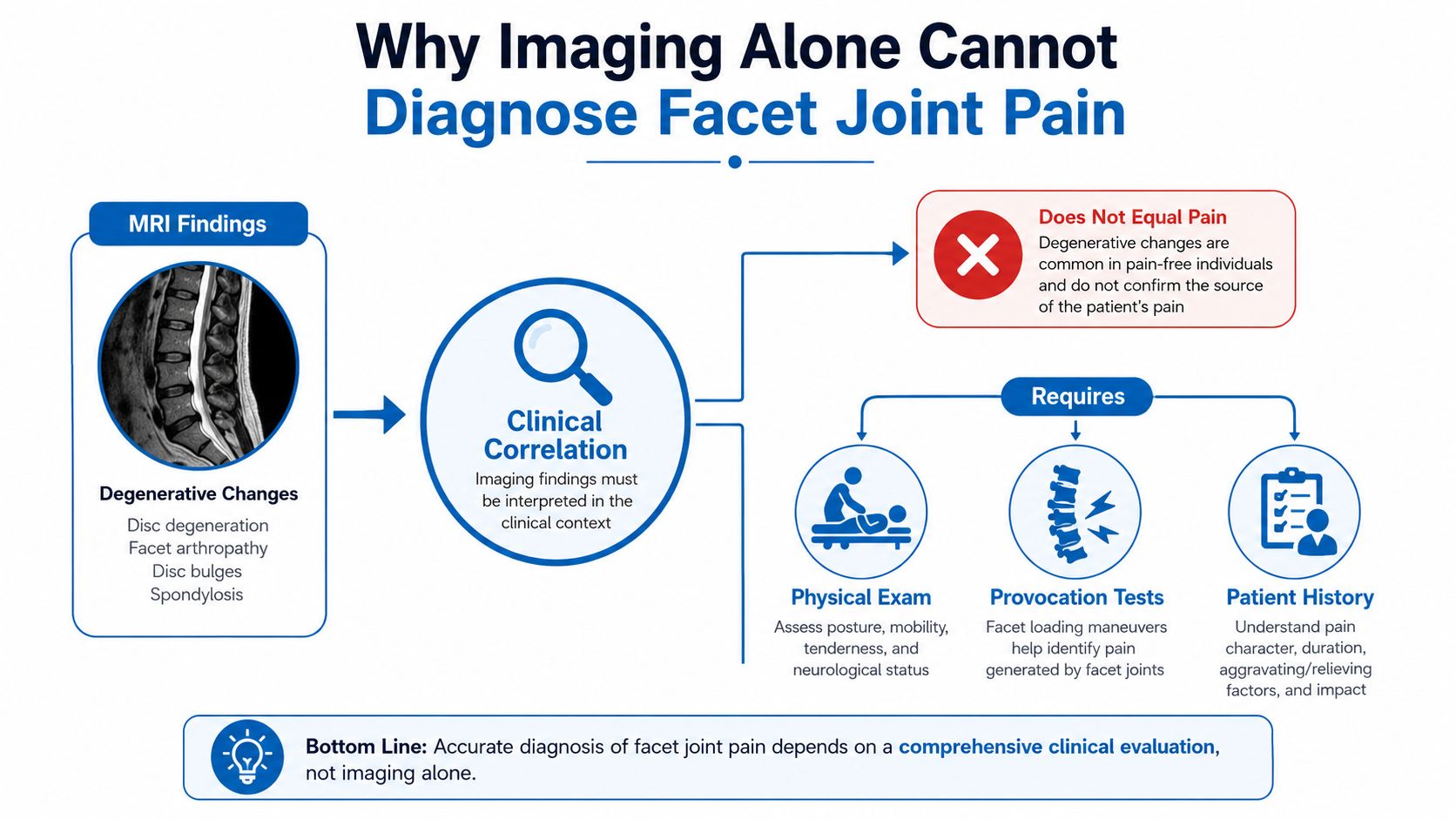
Imaging creates another source of confusion. An MRI or CT scan can show structural changes, but it can't reliably establish whether a particular facet joint is causing the patient's symptoms. Some patients have substantial degeneration with little pain, while others have severe symptoms without a dramatic imaging finding.
Clinical perspective: The scan describes anatomy. The diagnostic block tests whether a suspected pain pathway is actually active.
A precise diagnosis matters because advanced procedures target specific structures. If the facet joint has never been properly evaluated, a patient may continue cycling through general treatments that don't address the relevant pain generator. The most useful next step is usually not another scan, but a consultation that connects the symptom pattern, physical examination, imaging, and controlled diagnostic testing.
Understanding Cervical Facet Joint Anatomy and Function
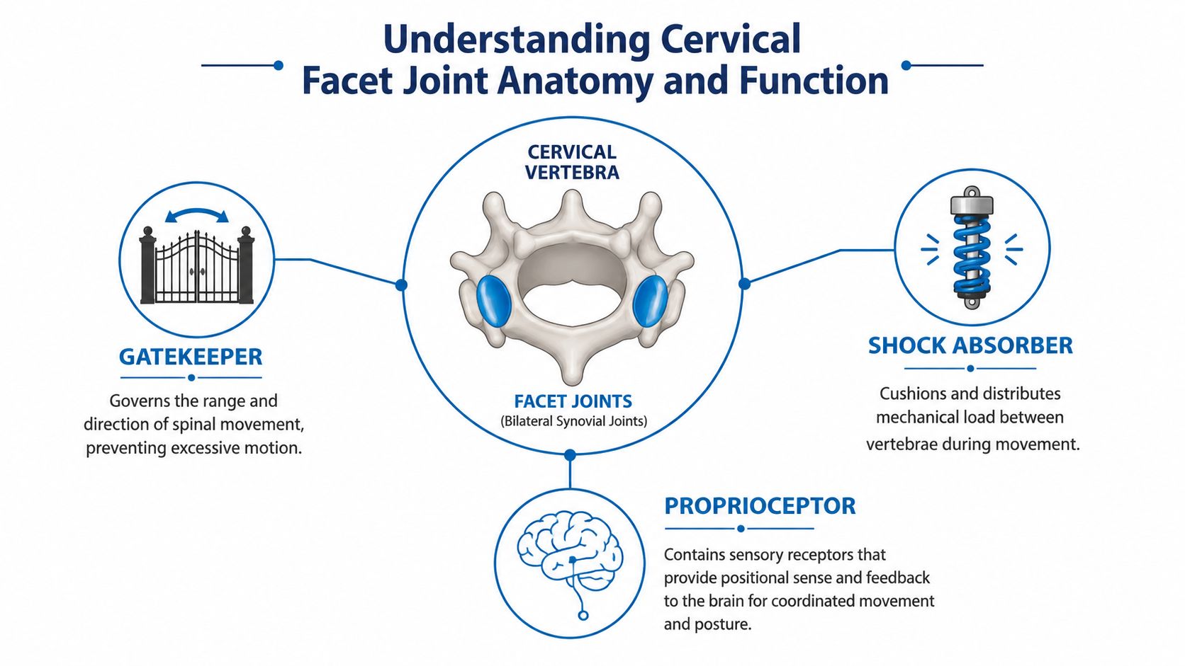
The cervical facet joints work like small paired hinges behind the vertebral bodies. They guide movement between neighboring vertebrae, help limit excessive motion, and contribute to stability. Like a door hinge, each joint allows movement along a controlled path while preventing the adjoining bones from shifting too freely.
Each facet joint is a synovial joint with a cartilage-covered surface and a surrounding capsule. The capsule contains sensitive tissues, and the joint's pain signals travel through medial branch nerves. Those nerves are important because clinicians can temporarily interrupt their signals with a medial branch block and, when appropriate, treat them with radiofrequency ablation.
How the joints support movement
The neck contains seven cervical vertebrae. The facet joints from C2–3 through C7–T1 are innervated by medial branches of the dorsal rami, providing the anatomical basis for medial branch blocks and radiofrequency ablation (2021 multispecialty guideline). Clinically, the C2–3 and C5–6 joints are identified as the most commonly implicated levels in neck pain in that guideline.
During flexion, the facet joints help guide the vertebrae as the neck bends forward. During extension, rotation, and lateral bending, they control how far and in what direction the neck moves. Their role isn't merely to permit motion. They also help the nervous system sense neck position, allowing the brain to coordinate posture and movement.
Wear, injury, or repeated loading can change that balance. Cartilage may thin, the capsule may become irritated, and bony overgrowth can develop. A joint that once guided smooth movement may then hurt when compressed, particularly during extension or rotation.
Why targeting requires anatomical precision
The nerve supply isn't a simple one-nerve-to-one-joint arrangement. A facet joint is typically supplied by medial branches from the same level and the level above. The C2–3 joint receives its supply from the third occipital nerve. Anatomical research has also identified multiple medial branches at some levels, plexus-like patterns in the upper cervical region, and direct branches to the facet joints (cervical medial branch anatomy study).
That complexity explains why image guidance and careful procedural planning matter. A useful visual reference for neighboring structures is this anatomical guide for neck and jaw from Bayside Osteopathic Health.
Symptoms and Warning Signs of Facet-Mediated Neck Pain
Facet-mediated pain commonly feels like a deep, dull ache at the back or side of the neck. It may refer toward the base of the skull, the top of the shoulder, or the area between the shoulder blades. That referral pattern is usually non-dermatomal, meaning it doesn't follow the narrow, predictable path associated with a specific nerve root.
Pain often increases when the neck extends or rotates. Looking upward, turning to check a blind spot, holding the head in one position during desk work, or sleeping with the neck rotated may aggravate symptoms. Rest or gentle flexion can reduce discomfort for some patients, although no single pattern confirms the diagnosis.
Comparing common sources of neck pain
| Feature | Facet Joint Pain | Disc-Related Pain | Muscular Pain |
|---|---|---|---|
| Typical location | Deep pain in the back or side of the neck | Neck pain that may accompany disc irritation | More diffuse soreness across muscles |
| Referral pattern | Base of skull, shoulder, or shoulder blade, often non-dermatomal | May travel into the arm in a nerve-related distribution | Broad discomfort near tight or tender muscles |
| Common aggravators | Extension, rotation, and prolonged static posture | Certain neck positions, lifting, or nerve irritation | Activity, overuse, stress, or direct muscle loading |
| Examination clues | Pain with selected movements and facet-loading maneuvers | Possible sensory, reflex, or strength changes | Tenderness and tightness in the involved muscles |
| Diagnostic limitation | Symptoms can overlap with other sources | Imaging may show changes without proving pain causation | Muscle tenderness doesn't exclude a joint problem |
A symptom pattern helps a clinician choose the next diagnostic step, but it isn't a substitute for confirmation. The neck pain clinical resource provides broader context for how different causes can overlap.
Warning signs that need prompt evaluation
Certain symptoms point beyond a routine mechanical neck problem. Urgent medical assessment is appropriate when neck pain occurs with:
- Progressive neurological deficits: Increasing weakness, loss of coordination, or worsening numbness requires timely evaluation.
- Systemic symptoms: Fever or unexplained weight loss can signal an illness that needs a broader workup.
- Cancer history: A personal history of cancer changes the level of concern.
- Unrelenting pain: Constant pain that doesn't vary with position, rest, or activity deserves prompt attention.
Why Imaging Alone Cannot Diagnose Facet Joint Pain
An MRI report can mention facet arthropathy, cartilage wear, joint-space changes, or bone spurs. Those findings describe what the joints look like, not whether they're producing the patient's pain. Structural degeneration is common in adults who may have little or no neck discomfort, so an abnormal scan can be a false positive for the pain source.
The opposite problem also occurs. A patient may have a relatively modest imaging finding while experiencing significant facet-mediated pain. Imaging can miss the functional and inflammatory aspects of a painful joint, and it can't reproduce the movement or loading that triggers symptoms during everyday life.
The role of controlled medial branch blocks
Recent consensus guidance emphasizes that imaging alone shouldn't establish cervical facet pain. It also recommends a six-week conservative-care trial before prognostic cervical facet blocks in acute neck pain, reinforcing that an invasive diagnostic procedure should follow clinical evaluation and appropriate initial care (recent consensus guidance).
A medial branch block places a small amount of local anesthetic near the nerves that carry sensation from the suspected facet joint. The clinician then compares the patient's familiar pain and functional ability before and after the block. If the expected pain disappears during the anesthetic's effective period and returns as it wears off, the result is more informative than an imaging finding alone.
Some practices use controlled or comparative blocks with different local anesthetics. The purpose is to see whether the timing and degree of relief correspond to each medication's expected duration, rather than relying on a nonspecific temporary improvement. This approach also recognizes the possibility of placebo response and technical variability.
Practical rule: A candidate for radiofrequency ablation should first demonstrate meaningful, reproducible, time-concordant relief from properly performed diagnostic blocks.
The process can feel like a bottleneck, but that's a safety feature. It reduces the chance of treating the wrong joint and helps identify patients who are more likely to benefit from denervating the medial branches. Patients can learn more about the general purpose of diagnostic injections through this nerve block treatment overview.
Treatment Options From Conservative Care to Advanced Procedures
Treatment should progress from the least invasive reasonable option toward procedures that require stronger diagnostic evidence. The right choice depends on the symptom pattern, examination, duration of pain, medical history, and whether controlled blocks confirm a facet-mediated source.
Conservative care
Physical therapy can focus on cervical stabilization, posture, movement control, and strengthening of the muscles that support the neck. Activity modification may reduce repeated extension or prolonged static positioning, while anti-inflammatory medication may help selected patients who can take it safely.
These measures are appropriate for many people, particularly when symptoms are recent or the diagnosis remains uncertain. They may not fully control chronic facet pain, but they can improve movement tolerance and help a patient determine which activities consistently trigger symptoms.
Sleep positioning also matters. A pillow should support a neutral neck position rather than forcing the head sharply forward or sideways. A practical cervical pillow guide can help patients think through positioning, although a pillow change won't diagnose or eliminate a painful facet joint.
Interventional options
An intra-articular facet injection places local anesthetic and often corticosteroid directly into the joint under image guidance. It may provide temporary symptom relief and can offer information about the joint, but current guidance says routine intra-articular injections shouldn't be the standard treatment because their benefit is limited. Medial branch blocks are often more predictive when radiofrequency treatment is being considered (2026 systematic review and guidance).
Radiofrequency ablation uses controlled thermal energy to interrupt pain transmission through the targeted medial branch nerves. It doesn't repair cartilage or reverse arthritis. Instead, it aims to reduce pain from a confirmed facet pathway, and it's generally considered only after appropriate diagnostic blocks. A plain-language overview of the procedure is available in this radiofrequency ablation resource.
Pulsed radiofrequency is a less destructive technique that uses intermittent energy rather than the same continuous thermal lesion used in conventional ablation. Its role depends on the individual case and the treating physician's approach.
Surgery isn't a routine treatment for isolated facet-mediated pain. Fusion or another structural procedure may be considered only when there's a separate indication such as instability, deformity, or significant nerve or spinal cord compression.
| Treatment | Approach | Ideal Candidate | Duration of Relief | Key Considerations |
|---|---|---|---|---|
| Physical therapy | Stabilization, posture, and movement retraining | Recent or mechanically influenced pain | Variable | Requires consistent participation and reassessment |
| Medication and activity modification | Reduces inflammation or limits aggravating load | Patients who can use medication safely | Variable | Doesn't confirm the pain generator |
| Intra-articular injection | Medication placed inside the facet joint | Selected patients needing joint-directed care | Often temporary | Not considered routine standard treatment |
| Medial branch block | Temporary anesthetic interruption of facet nerve signals | Patients being evaluated for facet-mediated pain | Short diagnostic window | Technique and interpretation are critical |
| Radiofrequency ablation | Lesioning of confirmed medial branch pain pathways | Patients with reproducible block response | Can be extended, but varies | Doesn't reverse joint degeneration |
| Surgery | Structural correction or stabilization | Patients with a separate surgical indication | Depends on procedure | Not appropriate for isolated facet pain in most cases |
The central decision point remains diagnostic confirmation. A procedure aimed at the facet nerves is unlikely to help if the pain comes from a disc, nerve root, muscle, or another structure.
Expected Outcomes and Recovery Timelines
Recovery depends on the treatment and the accuracy of the diagnosis. Conservative care often requires steady participation in exercises, posture work, and activity changes before the neck feels more dependable. Progress may be gradual, and persistent symptoms should prompt reassessment rather than an automatic assumption that more of the same treatment is needed.
Facet injections may begin helping after the procedure, but the response can vary widely. Some patients receive short-term improvement, while others get little meaningful benefit. Repeat procedures shouldn't be scheduled solely because time has passed. The clinician should first confirm that the earlier response was useful and clinically appropriate.
What recovery after ablation may feel like
Radiofrequency ablation can cause temporary soreness near the treatment sites. Pain relief may not be immediate, and some patients experience a gradual change rather than a sudden switch from pain to no pain. The treated nerve can eventually recover, so relief isn't necessarily permanent and repeat treatment may be considered when symptoms return and the original diagnosis remains sound.
Possible complications include infection, bleeding, temporary numbness, irritation of nearby nerves, and nerve injury. These risks are reasons to use image guidance, careful positioning, appropriate patient selection, and clear post-procedure instructions. A patient should contact the treating office promptly for concerning weakness, spreading numbness, fever, severe swelling, or worsening symptoms.
Incomplete relief doesn't always mean the procedure was performed incorrectly. The original diagnosis may have been incomplete, more than one pain generator may be present, or the targeted level may not account for all symptoms. The next step may involve renewed examination, treatment of another confirmed source, or a return to rehabilitation.
When to See an Interventional Pain Specialist
Neck pain doesn't always require an interventional consultation. A specialist evaluation becomes more reasonable when symptoms persist despite appropriate conservative care, when pain repeatedly spreads toward the shoulder or upper back without a clear nerve pattern, or when sleep, work, and routine movement remain significantly limited.
Patients with imaging that shows degeneration but no clear surgical target may benefit from an assessment focused on pain-source identification. The consultation should connect the scan with the examination rather than treating the radiology report as a diagnosis.
Useful questions include:
- Diagnostic method: Does the practice perform controlled medial branch blocks when facet pain is suspected?
- Ablation criteria: What degree and timing of relief are required before radiofrequency ablation is considered?
- Alternative explanations: Could the symptoms arise from a disc, nerve root, muscle, or more than one structure?
- Care coordination: How will physical therapy, medication management, and surgical consultation fit into the plan if needed?
A qualified specialist should explain why a particular level is being tested, what the block can and cannot prove, and what happens if the result is negative. That transparency helps patients avoid unnecessary procedures and move beyond repeated treatment cycles that never address the actual pain generator.
Interventional Pain Management evaluates neck pain through medical history, physical examination, imaging review, and targeted procedures when clinically appropriate. Patients with persistent symptoms can visit Interventional Pain Management to learn about diagnostic and treatment options for cervical facet joint pain and request an evaluation.