
You followed the instructions, completed rehabilitation, waited through recovery, and expected the original pain to fade. Instead, sitting still hurts, walking is limited, sleep is broken, or a new burning sensation travels down a leg. The next appointment often brings a difficult question: What should happen when back surgery has healed, but the patient hasn't improved?
Failed back surgery syndrome treatment shouldn't begin with an automatic march from medication to injections to another operation. FBSS is a description of persistent or returning pain after spinal surgery, not a diagnosis of its cause. The safest pathway starts by identifying what is generating pain today, then matching treatment to that finding.
Table of Contents
- When the Surgery Heals but the Pain Stays
- Finding the Real Source of Persistent Pain
- Interventional Options Beyond the Operating Room
- How Spinal Cord Stimulation Fits Into Treatment
- When Revision Surgery Is Worth Considering
- Building Your Own Treatment Pathway
- What Lasting Relief Actually Looks Like
When the Surgery Heals but the Pain Stays
A patient may do everything expected after lumbar surgery. The incision closes, physical therapy is completed, activity increases gradually, and follow-up imaging appears reassuring. Yet the patient still can't sit through a meal, walk around a store, or sleep without waking from back or leg pain.
That experience has a clinical name, failed back surgery syndrome, although many clinicians also use the term persistent spinal pain syndrome type 2. The label covers pain that continues, returns, or changes after spinal surgery. It doesn't prove that the operation was performed incorrectly, and it doesn't explain which structure is responsible.
The diagnosis describes the outcome. It doesn't identify the pain generator.
FBSS can follow a technically successful operation because the spine contains several possible sources of pain. A nerve may remain irritated, a neighboring segment may later become symptomatic, a facet joint may produce mechanical pain, or scar tissue may surround a nerve root. The original surgery may have corrected the problem it was designed to address while another pain source remained or developed.
The condition also deserves attention because it isn't rare after lumbar surgery. A review reported that the risk of developing FBSS after lumbar spinal surgery ranges from 4% to 50%, while its estimated frequency in the general population is about 0.02% to 2% (review of FBSS causes and treatment). These figures vary because studies define and measure FBSS differently, but they make one point clear: persistent pain after surgery requires a structured reassessment.
Some patients also need support for problems that overlap with spinal recovery. For readers seeking information about pelvic symptoms, mobility, or related rehabilitation concerns, prolapse care for aged care offers additional context from a physical therapy perspective.
The first question shouldn't be, “Which procedure comes next?” It should be, “What is causing the current pain, and does that cause have a treatable target?”
Finding the Real Source of Persistent Pain
A useful evaluation treats the FBSS label as the beginning of an investigation. The clinician compares the present symptoms with the symptoms that led to surgery, examines the patient, reviews the operative report, and looks for a pain pattern that points toward a specific structure.
Start with updated anatomy
Fresh imaging may be necessary even when an earlier postoperative scan looked acceptable. A new MRI can show recurrent disc material, narrowing around a nerve, changes at a level next to a fusion, or findings that were not present during the original recovery period. CT can add detail about bone, fusion status, hardware position, or subtle structural change.
The scan isn't interpreted in isolation. A visible abnormality matters only when it matches the patient's symptoms and physical findings. Many postoperative changes can appear dramatic but produce no pain, while a less obvious finding may be clinically important if it aligns with the patient's distribution of numbness, weakness, or radiating pain.
Map the symptoms, not just the pain score
A pain diary can help distinguish patterns that a single clinic conversation may miss. The patient can record:
- Location: Back, buttock, thigh, calf, foot, groin, or a broader area.
- Quality: Burning and electric symptoms often suggest nerve involvement, while aching linked to extension or rotation may suggest a mechanical source.
- Triggers: Sitting, standing, walking, bending, coughing, or lying down can each provide different clues.
- Timing: Constant pain, activity-related pain, and pain that appears mainly at night may lead the evaluation in different directions.
- Function: Walking distance, sleep continuity, stair use, and tolerance for sitting often reveal meaningful change better than a single rating.
A clinician may then use a diagnostic medial branch block when facet-mediated pain is suspected, or a selective nerve root block when a particular nerve appears responsible. These procedures can provide temporary information as well as relief. The important question is whether numbing the suspected structure changes the familiar pain in a way that matches the clinical examination.
Potential explanations include recurrent disc herniation, adjacent segment disease, hardware irritation, epidural fibrosis, residual foraminal narrowing, or a pain source outside the operated level. Muscle deconditioning, altered movement, and nervous-system sensitivity can also maintain symptoms after the original structural problem has been treated.
A treatment cannot be more precise than the diagnosis supporting it.
That principle determines every later choice. Radicular inflammation, facet pain, focal neuropathy, and widespread axial pain don't respond to the same intervention. If the workup hasn't identified a plausible generator, repeated procedures may add burden without clarifying the problem.
Interventional Options Beyond the Operating Room
Once the pain pattern and suspected source are clear, a procedure can answer a focused question. An epidural injection treats inflammation around an irritated nerve. A medial branch block tests whether facet joints are generating axial pain. Adhesiolysis addresses suspected scar-related restriction around a nerve root. The sequence should follow the diagnosis, not the availability of the next procedure.
Match the procedure to the pattern
An interlaminar epidural injection places medication into the epidural space from the midline. A transforaminal injection directs treatment nearer to a selected nerve root. A caudal injection enters through the sacral opening and may be useful when postoperative anatomy makes other routes difficult. These approaches fit best when leg pain, radiating pain, or nerve-root inflammation is prominent. The lumbar epidural steroid injection guide describes one commonly used approach.
Facet joint blocks and medial branch blocks address a different pattern. They can test whether the small joints at the back of the spine contribute to localized, mechanical back pain. If the temporary numbing produces a convincing change in the familiar pain, radiofrequency ablation may interrupt signals carried by the relevant medial branch nerves. It does not rebuild a damaged joint or remove a disc problem, so the suspected source must match the treatment.
Percutaneous adhesiolysis may be considered when scar tissue in the epidural space appears to restrict a nerve root and standard medication delivery has not provided enough benefit. Results are contradictory, so a scan showing scar tissue alone is not a sufficient reason to proceed. Sympathetic blocks or peripheral nerve blocks may fit symptoms that extend beyond a typical spinal distribution or suggest a regional pain mechanism outside the operated level.
| Procedure | Pain Pattern Treated | Typical Duration of Relief | Evidence Strength |
|---|---|---|---|
| Epidural steroid injection | Radiating or radicular pain associated with nerve irritation | Often short to intermediate | Useful in selected patients, but not a universal solution |
| Facet or medial branch block | Localized axial pain linked to suspected facet joints | Short-term diagnostic or therapeutic benefit | Strongest when the block confirms the suspected source |
| Radiofrequency ablation | Confirmed facet-mediated mechanical pain | Potentially longer than a diagnostic block | Supports carefully selected patients after diagnostic testing |
| Percutaneous adhesiolysis | Suspected scar-related nerve-root tethering | Variable | Contradictory results |
| Sympathetic or peripheral nerve block | Atypical regional or nerve-distribution pain | Variable and diagnosis-dependent | Depends heavily on the pain mechanism |
Relief does not need to be permanent to be useful. A temporary response may create enough comfort for rehabilitation, clarify whether the suspected generator matters, or improve movement and sleep. Repeating injections without a working hypothesis adds treatment burden while leaving the central question unanswered. At that point, the diagnostic pathway needs to be reconsidered.
Clear records become especially useful when several clinicians coordinate imaging, blocks, therapy, and medication changes. Teams can browse pain management resources for documentation considerations, while the treating physician remains responsible for interpreting findings and choosing care.
How Spinal Cord Stimulation Fits Into Treatment
Spinal cord stimulation, or SCS, is not the next procedure after injections fail. It is a trial-based decision point for selected patients whose pain remains chronic, neuropathic, and functionally limiting after appropriate evaluation and conservative care.
During a temporary trial, leads are placed near the spinal cord and connected to an external generator. The trial commonly lasts 5 to 7 days, and permanent implantation is considered when the patient experiences at least 50% pain reduction along with meaningful functional improvement (FBSS review and SCS evidence). The patient isn't committing to a permanent device before learning whether stimulation helps the relevant pain pattern.
What the comparative evidence established
SCS became a foundational option partly because comparative studies repeatedly found better long-term outcomes than repeat surgery in selected FBSS patients. In one summarized dataset, 47% of patients receiving SCS achieved long-term success at about 2.9 years, compared with 12% in the re-operation group. Another review reported that at 3 years, 47% of post-implantation SCS patients achieved at least 50% pain reduction and satisfaction, versus 12% in the re-operation cohort (long-term SCS comparison).
The PROCESS trial summary also reported at least 50% leg-pain reduction in 48% of SCS patients at 6 months, compared with 9% receiving conventional medical management. At 24 months, the response was maintained by 37% of the SCS group and 2% of the comparator group (SCS outcomes in FBSS). These results don't mean SCS works for everyone. They show why patient selection and the trial response matter.
Choose by phenotype, not novelty
Conventional low-frequency tonic SCS commonly produces paresthesia, or a tingling sensation, over the painful area. It has traditionally been used particularly for predominant radicular leg pain. High-frequency systems can provide paresthesia-free stimulation and may suit mixed axial and radicular patterns when tingling is poorly tolerated. Burst-style waveforms offer another stimulation strategy, but waveform choice should follow the patient's symptoms and trial response rather than marketing language.
Dorsal root ganglion stimulation targets a more specific nerve-root region and may be considered for focal neuropathic pain in a defined dermatome, such as the foot or groin. Recent guidance recognizes high-frequency SCS as an effective alternative in some FBSS settings, while still emphasizing carefully selected neuropathic pain cases (2025 coverage guidance_FINAL_V1.0.2025_pub03.19.2025_upd05.29.2025.pdf)).
A failed trial is useful information. It prevents permanent implantation when the system doesn't improve pain or function, and it redirects the team toward diagnosis, rehabilitation, medication strategy, or another treatment target. More information about the treatment process appears in this spinal cord stimulation overview.
When Revision Surgery Is Worth Considering
Another operation deserves a clear reason, not the persistence of pain. Revision surgery is defensible when new imaging identifies a structural problem that matches the patient's present symptoms and offers a realistic correction.
Findings that can justify reconsideration
Examples include a recurrent disc herniation at the operated level, hardware loosening with confirmed instability, adjacent-segment failure above or below a fusion, or foraminal stenosis that was missed or has progressed. In each case, the surgeon should be able to explain which structure is abnormal, which symptom it accounts for, and what the operation will change.
Revision is much less persuasive when imaging resembles the original preoperative picture, when the original fusion addressed mainly axial low back pain without a focal compressive lesion, or when the dominant complaint is widespread neuropathic pain without a surgically reachable target. Epidural scar can surround a nerve, but cutting through scar may create additional scar and may not restore normal nerve function.
The timing also matters. After the six-to-twelve-month period, neural scarring can make further decompression less attractive when no new structural lesion has emerged. The decision isn't based on the calendar alone, but a late operation needs especially strong anatomical justification.
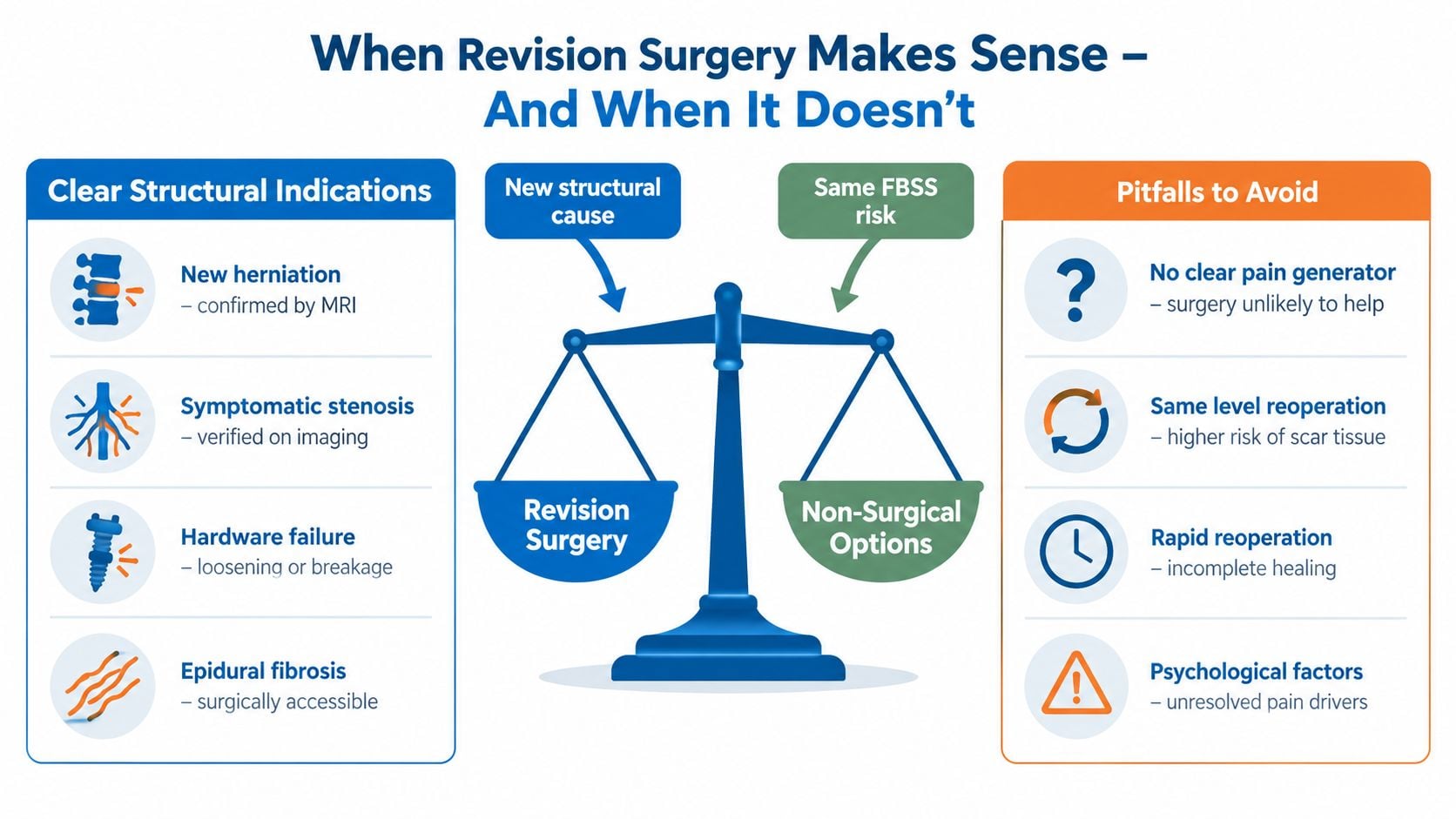
The risk of repeating the same FBSS pattern rises when surgery treats an unclear source. A patient considering spinal fusion should receive a specific explanation of the structural problem, the expected functional benefit, and the alternatives that would be lost or delayed by another operation.
Before agreeing to revision, the patient should ask: “What specific finding on the new scan will this operation correct, and what shows that it causes today's pain?”
That question separates a correctable lesion from an understandable but insufficient desire to do something more invasive. A second opinion is reasonable when the proposed target remains unclear.
Building Your Own Treatment Pathway
A patient may arrive after surgery with leg burning, back aching, poor sleep, and a scan that looks different from the last one. The next treatment should not be chosen from a preset ladder. FBSS care works better as a decision tree, where symptoms, examination findings, imaging, and prior responses determine the next branch.
The sequence of decisions
Start with diagnostic clarity. The team reviews how the pain behaves, examines strength and sensation, obtains appropriate imaging, and uses diagnostic blocks when they can test a specific structure. New weakness, bowel or bladder changes, fever, or other red-flag symptoms require prompt medical assessment rather than routine treatment escalation.
Then build active conservative care around the current problem. Physical therapy should reflect present movement limits and functional goals, rather than repeat exercises prescribed before surgery. Sleep improvement, activity pacing, conditioning, and a deliberate plan to reduce reliance on systemic opioids may support participation in rehabilitation. Medication can make movement and therapy more manageable, but increasing medication without a defined target rarely addresses the pain source.
Use an intervention to answer a specific question or change a specific symptom. Radicular inflammation may support an epidural approach. Confirmed facet pain may support medial branch treatment. A focal neuropathic pattern may call for a more selective nerve-targeting strategy. The result should either improve function or provide information that changes the working diagnosis.
Consider neuromodulation when neuropathic pain remains disabling despite appropriate care. A temporary spinal cord stimulation trial is a gate, not an automatic step toward implantation. Meaningful improvement in pain should be accompanied by better walking, sleep, sitting, or activity. Conventional stimulation is not the only option. High-frequency approaches may be discussed for persistent back and leg pain, while dorsal root ganglion stimulation may suit selected pain distributions. The choice depends on the pain pattern and the trial response, not on the device label alone.
Reserve revision surgery for a correctable structural target. A new lesion that matches the current symptoms matters more than symptom duration by itself. Axial back pain and dermatomal leg pain may require different diagnostic questions, rehabilitation goals, and treatment choices.
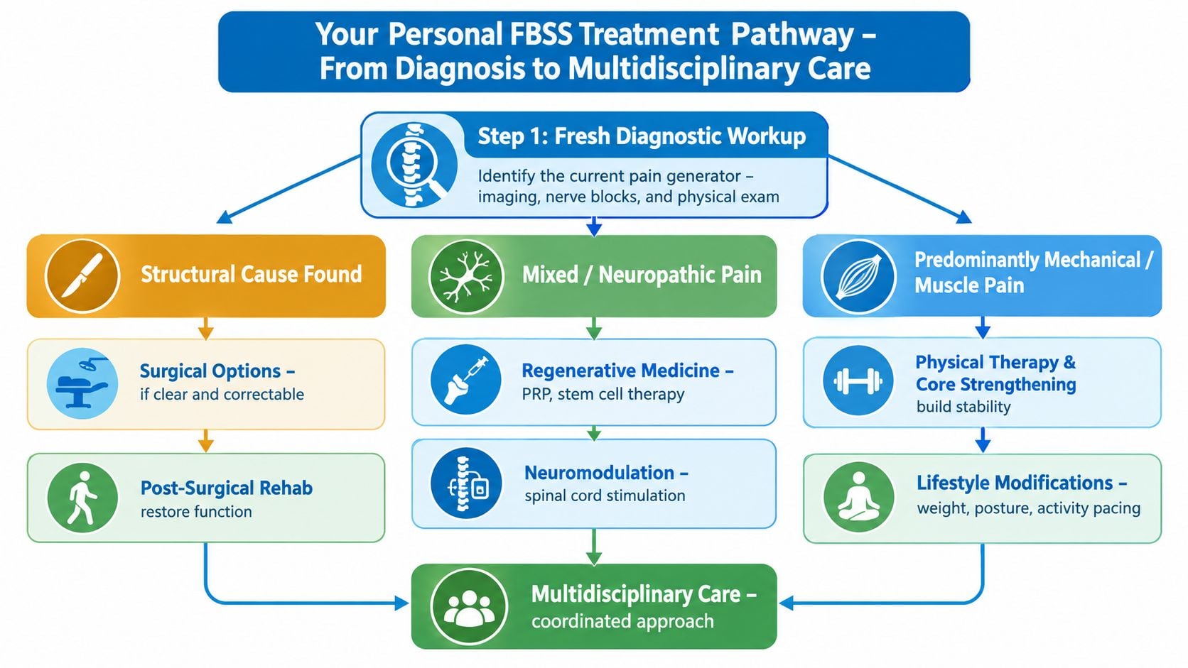
Three checkpoints help keep decisions grounded:
- Diagnostic checkpoint: Does the proposed treatment match a plausible pain generator?
- Trial checkpoint: If neuromodulation is considered, did the temporary trial improve symptoms and function?
- Structural checkpoint: Is there a lesion that surgery can correct and that fits the clinical picture?
This sequence limits procedure stacking when the diagnosis remains uncertain. It also makes an unsuccessful step useful, because the response can refine what comes next.
What Lasting Relief Actually Looks Like
A patient may still notice pain yet sleep through the night, walk to the store, sit through a meal, or take part in rehabilitation again. That is often a more useful measure of recovery than a pain score alone. Better function rather than zero pain should guide the conversation, alongside pain intensity, medication side effects, and the activities the patient wants to regain.
Evidence on traditional low-frequency spinal cord stimulation supports realistic expectations. A review of failed back surgery syndrome treatment reported approximately 50% pain relief in about 50% of patients over time (FBSS and neuromodulation review). Another systematic review reported mean pain relief of 58% across studies at an average follow-up of 24 months, with considerable variation between studies (systematic review of FBSS interventions). These figures describe groups, not a promise for one individual. A partial response may still be worthwhile if it restores activity, while a weak response should prompt review of the pain diagnosis, treatment target, or rehabilitation plan.
| Timepoint | Pain Reduction | Functional Goal | Medication Adjustment |
|---|---|---|---|
| Three months | Determine whether the early response is consistent and meaningful | Track walking, sitting, sleep, and therapy participation | Review benefit, side effects, and unnecessary escalation |
| Six months | Reassess whether improvement has plateaued or continued | Confirm progress toward daily activities and conditioning | Consider a deliberate reduction when function allows |
| Twelve months | Decide whether the current strategy still fits | Maintain gains and identify new or adjacent pain patterns | Revisit the medication plan and diagnostic pathway if progress has stalled |
These timepoints are review appointments, not guarantees or rigid rules. At each one, ask whether the treatment still matches the suspected pain generator. Someone who improves but remains limited may need a more focused rehabilitation plan, a different neuromodulation approach, or renewed diagnostic testing. New weakness, a changed pain distribution, worsening function, or an unexplained plateau should return the patient to diagnostic assessment rather than trigger automatic procedure stacking.
Lasting relief usually requires coordinated reassessment. It is less like finding one permanent switch and more like adjusting a map as symptoms, function, and examination findings change. Patients in New Jersey and Staten Island can discuss diagnostic evaluation, targeted procedures, neuromodulation, and surgical review with Interventional Pain Management, whose services include pain management and spine care matched to clinical findings. Visit Interventional Pain Management to request an evaluation and identify the next decision based on the current pain source.