
A desk-based professional may reach late afternoon with throbbing calves, a heavy feeling in both legs, and bulging veins that weren't there years ago. The natural fear is that treatment means hospital surgery, general anesthesia, and weeks away from work. In many cases, modern vein care follows a different path. An endovenous ablation procedure closes an unhealthy vein from inside the vessel through a small catheter, usually in an outpatient setting.
That convenience shouldn't be confused with a permanent cosmetic cure or a risk-free treatment. The useful questions are how the procedure works, who benefits, what recovery feels like, how long closure lasts, and what complications deserve prompt attention. The discussion below explains those points in the same practical order a vascular consultation would.
Table of Contents
- Why Your Legs Ache and What Modern Vein Care Can Do
- Understanding Venous Reflux and How Ablation Fixes It
- How the Endovenous Ablation Procedure Is Actually Performed
- Comparing Radiofrequency Ablation and Laser Techniques
- Real Risks and What Complication Rates Look Like
- Recovery Timeline from Day One to Full Activity
- How Long Results Last and Why Recurrence Happens
- Deciding If Endovenous Ablation Is Right for You
Why Your Legs Ache and What Modern Vein Care Can Do
After a long workday, your calves may ache, your ankles may swell, or itching and raised veins may become more noticeable. Sitting or standing for extended periods can bring on these symptoms, although the pattern varies from person to person. The underlying problem is often inefficient upward blood flow through the leg veins.
For many patients, the more useful questions are not only whether treatment can improve symptoms, but how long the result may last and what real risk looks like. Those answers depend on the vein involved, the ultrasound findings, and the treatment selected.
Older treatment commonly relied on vein stripping. Current care may use a slender catheter, ultrasound guidance, and local tumescent anesthesia instead. The catheter closes the vein responsible for abnormal backward flow, while other healthy veins continue carrying blood through the leg.
Practical rule: Treat the source of reflux, not only the visible vein that draws attention.
During an endovenous ablation procedure, a specialist places a catheter into a refluxing superficial vein and delivers radiofrequency or laser energy as the catheter is withdrawn. The heat seals the vessel from inside. Blood then uses healthier pathways, and the closed vein remains in the leg rather than being pulled out. Prominent surface branches may receive separate treatment when symptoms or appearance call for it.
The approach became established in clinical guidance. In 2013, NICE recommended endothermal ablation as the first-line interventional treatment for confirmed varicose veins with truncal reflux, before ultrasound-guided foam sclerotherapy and surgery, in the NICE recommendations for varicose veins.
That recommendation defines a role for ablation, not an automatic treatment for every enlarged vein. A sound decision connects symptoms, examination findings, and duplex ultrasound results. Patients can review general varicose and spider vein care, while recognizing that individual suitability and risk require clinical assessment.
Understanding Venous Reflux and How Ablation Fixes It
Healthy leg veins contain one-way valves. These valves open as blood moves toward the heart and close to prevent it from falling back toward the feet. When the valves in a superficial trunk vein fail, blood flows downward under gravity. That backward movement is called venous reflux.
A plumbing analogy helps. A properly functioning pipe has a valve that keeps fluid moving in the intended direction. If the valve no longer seals, pressure builds in the lower section. The vein can stretch, branch veins can enlarge, and the leg may develop heaviness, swelling, or visible varicosities.
Duplex ultrasound identifies this problem and maps the vein before treatment. Clinical protocols commonly consider reflux of more than 500 milliseconds on ultrasound when evaluating superficial venous disease, with treatment decisions also shaped by the patient's symptoms and clinical classification. The relevant disease spectrum can include CEAP classes C2 through C6, ranging from visible varicose veins to more advanced skin changes and ulceration.
What the catheter changes
Thermal ablation treats the refluxing trunk, often a saphenous vein, rather than directly removing every surface branch. Heat injures the vein wall in a controlled manner, causing the vessel to collapse and close. Over time, the body absorbs the closed segment, while circulation uses alternative veins.
Who may not be ready
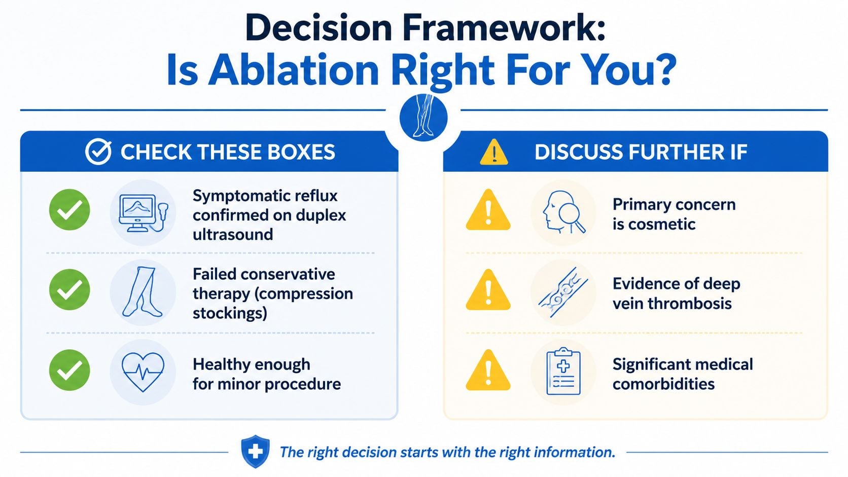
A person with acute deep vein thrombosis generally needs a different immediate plan, because the deep circulation is affected. Pregnancy can also change the timing and treatment approach. Compression therapy may be recommended before an intervention, especially when symptoms are mild, the diagnosis remains uncertain, or a clinician wants to assess response to conservative care.
Spider veins and isolated reticular veins usually don't represent the same trunk-vein reflux treated by thermal ablation. They're often managed with a different technique, such as sclerotherapy, after the clinician confirms that a deeper reflux source isn't driving them.
How the Endovenous Ablation Procedure Is Actually Performed
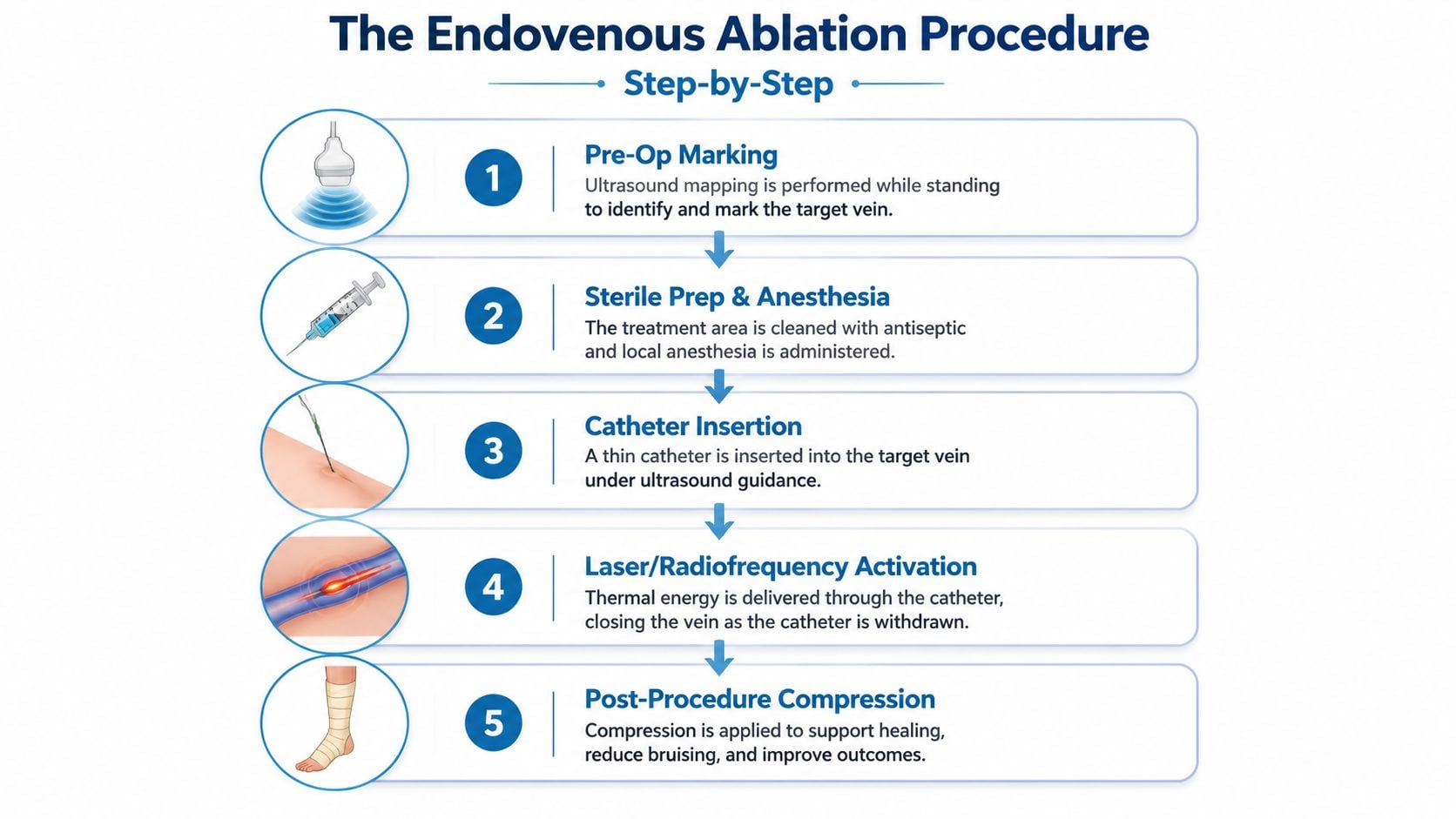
The visit usually begins before the patient lies on the procedure table. Duplex ultrasound helps the clinician trace the refluxing vein, identify its branches, and mark the skin. Standing can make the abnormal pathway easier to map because gravity fills the veins and reveals their course.
After the patient lies down, the leg is cleaned with an antiseptic solution. A small amount of local anesthetic numbs the access point, often near the knee or calf. The clinician then uses ultrasound to guide a needle and thread a thin catheter into the target vein through a tiny puncture. This doesn't require the long incision associated with open stripping.
Why tumescent anesthesia matters
The next step is infiltration of tumescent anesthesia, a dilute anesthetic solution placed along the length of the vein. It numbs the treatment corridor, separates the vein from nearby skin and nerves, and compresses the vessel around the catheter. That cushioning effect helps the clinician deliver energy to the vein wall while limiting unwanted heat exposure to surrounding tissue.
The catheter delivers either radiofrequency energy or laser energy as it's withdrawn. The heat causes the vein wall to contract and seal. Technical settings vary according to the device, vein size, depth, fiber design, and the segment being treated. For contemporary great saphenous vein laser protocols, the energy is adjusted rather than applied as a single fixed setting, as detailed in the technical review of endovenous laser and radiofrequency treatment.
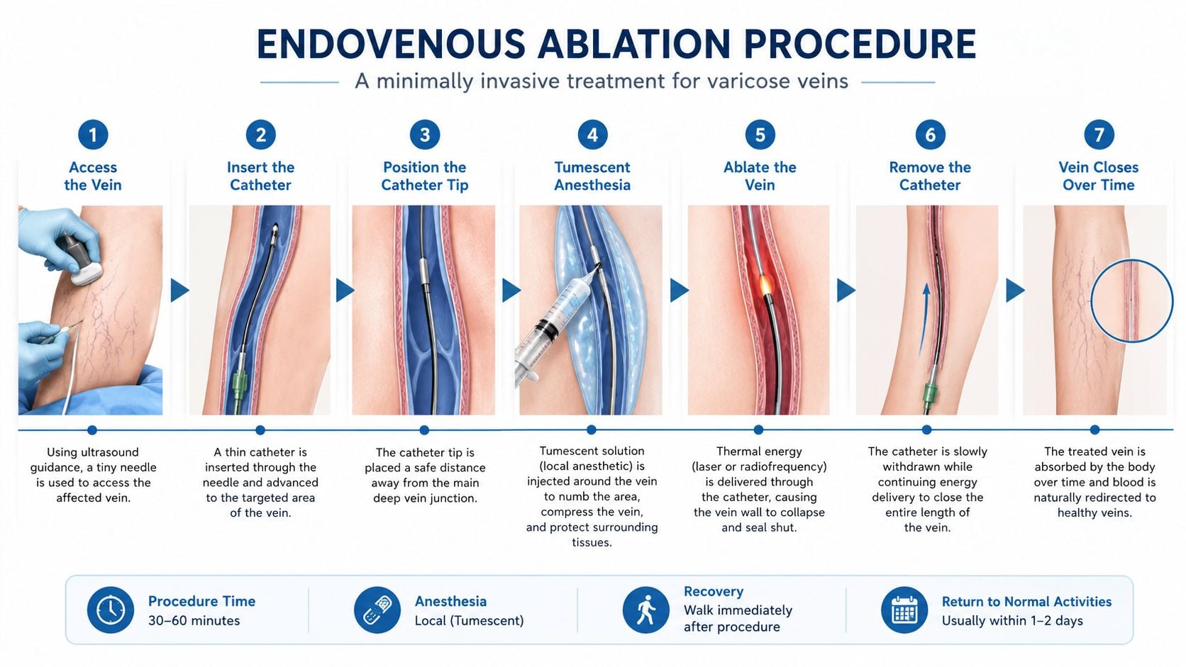
Once the catheter is removed, the puncture site receives a small dressing. A compression wrap or stocking supports the leg, and walking begins soon afterward. The room time is commonly described as less than a prolonged surgical admission, but exact timing depends on the vein map, the number of segments treated, and whether additional procedures are performed.
The patient may feel pressure during catheter placement and warmth or pulling as the energy is delivered. Sharp pain isn't something to ignore, so the treatment team should know immediately if local anesthesia doesn't feel adequate.
Comparing Radiofrequency Ablation and Laser Techniques
A patient choosing between radiofrequency ablation and endovenous laser ablation is usually asking two practical questions: which method fits my vein, and how long is the result likely to last? Both treatments use heat to close a refluxing vein, but the heat reaches the vein through different systems. RFA uses a catheter that heats segments in a controlled pattern. EVLA uses a laser fiber, whose wavelength and design affect how energy interacts with water and tissue inside the vein.
The choice depends on more than whether one technique sounds newer. Vein diameter, winding anatomy, depth, access, clinician training, available equipment, and the plan for treating tributaries can all change the recommendation. A relatively straight trunk may permit a different approach from a vein that curves sharply.
| Feature | Radiofrequency, RFA | Endovenous laser, EVLA |
|---|---|---|
| Energy source | Controlled radiofrequency heat delivered through a segmental catheter | Laser energy delivered through a fiber |
| Treatment effect | Contracts collagen and seals the vein wall | Produces thermal injury and vein-wall collapse |
| Technical range | Device-specific controlled heating | Wavelength and fiber-specific energy delivery |
| Recovery pattern | Often associated with less bruising and tightness | May produce more bruising or a pulling sensation in some patients |
| Main selection factors | Vein anatomy, device access, operator experience, treatment plan | Vein anatomy, fiber design, energy settings, operator experience |
Both methods can close the intended vein effectively, but closure is only one part of the outcome. NICE's evidence review reported 92% occlusion for the great saphenous vein, 84% for the small saphenous vein, and 94.5% for the great saphenous vein at six weeks in cited evidence sets (NICE surveillance commentary). These figures describe treated-vein closure, not guaranteed symptom relief or permanent prevention of new varicose veins.
Long-term expectations also depend on untreated tributaries, other refluxing veins, anatomy, and follow-up. A comparative trial summarized by NICE found procedural complications of 1% with laser ablation, 7% with foam sclerotherapy, and 8% with surgery. Those findings support a lower immediate complication burden for thermal catheter treatment in that comparison, but they do not make the two thermal techniques identical or risk-free.
Patients can review the practical mechanics in this radiofrequency ablation treatment resource, then ask why one modality suits the vein map, treatment goals, and expected follow-up.
Real Risks and What Complication Rates Look Like
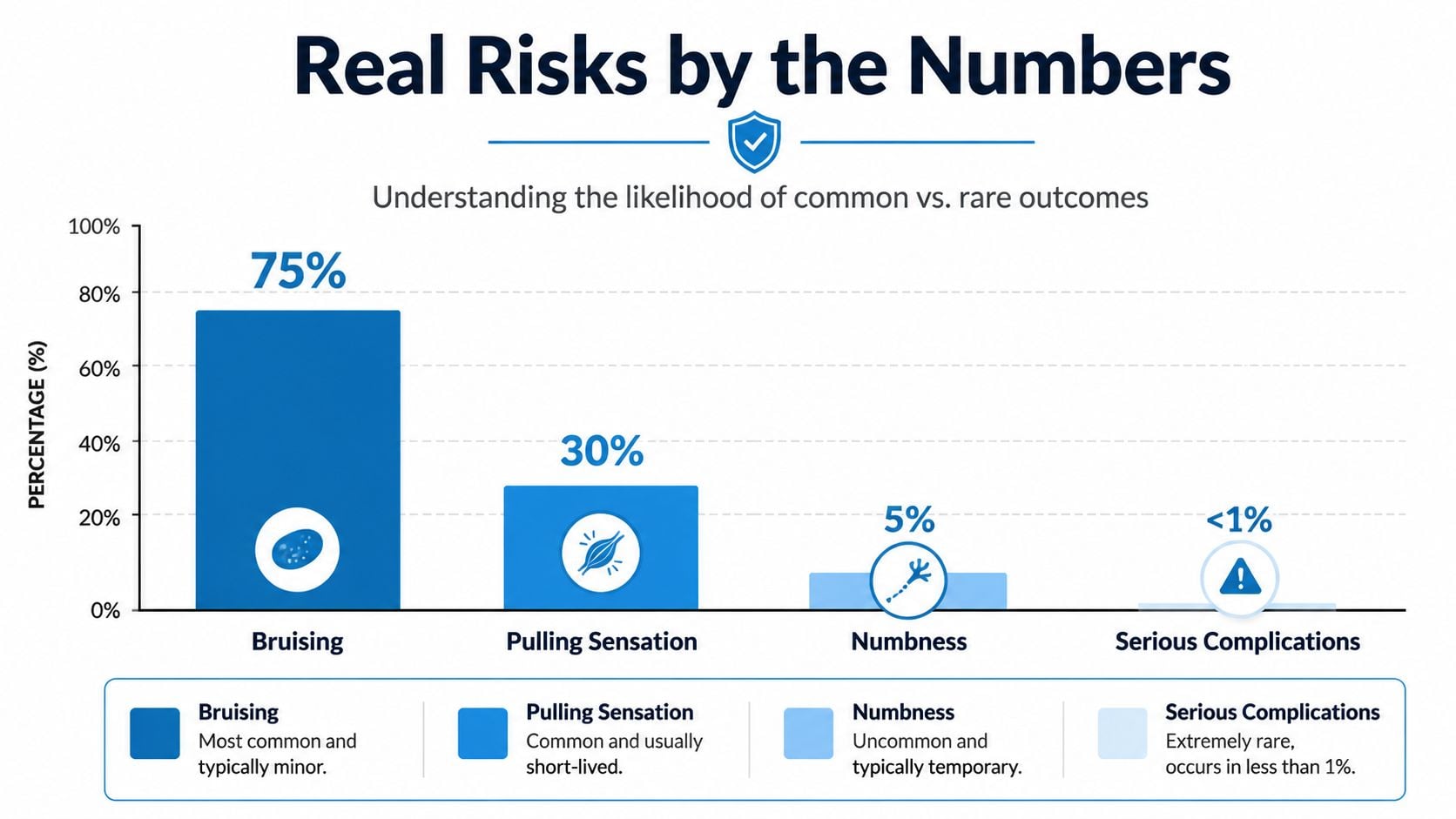
A consent discussion should separate expected healing from complications that need assessment. Bruising, tenderness, firmness, and a pulling sensation along the closed vein can occur as treated tissue reacts. These symptoms may be uncomfortable without indicating a deep clot or treatment failure.
More serious events are uncommon, but they still require attention. A pooled guideline analysis reported EHIT II through IV or DVT at 1.7%, DVT alone at 0.3%, and pulmonary embolism at 0.1% after endovenous procedures (SVS, AVF, and AVLS guideline evidence). These pooled estimates do not predict an individual result. Medical history, vein anatomy, procedure type, and follow-up practices can change risk.
Symptoms that deserve a call
A tender, firm line near the treated vein can reflect local inflammation. A rapidly enlarging calf, marked one-sided swelling, increasing warmth, or pain that escalates instead of settling warrants prompt contact with the clinic. Chest pain or unexplained shortness of breath requires emergency assessment.
Skin and nerve effects also belong in the consent discussion. A pooled review reported paresthesia in 2.6% after EVLA, while a separate clinical guidance source reported a 2.6% pooled incidence of skin burns in relevant laser-ablation data, as summarized in the evidence reviewed by the clinical policy (clinical policy and evidence summary). Sensory symptoms may improve gradually. Persistent or worsening numbness should be assessed by the treating clinician.
The key question is whether symptoms after treatment fit normal local healing or suggest thrombosis, infection, nerve irritation, or excessive tissue injury.
Recovery Timeline from Day One to Full Activity
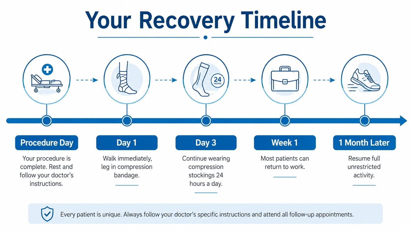
Recovery is usually organized around movement, compression, and gradual return to exertion. Walking soon after treatment helps maintain circulation, while compression supports the treated area and may reduce swelling. The treating clinic's instructions take priority because the plan can change when multiple veins or additional procedures are involved.
The first week
On procedure day, the leg may be wrapped, and walking generally begins before discharge. Rest at home is sensible, but prolonged immobility isn't. Mild soreness can often be managed with the medication plan supplied by the clinical team.
During the next few days, the leg may feel stiff or tight rather than severely painful. Desk work may be possible relatively soon for some patients, while jobs involving prolonged standing, lifting, or extensive walking may require more adjustment.
- Early movement: Short, frequent walks are generally encouraged.
- Compression: Wear the stocking or wrap for the duration prescribed by the treating clinician.
- Skin care: Keep puncture sites clean and follow instructions about showering, soaking, and heat exposure.
- Pain changes: Contact the clinic if pain becomes severe, sudden, or disproportionate.
The following weeks
Bruising can become more visible before it fades. Light walking is usually preferable to complete rest, but strenuous leg exercise and heavy lifting should wait until the clinician gives clearance. A follow-up duplex ultrasound is often arranged during early follow-up to confirm closure and assess the deep veins.
Full activity returns progressively. Residual firmness along the treated pathway can soften over time, and visible vein changes may lag behind functional closure. That difference explains why the leg may still look unchanged shortly after an technically successful procedure.
How Long Results Last and Why Recurrence Happens
An endovenous ablation procedure is durable, but it isn't a guarantee that venous disease will never appear elsewhere. The treated segment can reopen, a process called recanalization, or a different vein can later develop reflux. Those are separate mechanisms, and combining them under the single label “failure” can create unnecessary confusion.
The evidence summarized by NICE includes five-year recurrence rates of 18.7% for radiofrequency ablation, 38.6% for EVLA, and 34.6% for surgery (NICE overview of endovenous ablation evidence). These results vary by technique, patient selection, follow-up duration, and the definition of recurrence. They don't mean that every patient with a recurrent visible vein has an open treated trunk.
Two different pathways
Recanalization means the treated vein partially or fully reopens. It may occur earlier and can relate to anatomy, incomplete energy delivery, insufficient wall injury, or challenging access. Follow-up imaging helps distinguish this from a new refluxing pathway.
New reflux develops in an accessory vein, another superficial trunk, or a previously less important pathway. This can reflect progression of the underlying venous condition rather than a problem with the original closure. Family history, body weight, occupation, prior thrombosis, and the pressure placed on the legs over time can all influence the broader disease course, although they don't determine an individual outcome by themselves.
| Time horizon | Closure rate, RFA | Closure rate, EVLA | Most common failure mode |
|---|---|---|---|
| Early follow-up | High technical closure in appropriate anatomy | High technical closure in appropriate anatomy | Incomplete closure or early recanalization |
| Several years | Durable benefit, with recurrence possible | Durable benefit, with recurrence possible | New reflux in an adjacent or alternate pathway |
| Long-term care | Depends on anatomy and disease progression | Depends on anatomy and disease progression | Recurrent symptoms or untreated tributary disease |
Success should therefore mean less aching, reduced swelling, improved function, and protection against complications such as venous ulceration, not a promise that every surface vein will remain cosmetically absent forever.
Deciding If Endovenous Ablation Is Right for You
The practical starting point is symptomatic reflux confirmed by duplex ultrasound. Cosmetic concern alone usually doesn't establish a reason to close a trunk vein, because spider veins and isolated surface vessels may need a different approach.
A clinician will also consider skin changes, ulcer history, prior thrombosis, medication use, mobility, and whether compression therapy has been tried. Endovenous treatment can be one stage in chronic vein care, not a standalone event that removes every future risk.
Questions for the consultation
A productive consultation should explain the ultrasound map in plain language. Patients can ask:
- What vein is refluxing? The answer should identify the vessel and explain how it relates to the symptoms.
- Which technique fits the anatomy? RFA, EVLA, foam, or another option may be appropriate depending on vein size, depth, and shape.
- Will surface branches need separate treatment? Ablation may close the source while visible tributaries remain.
- What follow-up is planned? The patient should know when imaging will confirm closure and what symptoms require an earlier call.
- What activity restrictions apply? Guidance should reflect the patient's job, exercise routine, travel plans, and medical history.
- How are complications handled? The clinic should explain access to urgent assessment and ultrasound if swelling or pain changes.
Provider setting matters less than whether the team performs a careful ultrasound evaluation, explains alternatives, uses appropriate monitoring, and offers clear follow-up. A dedicated vein practice may focus heavily on venous imaging and staged treatment, while a broader vascular or multispecialty practice may coordinate vein care with other medical needs. The important issue is the quality and continuity of the clinical pathway.
Compression when prescribed, regular walking, attention to weight and mobility, and treatment of remaining reflux can extend the practical value of the procedure. Interventional Pain Management evaluates and treats varicose and spider vein conditions alongside other musculoskeletal and interventional services, with care plans based on clinical findings. Patients can review the practice's broader treatment services before arranging an evaluation.
Interventional Pain Management can assess symptomatic varicose veins, review duplex ultrasound findings, and discuss whether radiofrequency or another treatment fits the patient's anatomy and goals. Visit Interventional Pain Management to request an evaluation and receive a personalized plan for treatment, recovery, and long-term follow-up.