
The most popular advice about disc extrusion vs herniation is often wrong: a frightening MRI label doesn't automatically mean surgery. An extrusion is a more specific form of disc herniation, and it can cause significant nerve irritation, but the treatment decision depends on the full clinical picture, including pain, weakness, sensation, function, and urgent warning signs.
A scan shows anatomy. It doesn't measure suffering, predict recovery by itself, or determine whether an operation is necessary. In fact, evidence on disc behavior can be surprisingly reassuring. A systematic review found spontaneous regression in about 70% of disc extrusions, compared with 41% of protrusions (systematic review and meta-analysis). That doesn't mean every extrusion should be watched indefinitely, but it does mean the word “extrusion” shouldn't be treated as a surgical verdict.
| Disc finding | What the term describes | Usual clinical meaning |
|---|---|---|
| Bulging disc | Disc tissue extends broadly beyond its usual margin | May be incidental or may contribute to narrowing and irritation |
| Protrusion | A focal herniation in which the base is wider than the outward portion | A contained morphology in many cases |
| Extrusion | A herniation with a narrower base than its outward extension, or material extending above or below the disc level | A more specific, often noncontained morphology |
| Sequestration | Displaced disc material loses continuity with the parent disc | A free fragment that may migrate and can also resorb |
The key question isn't just, “Is an extrusion worse than a herniation?” The more useful questions are: Which nerve is affected? Is strength changing? Can the person function? Are symptoms improving or progressing?
Table of Contents
- Rethinking Your MRI Results
- The Anatomy of Disc Displacement
- How Radiologists Classify Disc Morphology
- The Counterintuitive Natural History of Extrusions
- Connecting Imaging Findings to Real-World Symptoms
- Targeted Treatment Pathways for Disc Pathology
- Choosing the Right Multidisciplinary Spine Team
Rethinking Your MRI Results
Many patients see “disc extrusion” on an MRI report and immediately think surgery is next. That conclusion is understandable, but the label does not carry that meaning. Disc extrusion describes the shape and containment of displaced disc material. It does not, by itself, determine treatment.
Disc herniation is the broader category. In standardized spine-imaging language, it means focal displacement of disc material beyond the disc space involving less than 25% of the disc circumference. Disc extrusion is one type of herniation. Its base is narrower than the material extending outward in at least one plane, or the material may lose continuity with the original disc, according to the standardized lumbar disc terminology.
The distinction helps describe what appears on the scan, but symptoms and examination determine its clinical importance. An extrusion may allow disc material to migrate and contact the thecal sac or a nerve root. A patient with a large-looking extrusion and steadily improving leg pain may need a different plan from someone with a smaller protrusion and progressive weakness.
What the label can and cannot tell
An MRI can show the disc level, direction, and apparent relationship to nearby nerves. It may describe the material as central, paracentral, foraminal, or far lateral. These location details help a spine specialist judge whether the image matches the pattern of pain, numbness, or weakness.
The scan cannot measure pain intensity or predict recovery on its own. Some disc findings produce no symptoms. A less extensive finding can still irritate a nerve if it lies in a confined space. That is why a clinical consultation includes strength, reflexes, sensation, walking ability, and the effect of symptoms on daily activities.
Practical rule: An MRI label starts the conversation. It does not finish the treatment decision.
Prompt medical assessment matters when neurological function is worsening. New or increasing weakness, marked difficulty walking, or changes in bladder or bowel control require urgent evaluation. These findings have greater immediate significance than the distinction between protrusion and extrusion alone.
The reassuring point is not that every extrusion is harmless. Disc material can cause substantial pain and functional limitation. However, extrusion is not synonymous with emergency surgery. Many cases improve as inflammation settles and the displaced material resorbs. Careful follow-up, symptom-guided treatment, and targeted interventional pain management can be appropriate when nerve irritation persists, while surgery is considered when the clinical picture requires it.
The Anatomy of Disc Displacement
A spinal disc works much like a jelly donut, although the analogy has limits. The tough outer ring is the annulus fibrosus, and the softer inner material is the nucleus pulposus. The disc sits between vertebrae, helping the spine move while distributing mechanical load.
A disc problem develops when the outer ring changes shape or develops a defect, allowing the inner material to move beyond its usual boundary. The terms used in an MRI report describe how far that material has traveled and whether it remains connected to the parent disc. A broader degenerative disc disease overview can help place these changes in the wider context of disc wear and spinal mechanics at Interventional Pain Management's degenerative disc resource.
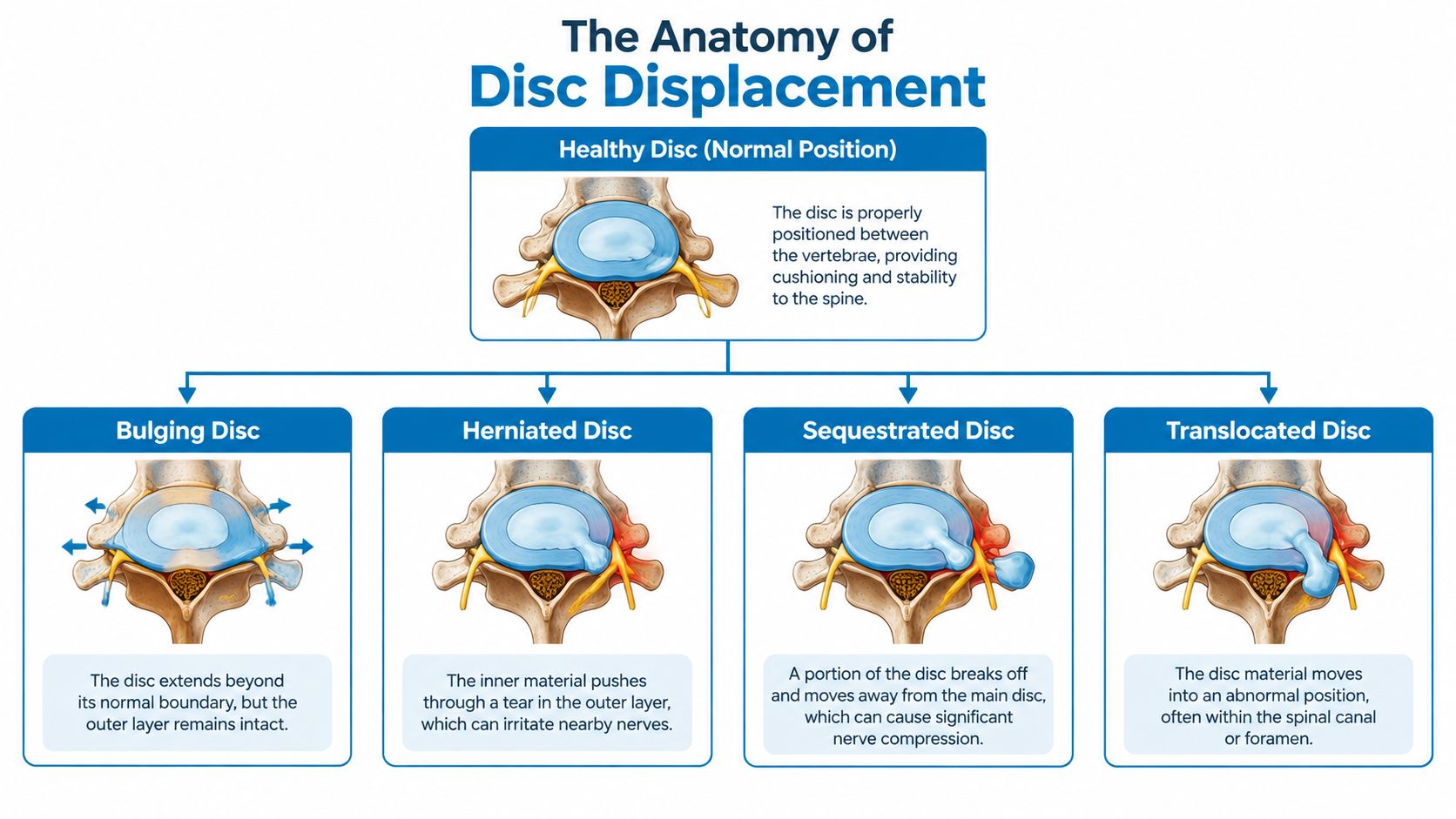
The displacement spectrum
A bulge generally describes a broad extension of disc tissue around a substantial portion of the disc margin. It may reflect age-related change and may not be the source of pain. A bulge can still matter if it contributes to narrowing around a nerve, but the word itself doesn't prove nerve compression.
A protrusion is focal. The disc material extends outward, but the base attached to the parent disc is broader than the displaced portion. The outer ring may be stretched or damaged, but the displaced material remains relatively contained.
An extrusion has a different geometry. Its base is narrower than the outward portion in at least one imaging plane, or the material extends above or below the level of the disc. This configuration suggests a more substantial annular defect and gives the material greater potential to migrate along the spinal canal.
A sequestration occurs when the displaced fragment loses continuity with the parent disc. The free fragment may move away from the original disc space. That sounds alarming, but loss of continuity can also expose the material to the body's inflammatory and immune processes, which helps explain why some larger or more separated fragments resorb.
These terms describe morphology, not a simple ladder from “mild” to “severe.” A radiologist may call one finding an extrusion because of its shape, while the patient's symptoms remain modest. Another patient may have a smaller focal finding that directly affects a nerve root and produces intense sciatica.
How Radiologists Classify Disc Morphology
Radiologists don't choose “protrusion” or “extrusion” as casual synonyms. The terminology follows a standardized framework developed to make reports more consistent across MRI and CT interpretation. The framework formalized distinctions that had previously been blurred by loosely used words such as prolapse or rupture (radiology terminology reference).
The first point is the 25% circumference rule. In widely used nomenclature, a disc herniation is focal displacement beyond the disc space involving less than 25% of the disc circumference. This separates a focal herniation from a generalized disc contour change.
The base and dome rule
The central imaging question is the relationship between the base and the outward portion, sometimes described clinically as the dome. With a protrusion, the base where the material remains attached to the disc is wider than the outward extension. With an extrusion, the base is narrower than the displaced portion in at least one plane.
MRI also shows whether material extends above or below the original disc level. That craniocaudal extension supports the diagnosis of extrusion even when the shape isn't easy to understand from a single image. If the material no longer connects to the parent disc, the report may describe sequestration.
A patient reading a report should also look beyond the morphology word. The most useful details often include:
- Level: Such as a lower-lumbar disc or a cervical disc.
- Direction: Central, paracentral, foraminal, or far lateral.
- Nerve relationship: Whether the report describes contact, displacement, or compression.
- Mass effect: Whether the thecal sac or a specific nerve root appears crowded.
- Migration: Whether material extends above or below the disc space.
Why standardized wording affects care
Objective wording improves communication among the radiologist, pain specialist, physical therapist, and surgeon. It helps the team compare the same anatomy over time and distinguish a contained protrusion from a potentially noncontained extrusion within one diagnostic system.
A report should still be read in context. “Nerve contact” isn't always the same as clinically meaningful compression, and compression on the side opposite the patient's symptoms may not explain the complaint. The physical examination supplies the missing information by testing whether the suspected nerve is malfunctioning.
The practical takeaway is simple: the morphology label identifies what the disc looks like. The treatment plan depends on what that anatomy is doing to the patient.
The Counterintuitive Natural History of Extrusions
A more dramatic MRI label does not automatically mean surgery is required. In some cases, an extrusion has a greater chance of shrinking than a smaller protrusion because the body can recognize exposed disc material and begin clearing it.
Macrophages, specialized immune cells, may help remove displaced nucleus pulposus material. A clinical study found that extruded lumbar discs reduced faster than bulging discs during follow-up and associated macrophage infiltration with greater spontaneous reduction (clinical review of spontaneous disc reduction).
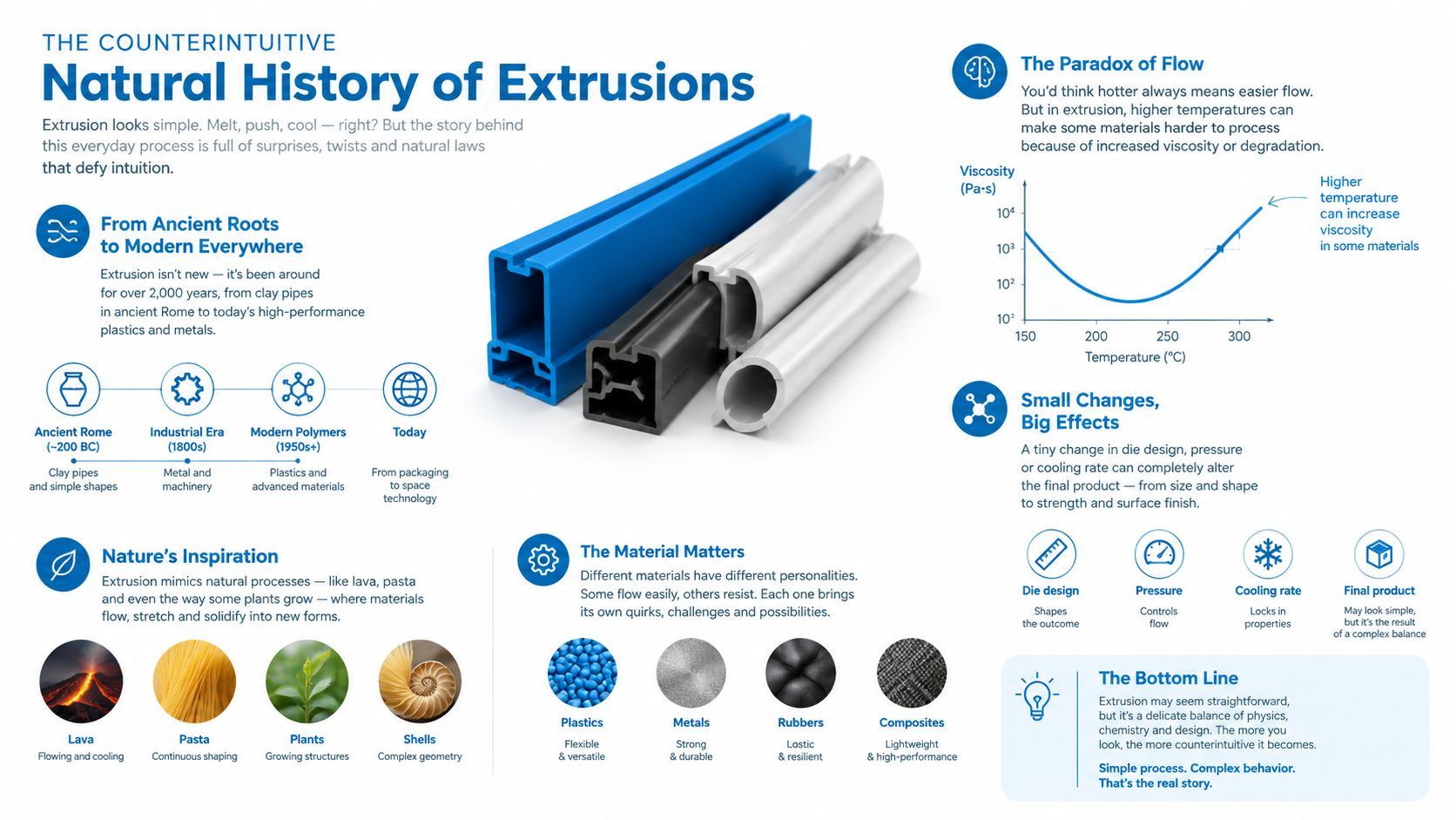
Comparing how morphologies change
A systematic review and meta-analysis reported spontaneous regression in about 70% of disc extrusions, compared with 41% of protrusions. It also reported complete resolution in 15% of extruded discs and 13% of bulging discs (natural-history data on lumbar disc herniation).
Another review reported regression probabilities of 52.5% for protrusions, 70.4% for extrusions, and 93.0% for sequestrations, with greater nucleus pulposus extrusion and stronger gadolinium enhancement associated with a higher likelihood of regression. These figures describe group patterns, not a personal guarantee.
The anatomy helps explain why an apparently larger fragment can improve:
- Contained material may remain partly shielded from the body's immune response.
- Noncontained material is more exposed to surrounding tissue and inflammatory cells.
- Migration can increase the surface area available for resorption.
- Inflammation may cause early pain while also helping clear disc fragments.
Why waiting can be active care
Observation means monitoring the condition while supporting recovery. A specialist may recommend medication, movement modification, physical therapy, and follow-up visits that track pain, strength, sensation, and function. An image-guided injection may reduce nerve inflammation and create a useful period for rehabilitation when pain prevents normal movement. This targeted form of interventional pain management can support recovery without treating the MRI label alone.
Patience becomes inappropriate when neurological function worsens or emergency warning signs appear. Progressive weakness, substantial functional decline, or cauda equina symptoms require prompt assessment. Without those concerns, the higher regression tendency of extrusions supports a measured plan rather than reflexive surgery.
A larger fragment can look worse on a scan and still have a meaningful chance of shrinking without an operation.
Spontaneous resorption may reduce mechanical and inflammatory burden, but it does not guarantee immediate relief, complete recovery, or resolution of every pain generator. Treatment should follow the patient's clinical course and neurological findings, not the emotional impact of a radiology word.
Connecting Imaging Findings to Real-World Symptoms
An MRI shows structure, but the patient experiences pain, numbness, tingling, weakness, and loss of function. Those experiences depend heavily on where disc material sits relative to a nerve. A large central extrusion may occupy space without affecting a specific nerve root, while a smaller paracentral or foraminal lesion may contact a nerve in a tight passage.
A 2025 review of lumbar-disc-herniation modeling reported that extrusions represented about 50% of lumbar disc herniations and protrusions about 34% in its patient synthesis. It also reported that paracentral herniations comprised 61% of cases, with the L4-L5 and L5-S1 levels together representing 91% of reported paracentral cases in that synthesis (2025 review of lumbar disc herniation modeling.)
Location often matters more than appearance
A lower-lumbar disc finding may irritate a nerve traveling into the buttock, thigh, calf, or foot. Patients often describe this as radicular pain or sciatica, especially when the pain follows a recognizable path rather than staying only in the low back. Numbness, tingling, and weakness can provide additional clues about nerve involvement, while a sciatica and pinched-nerve guide offers further patient-focused context.
| Disc condition | Typical nerve impact | Common symptom profile |
|---|---|---|
| Bulging disc | May narrow a nerve passage without a focal root injury | Local back or neck pain, or no symptoms |
| Protrusion | Can irritate or compress an adjacent nerve root | Radiating pain, tingling, numbness, or weakness |
| Extrusion | May create stronger focal mass effect or migrate near a nerve | Radicular pain, sensory change, and possible motor deficit |
| Sequestration | A free fragment may move along the canal and affect a nerve at a different position | Variable nerve pain, numbness, or weakness depending on migration |
The table describes patterns, not rules. A person can have an extrusion without leg pain, or severe leg pain from a smaller protrusion. The examination determines whether the suspected nerve is losing function.
What a specialist correlates
A spine clinician compares the side of the pain with the side and level of the MRI finding. Strength testing can reveal difficulty lifting the foot, extending the knee, or performing other movements controlled by specific nerve pathways. Sensory testing and reflexes add further evidence.
Functional impact also matters. Trouble standing, walking, sleeping, working, or managing ordinary activities may justify more active treatment even when strength remains intact. The best plan connects the scan to the lived problem rather than treating the most dramatic image in isolation.
Targeted Treatment Pathways for Disc Pathology
Treatment usually progresses according to symptoms, neurological findings, and response to care. The presence of an extrusion may influence the discussion, but it doesn't dictate one universal sequence. A patient with improving pain and stable strength may continue rehabilitation, while a patient with persistent nerve pain may benefit from a targeted intervention.
Starting with symptom control and movement
Early care often focuses on reducing aggravating activity without prolonged immobilization. A clinician may recommend medication when appropriate, guided physical therapy, gradual strengthening, posture and movement changes, and practical adjustments at work or home.
Physical therapy has a specific purpose. It can help restore movement, improve trunk and hip control, and rebuild tolerance for activity. The program should account for the patient's pain distribution and neurological status rather than relying on generic exercises.
Adding image-guided interventions
When radicular pain remains disruptive, an epidural steroid injection may reduce inflammation around an irritated nerve. A selective nerve block can also help identify whether a particular nerve root is generating the symptoms while providing temporary relief that supports rehabilitation.
These procedures don't remove the disc fragment. Their role is more targeted. By calming inflammatory pain, they may create a window in which the person can walk, sleep, exercise, and participate in recovery while the body's natural resorption process continues.
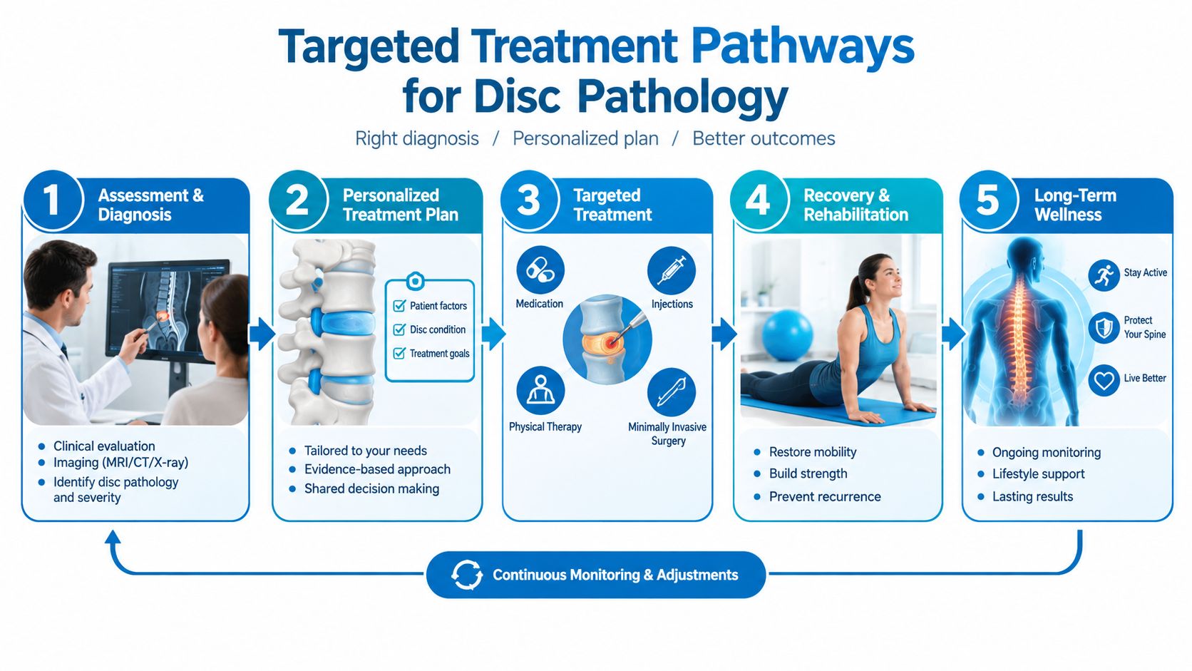
Considering advanced options
Persistent neuropathic pain after appropriate treatment may lead to a discussion of neuromodulation, including spinal cord stimulation, particularly in selected chronic or postsurgical pain syndromes. Surgery becomes more relevant when symptoms remain disabling, a matching compressive lesion persists, or neurological deficits progress.
Minimally invasive decompression or endoscopic procedures may be considered when the anatomy and clinical findings support removal of the offending material. Information about discectomy treatment options can help patients understand that “surgery” includes different approaches, each selected according to the level, fragment location, stability, and surgeon assessment.
A practical pathway may look like this:
- Confirm the pain generator: Match symptoms and examination findings to the MRI.
- Protect function: Use movement-based care and symptom control.
- Target nerve inflammation: Consider an image-guided injection when appropriate.
- Reassess objectively: Track pain, walking, sensation, strength, and daily activity.
- Escalate selectively: Discuss decompression or advanced pain procedures when the clinical indication is clear.
Urgent surgery is a separate conversation when bladder or bowel control changes, saddle-area numbness, or rapidly worsening weakness suggests serious nerve compromise. Those symptoms need immediate medical evaluation rather than routine scheduling.
Choosing the Right Multidisciplinary Spine Team
Disc extrusion care works best when the team can evaluate both sides of the decision. One clinician may focus on pain control, another on rehabilitation, and a spine surgeon on decompression or reconstruction when necessary. Coordinated review reduces the risk of treating an MRI label without understanding the patient's function and neurological status.
A useful practice should offer access to clinicians trained in pain management, anesthesiology, orthopedics, and spine surgery. That range matters because the appropriate plan may involve noninvasive care, an image-guided injection, neuromodulation, minimally invasive decompression, or a more complex operation. The team should explain why a recommendation fits the anatomy rather than presenting surgery as the default response to an alarming report.
Questions worth asking
Patients can make the consultation more productive by asking:
- What exactly is extruded? The report should be translated into plain language.
- Which nerve is affected? The side and level should match the symptoms.
- Is there objective weakness? Strength and reflex findings can change urgency.
- What happens if conservative care continues? The clinician should explain monitoring and follow-up.
- What would justify an injection or operation? Clear thresholds reduce uncertainty.
- Who coordinates the imaging and treatment plan? Patients shouldn't have to assemble disconnected opinions.
Location and access also influence continuity. For adults in New Jersey and Staten Island, a practice with multiple offices, coordinated diagnostic review, and scheduling availability across the week may make follow-up easier. Insurance guidance, transportation support, and case coordination can also matter when pain interferes with work or mobility.
Interventional Pain Management offers evaluation and treatment across noninvasive, interventional, and surgical pathways, including epidural injections, nerve blocks, spinal cord stimulation, and spine procedures. The central principle remains the same: the MRI should inform the plan, while symptoms, neurological function, and personal goals determine its direction.
Patients with new weakness, bladder or bowel changes, saddle numbness, or rapidly worsening symptoms should seek urgent medical assessment. Others can arrange a structured spine evaluation, bring the MRI images and report, and ask for a plan that explains both why surgery may be needed and why it may not be necessary.
For help interpreting an extrusion, persistent sciatica, or nerve-related weakness, schedule a coordinated evaluation with Interventional Pain Management. The practice can review imaging, assess neurological function, and discuss targeted injections, rehabilitation, neuromodulation, or minimally invasive spine care when clinically appropriate.