
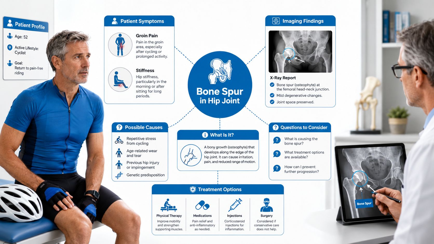
A 52-year-old recreational cyclist has spent months managing deep groin pain, stiffness after sitting, and discomfort when climbing out of the saddle. An x-ray finally provides an answer that sounds alarming: “bone spur in hip joint.” The immediate fear is understandable. Many patients assume the spur is a loose piece of bone that must be removed.
Usually, that isn't what the report means. A hip bone spur, medically called an osteophyte, is generally a bony overgrowth along the edge of the hip joint. It's often a sign of an underlying process, especially hip osteoarthritis or femoroacetabular impingement, rather than a complete diagnosis by itself. The important question isn't whether a spur exists. It's whether the finding matches the patient's symptoms, examination, and other joint changes.
Table of Contents
- What a Bone Spur in the Hip Joint Really Means
- How Hip Osteophytes Form Over Time
- Why a Bone Spur in the Hip Joint Causes Pain
- Imaging Options for Detecting Hip Osteophytes
- Conservative Treatment for a Hip Bone Spur
- Injection Options for Symptomatic Hip Spurs
- Surgical Options When Conservative Care Fails
- Your Next Steps After a Hip Bone Spur Diagnosis
What a Bone Spur in the Hip Joint Really Means
The hip is a ball-and-socket joint. The rounded femoral head fits into the acetabulum, the cup-shaped part of the pelvis. A hip osteophyte forms near the joint margins, often around the acetabular rim or where the femoral head meets the neck. It develops as the bone and surrounding tissues respond to altered loading, cartilage damage, or abnormal contact.
A useful analogy is a riverbank. When water flows smoothly, the bank stays relatively stable. Where the current becomes turbulent, sediment collects along the edges. An osteophyte is somewhat like that buildup. It reflects a region where the joint has experienced repeated mechanical stress or remodeling. The analogy isn't perfect, but it helps explain why the spur is often a marker of changing joint mechanics rather than an isolated object causing every symptom.
Hip osteophytes are strongly associated with hip osteoarthritis. A birth-cohort study of adults aged 63 years found ultrasound features of osteoarthritis were common, with hip osteoarthritis estimated at 41% in that cohort, as reported in the hip osteoarthritis birth-cohort study. A separate UK Biobank analysis found radiographic hip osteoarthritis in 18.8% of participants and osteophytes in 10% overall, with the lateral acetabulum the most frequent reported site in that analysis, according to the UK Biobank osteophyte and hip osteoarthritis research.
The questions that matter after the report
A patient shouldn't stop at the phrase “bone spur.” The next clinical questions are:
- What caused it? The answer may involve osteoarthritis, impingement, prior injury, or a combination of factors.
- Does it explain the pain? A spur can be incidental, while the actual pain generator may be cartilage, labral tissue, the joint capsule, or nearby soft tissue.
- Is more imaging needed? A plain x-ray may show bone but not the labrum or early cartilage damage.
- Which treatment addresses the problem? Therapy, medication, injections, or surgery should target the mechanical and inflammatory drivers, not merely the word on the report.
Historically, osteophytes became central to hip osteoarthritis diagnosis because radiographic systems used them alongside joint-space narrowing and other bony changes. A major review reported that osteophytes alone, and the combination of osteophytes with joint-space narrowing, had about 89% sensitivity and 90% specificity in the cited diagnostic context, as summarized in the review of hip osteoarthritis diagnosis and pathology. For broader guidance on managing the underlying condition, patients can also review these osteoarthritis treatment options.
How Hip Osteophytes Form Over Time
An osteophyte doesn't appear overnight. It usually develops through a gradual interaction between cartilage stress, bone remodeling, and altered movement.
From cartilage stress to new bone
Cartilage contains cells called chondrocytes. When the joint experiences repeated overload or injury, these cells respond to changes in their environment. The cartilage may lose some of its normal resilience, while the bone beneath it, called subchondral bone, undergoes remodeling. Chemical signals from stressed cartilage and bone can encourage new tissue formation near the joint margins.
Mechanical factors then reinforce the process. If cartilage becomes thinner or the joint becomes less stable, pressure may spread unevenly across the femoral head and acetabulum. The tissues around the joint respond to that altered loading, and the periosteum, the membrane covering bone, can contribute to new bone formation at the edges.
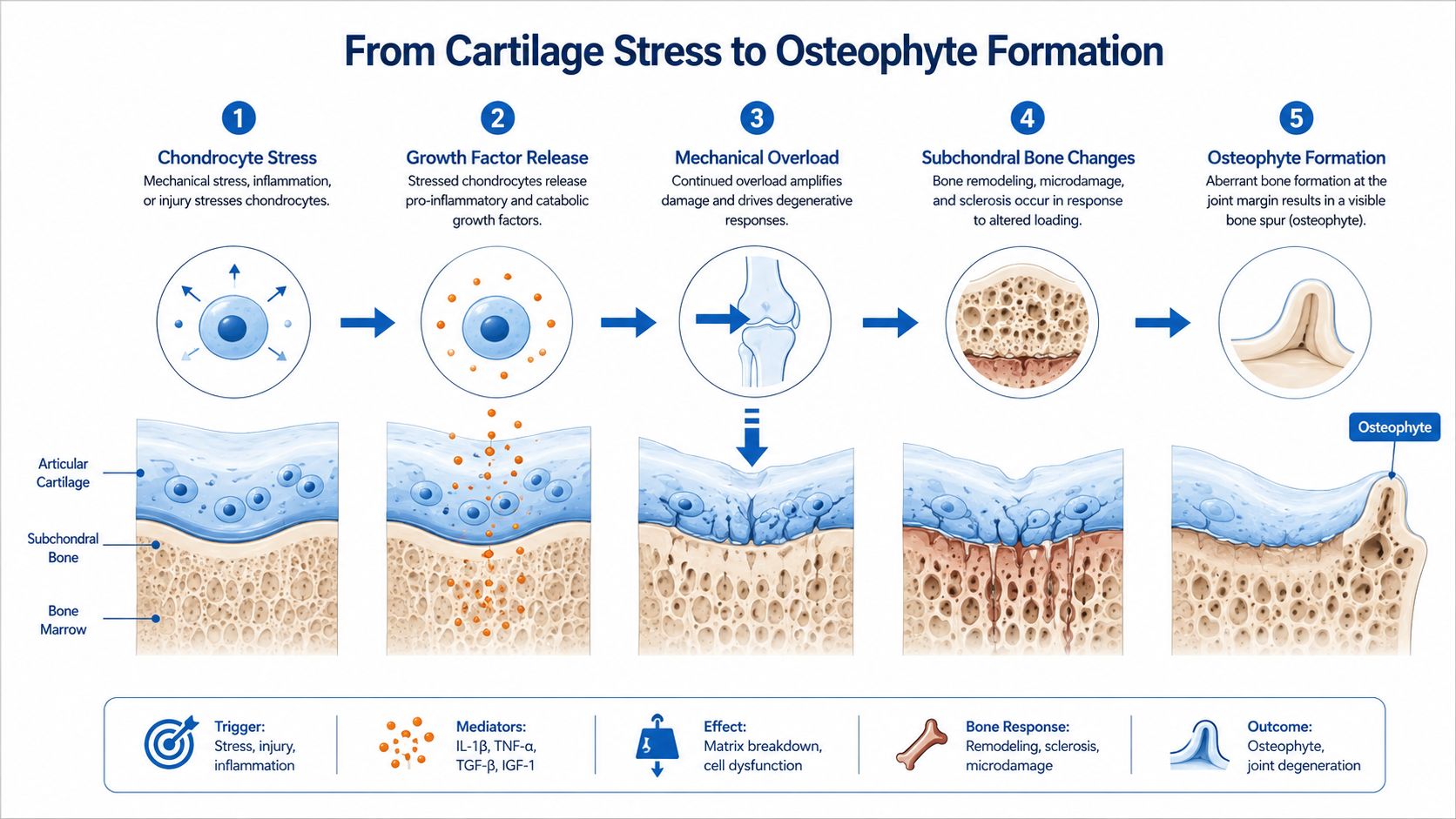
The result is a visible bony projection. Its location can provide useful information, but it still needs to be interpreted with the rest of the examination.
Two common mechanical pathways
Hip osteoarthritis commonly produces marginal osteophytes around the acetabular rim and the femoral head-neck junction. As cartilage wears and the joint surface changes, the body remodels the margins. Osteophytes may increase the apparent stability of the joint, but they can also reduce available space and restrict motion.
Femoroacetabular impingement, or FAI, follows a different pattern. A cam-type shape creates extra bone at the femoral head-neck junction, while a pincer-type shape reflects excessive coverage or abnormal contour at the acetabular rim. Repeated contact during flexion and rotation can stress the labrum and cartilage. In this setting, a bony prominence may be part of the impingement pattern rather than a consequence of diffuse cartilage wear.
Location therefore matters. Marginal osteophytes may support an osteoarthritis pattern, while an anterolateral bump may raise concern for cam-related mechanics. Neither finding identifies the pain source by itself. Clinicians still need to match the image with movement limitations, provocative tests, and the patient's daily activities. More background on joint degeneration is available through this osteoarthritis clinical resource.
Why a Bone Spur in the Hip Joint Causes Pain
A hip osteophyte becomes clinically relevant when it alters motion, irritates nearby tissue, or coincides with other structural damage. Its location and relationship to movement help determine whether it is part of the pain source.
During hip flexion and internal rotation, a spur can narrow the space between the femoral head-neck region and the acetabular rim. Contact in this area may create pinching groin pain during cycling, squatting, getting into a car, or sitting low. The same movement can place traction and shear stress on the labrum, the ring of tissue that deepens the socket. Catching, clicking, or pain with pivoting may follow. A related discussion is available in this resource on hip labral tears.
The joint capsule and nearby soft tissues may also become irritated. Inflammation can make the hip feel stiff and amplify pain even when the bony change is modest. Altered anatomy can occasionally affect nearby neurovascular structures or refer discomfort into the thigh. Groin, thigh, buttock, and lateral hip pain have different possible sources, so the examination must identify the pattern rather than rely on the x-ray alone.
Clinical rule: The x-ray identifies an anatomical finding. The examination determines whether that finding behaves like the patient's pain generator.
Research supports a measured interpretation. In a UK Biobank analysis, acetabular and femoral osteophytes were associated with increased odds of hip pain. The likelihood of pain exceeded 50% when total osteophyte area reached 150 mm², according to the study of osteophyte area and hip pain. The finding becomes more actionable when it matches restricted motion, joint-space loss, cartilage or labral injury, and a reproducible painful movement.
How Hip Osteophytes Produce Symptoms
| Mechanism | Structure Affected | Typical Symptom Pattern |
|---|---|---|
| Mechanical impingement | Femoral head-neck junction and acetabular rim | Groin pain with flexion, rotation, sitting, or squatting |
| Traction and shear | Acetabular labrum | Catching, clicking, sharp pain during pivoting |
| Local irritation | Joint capsule and adjacent soft tissues | Aching, stiffness, and pain with repeated movement |
| Referred or neurovascular irritation | Nearby nerves and soft tissues | Thigh or groin discomfort, sometimes with altered sensation |
A diagnostic intra-articular injection can test whether the pain originates inside the hip joint. If a carefully performed injection temporarily relieves familiar groin pain during a previously painful movement, that response supports an intra-articular source. It does not by itself establish that the spur requires removal or that surgery is appropriate. Targeted treatment is most reasonable when the mechanical pattern is consistent, symptoms persist, and examination and imaging point to the same structure. For broader discussion of joint symptoms, see this Lifeworks Integrative Health joint pain episode.
Imaging Options for Detecting Hip Osteophytes
Plain radiography is usually the starting point because it shows the overall bony architecture. A clinician can assess osteophytes, joint-space narrowing, subchondral sclerosis, alignment, and more advanced structural changes. The limitation is equally important: an x-ray shows bone and indirect evidence of cartilage loss, but it doesn't directly show the labrum and may miss early cartilage injury.
A study comparing radiographs with MRI found that radiographs had specificity ranging from 0.76 to 0.90 but variable sensitivity ranging from 0.44 to 0.78 for several forms of cartilage and bony pathology. Acetabular osteophytes had lower reported specificity, ranging from 0.42 to 0.58, in that comparison. These findings are detailed in the radiography and MRI comparison of hip joint pathology. In practical terms, a visible abnormality may be meaningful, but a normal or subtle x-ray doesn't reliably exclude clinically important disease.
Choosing the modality by the question
Ultrasound can assess the anterior capsule, fluid, tendons, and some superficial bony contours. It can also examine movement dynamically. Its usefulness depends heavily on the operator and the specific structure being evaluated.
MRI is valuable when symptoms suggest labral injury, cartilage defects, bone-marrow changes, or another soft-tissue pain generator. Standard MRI may answer the clinical question, while an arthrogram can be considered when detailed labral assessment is needed. The choice depends on the examination, prior imaging, and local protocol.
CT provides highly detailed information about bone shape. It can clarify cam or pincer morphology, define complex osteophytes, and support surgical planning. CT is less useful than MRI for directly evaluating the labrum and cartilage.
Imaging modalities for hip osteophytes
| Modality | What it shows best | Key limitation |
|---|---|---|
| X-ray | Osteophytes, joint-space narrowing, sclerosis, alignment | Misses many soft-tissue lesions and early cartilage changes |
| Ultrasound | Anterior soft tissues, fluid, dynamic movement, selected superficial spurs | Operator-dependent and limited for deep joint structures |
| MRI | Labrum, cartilage, marrow, capsule, and surrounding soft tissue | May not define complex bone morphology as clearly as CT |
| CT | Cam and pincer morphology, detailed osteophyte shape, surgical anatomy | Less informative for labral and cartilage tissue |
A persistent mechanical symptom pattern deserves reevaluation even when the first radiograph looks reassuring. The next test should answer a specific question rather than just provide more images.
Conservative Treatment for a Hip Bone Spur
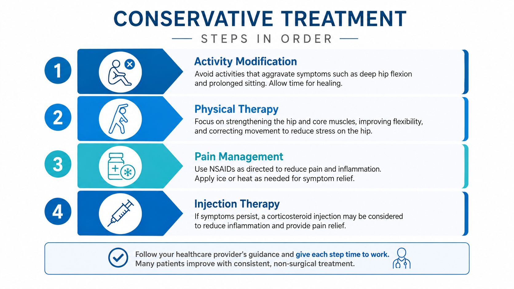
Conservative care targets the forces and inflammation around a hip osteophyte rather than removing the spur itself. The treatment plan should address the mechanical problem behind the image, such as osteoarthritis or femoroacetabular impingement, because the osteophyte is often a sign of that process rather than the entire diagnosis.
Start by changing the provoking load
Activity modification is often the first adjustment. Deep hip flexion, prolonged low sitting, repeated pivoting, and high-impact loading can aggravate impingement or an arthritic joint. A cyclist might raise the saddle, reduce resistance, shorten riding sessions, or temporarily choose flatter, lower-load routes. Low-impact aerobic activity helps preserve conditioning while reducing repeated force in the painful position.
Physical therapy should match the examination rather than follow a generic routine. A therapist can address hip flexor and adductor flexibility, gluteal and trunk strength, pelvic control, and movement retraining. The goal is better load sharing across the hip. Stretching that repeatedly forces painful impingement can irritate the joint, so exercises should stay within the patient's motion limits and be adjusted as symptoms change.
Add symptom control thoughtfully
Weight management can reduce the load transmitted through the hip, but it should be discussed without unrealistic promises. A clinician can consider an anti-inflammatory medication when appropriate. Kidney disease, gastrointestinal bleeding risk, cardiovascular disease, medication interactions, and age all affect that decision. Review the medical history and current medications before starting or continuing these drugs.
Supplements such as glucosamine, chondroitin, and omega-3 products do not remove an osteophyte or reliably rebuild damaged cartilage. Symptom responses differ, while structural benefit remains limited and product quality varies. Treat these products as optional adjuncts, not as a way to correct the underlying mechanical problem.
Practical rule: A treatment plan is working when walking, sleep, sitting, and daily movement improve, not merely when an image looks unchanged.
Conservative care also provides time to clarify the pain source. Improvement with movement retraining supports a mechanical contribution, while persistent pain despite appropriate rehabilitation calls for reassessing the diagnosis and treatment plan. Options include medication adjustment, an image-guided injection, advanced imaging, or specialist review.
Injection Options for Symptomatic Hip Spurs
An injection can serve two different purposes. It may reduce pain and inflammation, and it may help determine whether the hip joint itself is responsible for the symptoms. It won't remove the osteophyte.
Corticosteroid injections
An image-guided intra-articular corticosteroid injection places medication inside the hip joint. Ultrasound or fluoroscopy helps the clinician reach this deep structure accurately. The anti-inflammatory effect can provide short-term to intermediate relief, often lasting weeks to a few months, although individual responses vary.
The diagnostic value can be as important as the therapeutic value. If the injection temporarily reduces the patient's familiar groin pain during a provocative task, the result supports an intra-articular pain source. Relief doesn't prove that an osteophyte alone caused the pain, and it doesn't establish that surgery is required. More information about this procedure is available through corticosteroid injection care.
Hyaluronic acid and regenerative approaches
Hyaluronic acid aims to improve lubrication and alter the joint environment. Evidence in the hip is weaker and less consistent than in the knee, so it may be considered selectively, particularly when symptoms are mild to moderate and other measures haven't provided enough relief.
Platelet-rich plasma and bone-marrow aspirate concentrate are intended to influence the biological environment around damaged tissue rather than reshape bone. Current evidence is variable, and benefits may be modest. Cost, access, preparation methods, medical history, and the degree of osteoarthritis all affect whether these options make sense.
Hip injection options at a glance
| Injection type | Duration of benefit | Best candidate |
|---|---|---|
| Intra-articular corticosteroid | Often short-term to intermediate | Patient needing symptom relief or diagnostic clarification |
| Hyaluronic acid | Variable and less predictable in the hip | Selected patient with mild to moderate degenerative symptoms |
| PRP or bone-marrow concentrate | Variable, with inconsistent evidence | Carefully selected patient seeking a biologic option after informed discussion |
A responsible injection discussion includes alternatives, risks, expected duration, and what the result will change. Repeated procedures without a diagnostic or functional plan rarely address the underlying mechanical problem.
Surgical Options When Conservative Care Fails
Surgery is justified when hip pain remains functionally limiting despite structured conservative care and the imaging pattern matches the patient's symptoms. Activity modification, rehabilitation, medication management, and selected injections should have been used appropriately. A spur seen on an x-ray, without related loss of motion or mechanical pain, usually does not justify an operation. The spur is often a sign of the underlying mechanical problem, such as osteoarthritis or femoroacetabular impingement.
Hip arthroscopy and preservation procedures
Hip arthroscopy may help when the joint surface remains relatively preserved, but focal bone shape causes impingement, labral stress, or restricted movement. The surgeon may reshape the femoral head-neck junction, address the acetabular rim, repair or treat the labrum, and remove loose fragments when indicated.
The goal is to improve abnormal contact and protect remaining tissue. Arthroscopy cannot reliably reverse widespread cartilage loss. If diffuse osteoarthritis is driving the pain, removing one prominent spur may leave the main source of symptoms unchanged.
Open hip-preservation surgery may be appropriate when the deformity is substantial or cannot be corrected adequately through small portals. The decision depends on the anatomy, functional demands, potential to preserve the joint, and the surgeon's assessment.
Total hip replacement
Total hip replacement generally fits advanced osteoarthritis with broad cartilage loss, marked joint-space narrowing, subchondral changes, cysts, and extensive osteophyte formation. It replaces damaged joint surfaces rather than targeting one isolated bony projection. Stiffness, night pain, limited walking, and declining daily function often weigh heavily in the decision.
Age contributes to the discussion, but it does not decide the treatment by itself. Overall health, bone quality, activity goals, expectations, and the match between radiographic disease and pain also influence the recommendation. Arthroscopy is less invasive but still requires months of rehabilitation. Replacement is major surgery with staged recovery and activity precautions.
Surgical Options for a Symptomatic Hip Bone Spur
| Procedure | Best suited for | Primary goal | Recovery overview |
|---|---|---|---|
| Hip arthroscopy | Focal impingement with relatively preserved joint surfaces | Improve contact and address labral or loose-tissue problems | Structured rehabilitation over several months |
| Open hip preservation | Deformity not adequately treated arthroscopically | Correct substantial structural abnormality | More extensive recovery and rehabilitation |
| Total hip replacement | Advanced osteoarthritis with broad joint damage | Replace damaged joint surfaces | Staged recovery with activity precautions |
The treatment choice should follow the joint's actual condition. A surgeon can explain whether preserving the remaining joint or replacing damaged surfaces offers the better path, based on anatomy, cartilage status, symptoms, and day-to-day function.
Your Next Steps After a Hip Bone Spur Diagnosis
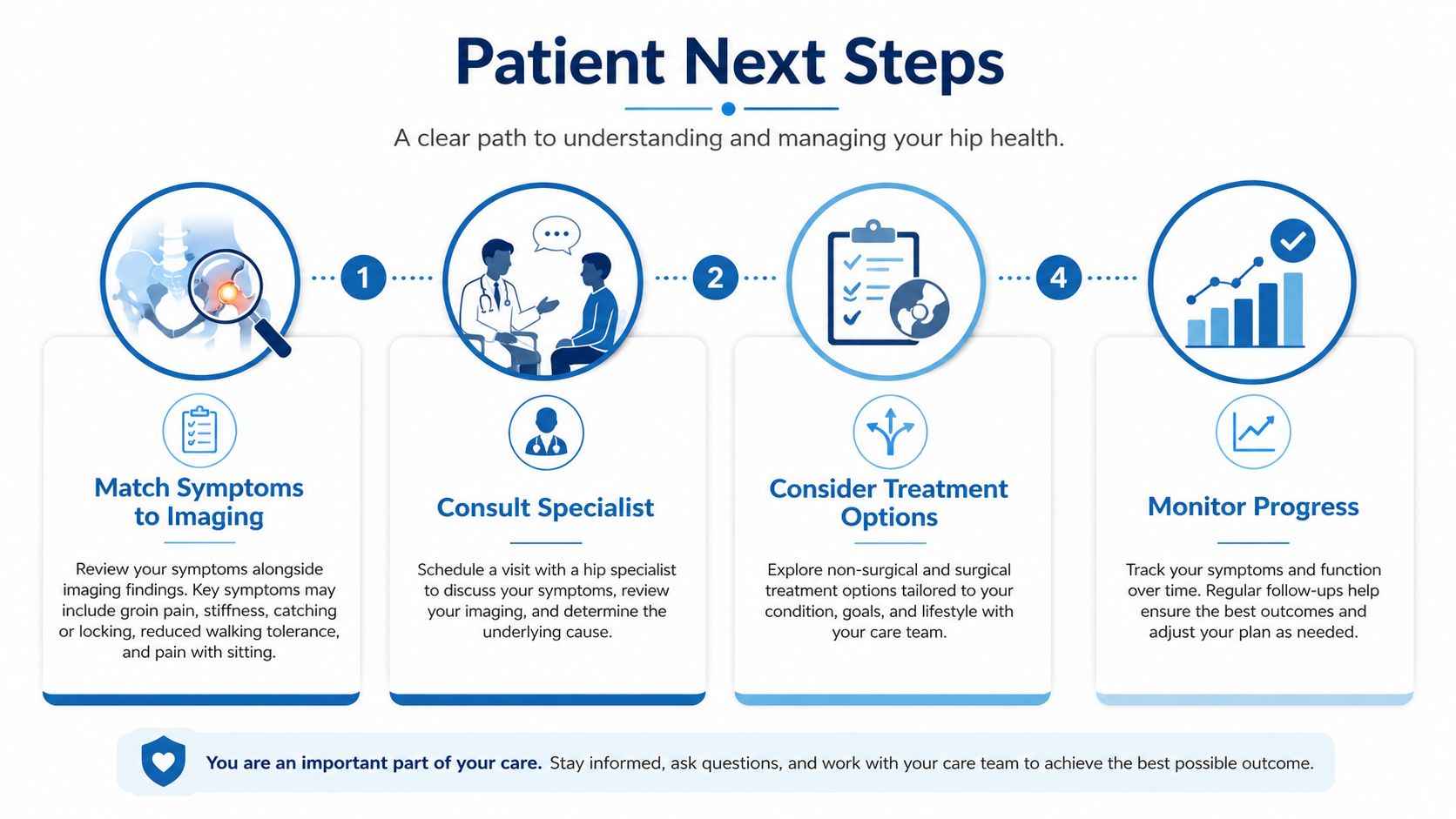
A hip osteophyte signals mechanical change and warrants comparison with your pain pattern before it receives diagnostic weight. Deep groin pain, stiffness, catching, reduced walking tolerance, or pain with sitting and pivoting may point to osteoarthritis or femoroacetabular impingement within the joint. Lateral hip pain can involve the greater trochanteric region, while buttock pain may come from tissues outside the joint.
Use a practical decision framework
Review the actual images. The clinician should examine joint-space narrowing, bone remodeling, alignment, and the spur's location. A radiology report helps, but the image, examination, and symptom pattern carry more meaning than the word “spur” alone.
Match symptoms to movement. Note whether flexion, rotation, stairs, cycling, prolonged sitting, or walking reproduces the pain. A consistent mechanical pattern helps identify the structure responsible.
Choose further evaluation when needed. If an x-ray appears relatively preserved but the examination raises concern for labral disease, impingement, fracture, or another deep-joint problem, MRI or CT may clarify the anatomy. The suspected problem should guide the imaging choice.
Use injections selectively. An image-guided intra-articular injection can sometimes reduce pain and provide diagnostic information. Temporary improvement supports the joint as a pain source, though it does not establish a need for surgery.
Escalate for persistent disability. Orthopedic hip evaluation is reasonable when pain continues to limit work, exercise, sleep, walking, or daily activities despite a structured conservative plan. Advanced joint damage may lead to a replacement discussion. Focal impingement with preserved cartilage may instead support hip-preservation treatment.
Know when symptoms need urgent attention
Sudden inability to bear weight, fever, or a hot, severely painful joint requires urgent medical assessment. These symptoms may indicate a problem that should not be automatically attributed to a chronic osteophyte.
For milder symptoms, a clinician may recommend avoiding provocative positions, maintaining comfortable low-impact activity, and beginning structured hip and trunk strengthening. Reassessment is appropriate if pain persists, worsens, disrupts sleep, or causes a limp. The aim is to identify the pain generator and restore safe, useful movement.
Interventional Pain Management evaluates hip pain through medical history, physical examination, imaging review, and image-guided non-operative treatments when appropriate. Patients with a bone spur in the hip joint can visit Interventional Pain Management to discuss whether osteoarthritis, impingement, labral disease, or another condition is driving symptoms and to plan the next step.