
Could chronic back pain be coming from a structure other than the lumbar spine, and could the success of treatment depend more on which nerves are targeted and how they're treated than on the procedure's name? That question matters for anyone considering sacroiliac joint radiofrequency ablation, because the procedure doesn't work identically for every patient or every technique.
The sacroiliac joint sits where the sacrum meets the pelvic bones. It transfers load between the spine and legs, so accurate diagnosis requires more than labeling every ache as disc pain. A careful evaluation can determine whether the posterior sacroiliac joint complex is the dominant pain source, whether diagnostic blocks are convincing, and whether the planned ablation matches the patient's anatomy and symptoms.
Table of Contents
- Is Your Chronic Back Pain Coming From the Sacroiliac Joint
- How Sacroiliac Joint Radiofrequency Ablation Works
- Clinical Outcomes and Evidence Behind the Procedure
- Realistic Recovery Timeline and What to Expect
- How Long Relief Lasts and What Patients Should Expect as Nerves Regenerate
- Comparing Options RFA Versus Other Pain Treatments
- Preparing for Your Consultation and Making the Decision
Is Your Chronic Back Pain Coming From the Sacroiliac Joint
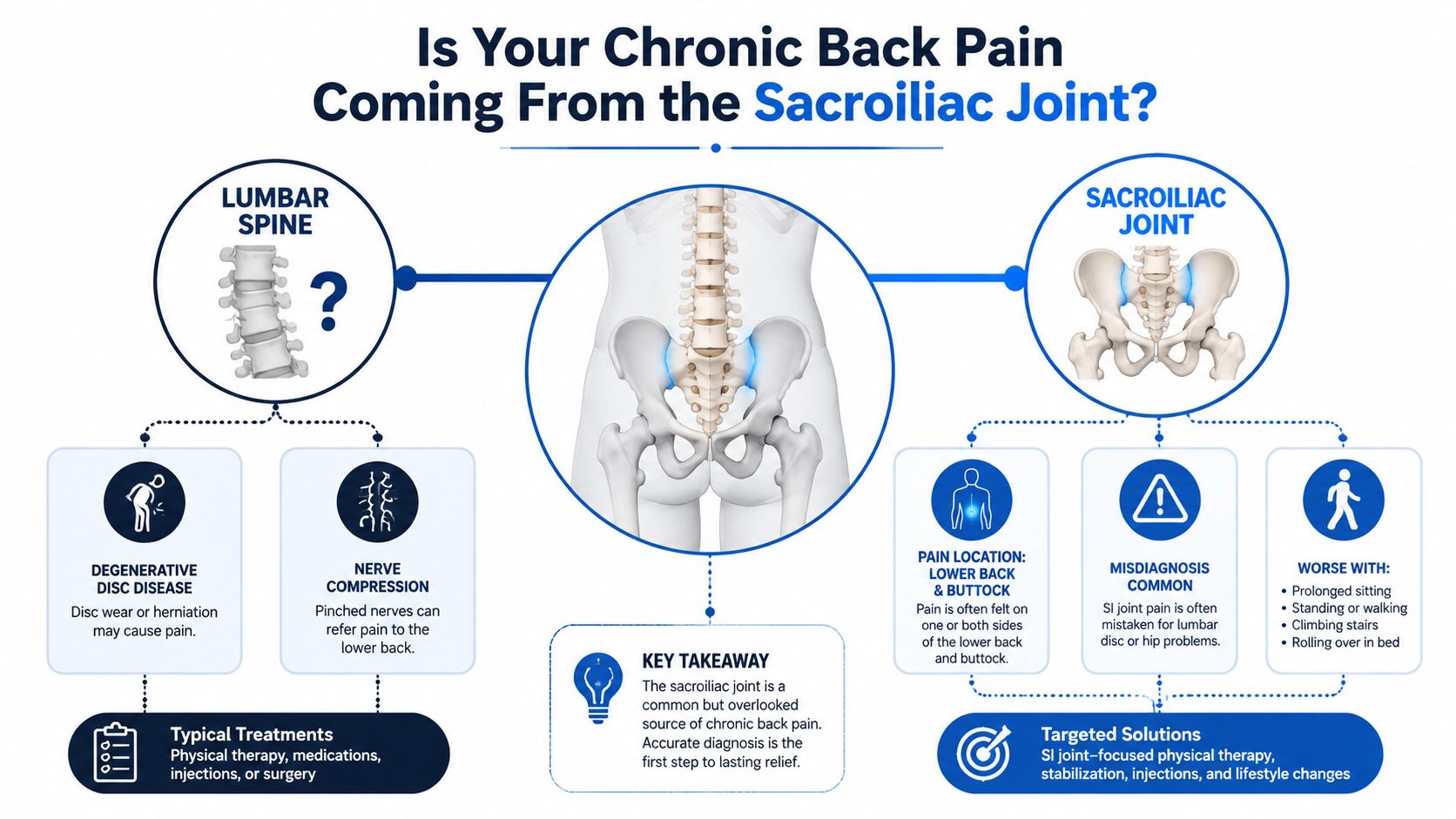
Diffuse low back pain is often blamed on lumbar discs, arthritis, or spinal stenosis. Those conditions can cause pain, but the sacroiliac joint is another important possibility. It functions like a heavy suspension bridge between the spine and pelvis, absorbing forces as the body bends, walks, climbs stairs, and changes direction. When the joint or its surrounding ligaments become irritated, the pain can feel deep, stubborn, and difficult to locate.
Sacroiliac joint pain commonly centers in the lower back or buttock. It may spread into the upper thigh, but it typically doesn't follow the classic pattern of true sciatica, which travels along an irritated nerve root into the leg. Symptoms can worsen with prolonged standing, turning in bed, climbing stairs, getting out of a car, or placing weight unevenly through the pelvis.
Why location alone isn't enough
Pain location provides a clue, not a diagnosis. A disc problem, hip disorder, muscle injury, sacroiliac joint disorder, or more than one condition can produce overlapping symptoms. A clinician will usually combine a medical history, physical examination, review of imaging when appropriate, and targeted diagnostic injections.
Patients often benefit from reviewing the differences between sacroiliac joint dysfunction and other causes of lower back pain before the consultation. The key issue is whether the joint is generating the patient's familiar pain, not whether the joint looks abnormal on an image.
The diagnostic turning point
A strong evaluation asks several practical questions:
- Pain pattern: Does discomfort remain concentrated near the posterior pelvis and buttock?
- Provocative movement: Do specific examination maneuvers reproduce the familiar pain?
- Alternative sources: Have lumbar, hip, and neurologic causes been assessed?
- Block response: Does a targeted anesthetic block temporarily reduce the usual pain and improve movement?
Clinical perspective: The right treatment starts with identifying the right pain generator. A procedure aimed at the wrong structure can be technically perfect and still disappoint.
This is why sacroiliac joint radiofrequency ablation shouldn't be selected merely because back pain has lasted a long time. It becomes more reasonable when the symptom pattern, examination, and diagnostic testing point toward the posterior sacroiliac joint complex and its pain-transmitting nerves.
How Sacroiliac Joint Radiofrequency Ablation Works
Radiofrequency ablation is a planned sequence, not a single injection. The process begins by confirming that the suspected nerves are carrying the patient's pain.
Step one, confirm the pain source
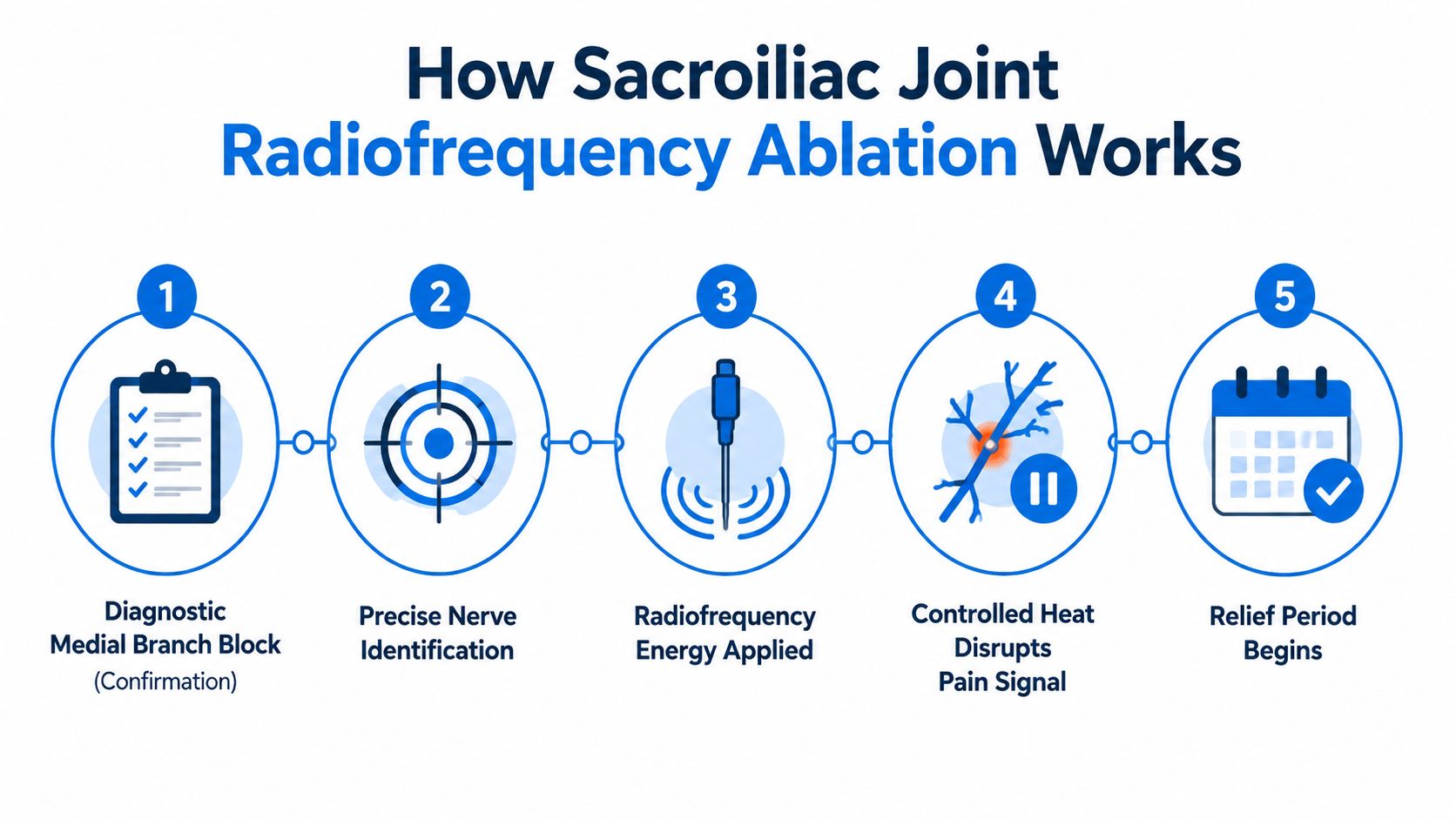
Before ablation, the treating physician generally performs diagnostic medial branch or lateral branch blocks, depending on the target pattern and practice protocol. A small amount of local anesthetic is placed near the nerves believed to transmit pain from the sacroiliac joint complex. Temporary improvement in the patient's usual pain, especially when it allows a previously painful activity, supports proceeding.
The block isn't intended to provide lasting treatment. Its purpose is diagnostic. If the result is unclear, absent, or inconsistent with the patient's typical symptoms, the physician may reconsider the diagnosis rather than move directly to ablation.
Step two, position the treatment needles
The procedure is usually performed as a minimally invasive, image-guided outpatient intervention. The patient lies in a position that allows the physician to visualize the sacral landmarks, the skin is cleaned, and local anesthetic numbs the entry points. Sedation may be offered for comfort, but the exact plan depends on health history, procedure complexity, and the center's protocol.
Fluoroscopy or another imaging method guides the needles toward the selected nerve targets. The physician may use brief electrical testing to confirm that the needle is close to the intended sensory nerve and away from structures that shouldn't be treated.
Step three, apply controlled heat
A radiofrequency probe passes through each correctly positioned needle. Electrical current creates controlled heat at the nerve, interrupting its ability to transmit pain signals. The objective isn't to remove the joint or repair every structural change. It's to reduce pain signaling from carefully selected nerves while preserving surrounding tissue.
The difference between techniques matters. Cooled radiofrequency ablation circulates fluid around the probe, changing the shape and size of the treated area. Conventional monopolar RFA creates a more focused lesion. Neither approach is automatically right for every patient. Placement must match the target nerves, anatomy, and treatment plan.
For a broader explanation of image-guided radiofrequency ablation, patients can review the procedure before discussing whether it fits their situation. Immediate soreness at the needle sites is common, and complete relief usually doesn't occur the moment the patient leaves the clinic.
Clinical Outcomes and Evidence Behind the Procedure
A 2022 systematic review of randomized controlled trials found that 15 of 16 trials reported positive results. The authors concluded that evidence supported RFA for chronic sacroiliac joint pain for up to one year after treatment. Pooled findings showed that about 50% of patients achieved more than 50% pain reduction at three months. Across studies, responder rates ranged from 32% to 89%, largely because patient selection, treatment technique, and outcome definitions differed (systematic review of sacroiliac joint RFA).
These findings describe groups, not guarantees. RFA works best when the sacroiliac joint is the main pain source, diagnostic testing supports the selected nerve targets, and the treatment method fits the patient's anatomy. If pain comes from another structure, or the treated nerves do not cover the dominant pain pathway, improvement may be limited.
Why cooled RFA receives attention
A multicenter randomized comparative-effectiveness study found that cooled RFA produced a mean Numeric Rating Scale pain score of 3.8±2.4 at three months, compared with 5.9±1.7 with standard medical management. Mean pain reduction was 2.5±2.5 points with cooled RFA versus 0.4±1.7 points, with p<0.0001 (ASPN best-practice guideline and comparative data).
| Treatment modality | Mean pain score at 3 months | Clinical outcome |
|---|---|---|
| Cooled RFA | 3.8±2.4 | Greater mean pain reduction than standard medical management |
| Standard medical management | 5.9±1.7 | Smaller mean pain reduction |
Cooled and conventional RFA should not be treated as interchangeable. Cooled RFA creates a larger treatment field, while conventional monopolar RFA creates a more focused lesion. Ask which nerves will be treated, why that target was chosen, and whether the published evidence matches the technique being offered.
Earlier evidence reported mean pain-relief estimates of 0.616 at three months and 0.499 at six months. A later systematic review and network meta-analysis included 9 randomized studies with 652 participants, while a 2024 meta-analysis rated the evidence as Level III with a fair recommendation for radiofrequency neurotomy (2023 systematic review and network meta-analysis).
The practical conclusion is measured optimism. RFA can help carefully selected patients, but results depend on the diagnosis, nerve targets, equipment, and technical execution.
Realistic Recovery Timeline and What to Expect
The first day often feels more procedural than therapeutic. A patient may leave with small bandages, localized tenderness, stiffness, or a bruised sensation near the treatment sites. Sedation can cause temporary grogginess, so transportation arrangements and the clinic's instructions matter.
The early period
During the first several days, most patients should protect the treated area from unnecessary strain. Light movement is usually preferable to remaining completely still, but heavy lifting, strenuous exercise, and repeated bending should wait until the treating clinician provides clearance.
The treated nerve doesn't stop transmitting pain in exactly the same way for everyone. Local anesthetic or sedation may briefly change symptoms, and post-procedure inflammation can temporarily make the area feel more sensitive. That early soreness doesn't necessarily mean the treatment failed.
Practical rule: Early tenderness measures the body's response to the needle and heat. It doesn't reliably predict the final pain-relief result.
The following weeks
As healing progresses, patients may notice that standing, walking, sleeping, or changing position becomes easier. The improvement may be gradual rather than dramatic. Physical therapy, gentle mobility work, and strengthening can help translate pain reduction into better function, but exercises should be adjusted to the patient's diagnosis and recovery status.
A return to desk work may be possible relatively soon for some patients, while physically demanding work may require more modification. There isn't one safe schedule for every person, because medication use, other spine or hip conditions, job demands, and the number of treated targets all matter.
New weakness, progressive numbness, loss of bladder or bowel control, fever, spreading redness, drainage, severe swelling, or pain that escalates sharply warrants prompt medical contact. The treating office should provide specific aftercare instructions and an emergency contact pathway before discharge.
How Long Relief Lasts and What Patients Should Expect as Nerves Regenerate
Sacroiliac joint radiofrequency ablation is not a permanent repair. It reduces pain signals from selected nerves, while the joint, ligaments, muscles, and movement patterns remain part of the clinical picture. The durability of relief therefore depends on more than the procedure alone.
Treated nerves can gradually regenerate. As signaling returns, pain may return as well, but the timing and intensity differ. Anatomy, ongoing mechanical stress, another pain generator, and the RFA technique can all influence the result. Patients should ask whether the clinician uses conventional or cooled RFA, which nerves will be targeted, and why that approach fits their anatomy. These methods create different treatment zones, so outcomes should not be assumed to be identical for every patient.
Why repeat treatment requires proof
Repeat ablation should follow a documented response, rather than an automatic schedule. The ASPN guideline recommends repeating SIJ RFA no more than every six months, and only when the prior procedure produced at least 50% pain relief plus functional improvement (ASPN best-practice guideline).
This criterion helps prevent repeating a procedure that did not provide meaningful benefit. Pain scores matter, but function offers another test: walking farther, sleeping more comfortably, completing work tasks, or relying less on rescue medication.
A durable plan includes reassessment, appropriate exercise, attention to lifting and movement habits, and evaluation of other conditions that may continue stressing the pelvis. Research may clarify how long relief lasts for selected patients, but counseling should remain grounded in the individual response and technique used.
Relief opens a window for restoring movement and function. Patients can use that window to address factors that keep pain active.
Comparing Options RFA Versus Other Pain Treatments
Which treatment makes sense depends first on the suspected pain generator, then on how clearly testing supports that conclusion. Physical therapy can improve hip mobility, trunk control, pelvic mechanics, strength, and tolerance for daily loads. Medication may reduce symptoms enough to make movement possible, but it does not selectively test or interrupt the nerves carrying sacroiliac joint pain.
A corticosteroid injection has a different role. It may calm local inflammation and offer diagnostic information, yet its relief can be temporary. RFA applies controlled heat to selected pain-transmitting nerves. That approach may fit patients whose diagnostic blocks produced meaningful, temporary relief in a pattern consistent with nerve-mediated sacroiliac joint pain.
How clinicians choose among options
| Option | Main purpose | Best fit when |
|---|---|---|
| Physical therapy | Improve movement, strength, and load tolerance | Movement impairment or deconditioning contributes to symptoms |
| Medication | Reduce pain enough to support daily activity | Symptoms need short-term or ongoing medical management |
| Injection | Deliver medication near a suspected painful structure | Inflammation or diagnostic clarification is relevant |
| RFA | Interrupt selected nerve pain signals | Diagnostic blocks support the targeted sacroiliac pain pathway |
Patient selection matters as much as the treatment label. A person with poorly localized pain may need further evaluation before RFA, while a person with consistent symptoms and supportive blocks may have a clearer target. The clinician should also consider hip disease, lumbar nerve compression, inflammatory conditions, fracture, infection, and structural instability, because these problems can require different care.
Technique can change the expected result. Conventional and cooled RFA create different treatment zones, so patients should ask which method is being proposed, which nerves will be targeted, and how imaging and anatomy guide probe placement. A nerve block may be used as a diagnostic or therapeutic step, but its meaning depends on the suspected source and the response afterward.
Patients reviewing nonsurgical care for another spinal condition may find these spinal stenosis non surgical approaches helpful, while remembering that spinal stenosis and sacroiliac joint pain require different diagnostic reasoning.
The more useful question is whether RFA addresses the confirmed source more directly than the alternatives being considered.
Preparing for Your Consultation and Making the Decision
Could the planned treatment be aimed at the right pain source, using a technique suited to your anatomy? A productive consultation should clarify why the sacroiliac joint is believed to be causing the pain, which nerves will be treated, and what improvement would count as success.
A patient with posterior SI complex dominant pain, clearly positive diagnostic blocks, and symptoms that continue despite appropriate conservative care may be a reasonable candidate. Further evaluation may be appropriate when pain location remains uncertain, hip disease has not been addressed, neurologic symptoms are progressing, or a block did not produce meaningful relief. The clinician should also consider lumbar nerve compression, inflammatory conditions, fracture, infection, and structural instability because each may require a different plan.
Questions worth bringing to the appointment
- Diagnosis: What findings show that the sacroiliac joint complex, rather than the lumbar spine or hip, is the dominant pain generator?
- Blocks: Which diagnostic nerves were tested, and how much pain relief and functional improvement followed?
- Technique: Will the procedure use cooled or conventional RFA, and why does that method fit the patient's anatomy?
- Targets: Which nerves will be treated, and how will imaging confirm safe probe placement?
- Expected result: What improvement in walking, sitting, sleeping, or work would indicate success?
- Risks: Which complications relate to the patient's medical history, medications, and previous procedures?
- Recovery: Which activities should be limited, and when might normal work or exercise resume?
- Long-term plan: What happens if relief is partial or temporary?
- Coverage: Does the insurer require specific documentation, diagnostic blocks, or authorization?
Technique deserves careful discussion. Cooled and conventional RFA create different treatment zones, and their probe-placement strategies may differ. Patients do not need to memorize the measurements. They should ask how the clinician chooses the target map, uses imaging, accounts for individual anatomy, and determines whether the selected method can reach the relevant nerves safely.
The procedure label alone cannot predict the result. Selection criteria and technical execution both matter.
Insurance approval may depend on the documented diagnosis, previous treatment, block results, medical necessity, and the payer's interpretation of the evidence. Some policies may still describe RFA as insufficiently supported or investigational despite favorable comparative findings. The office should verify requirements before scheduling. Ask about authorization, out-of-pocket responsibility, sedation, transportation, and follow-up.
The decision should also include rehabilitation and broader pain care. Depending on the confirmed pain source, an individualized plan may include exercise, medication review, behavioral support, orthopedic or spine evaluation, and other interventions. Interventional Pain Management provides diagnostic evaluation and image-guided pain procedures alongside orthopedic and spine services, so the plan can reflect the diagnosis rather than the procedure alone.
The most useful consultation ends with a shared decision based on diagnostic certainty, technical fit, realistic goals, and a plan for reassessment. Patients should ask why cooled or conventional RFA is being recommended, what evidence supports the target, and how success will be judged for their daily activities.
Interventional Pain Management evaluates suspected sacroiliac joint pain through medical history, examination, diagnostic review, and image-guided treatment options when appropriate. Patients in New Jersey and Staten Island can visit Interventional Pain Management to discuss whether sacroiliac joint radiofrequency ablation fits their diagnosis, goals, and long-term care plan.